Thrombosis of gallbladder vein presenting as acute peritonitis
2010-12-14ChunLinGeXiaoGuangLuoandYongFengLiu
Chun-Lin Ge, Xiao-Guang Luo and Yong-Feng Liu
Shenyang, China
Thrombosis of gallbladder vein presenting as acute peritonitis
Chun-Lin Ge, Xiao-Guang Luo and Yong-Feng Liu
Shenyang, China
(Hepatobiliary Pancreat Dis Int 2010; 9: 100-102)
thrombosis;gallbladder vein;deep vein thrombosis described. Even during laparotomic investigation it is hard to make a correct diagnosis. We here report a case of thrombosis in the gallbladder vein and attempt to analyze the causes of thrombosis at this unusual site. We conclude that tuberculosis is responsible for thrombosis in our case; this is not a well-known cause of deep vein thrombosis.
Introduction
Thrombosis of the gallbladder vein occurs rarely,usually with a combination of ascites, thickened gallbladder wall, and reversal of fl ow in the portal vein, suggesting vascular occlusive disease within the portal vein system.[1,2]However, very few clinical features of gallbladder vein thrombosis have been
Case report
On September 7, 2007, a 75-year-old man with a sudden continuous ache in the upper abdomen for 2 hours resorted to the emergency room. The patient had a history of chronic obstructive pulmonary disease, had been diagnosed with pulmonary tuberculosis one year ago, and had been under anti-tuberculosis therapy since then. He was a heavy smoker for over 20 years with more than 20 cigarettes a day.
The initial physical investigation identi fi ed panabdominal tenderness which was more severe at the right upper quadrant, muscle tension with rebound tenderness, bowel sounds at 1/min, and scattered dry rales in both lungs. No other meaningful signs were found. Laboratory investigations revealed a moderately increased white blood cell count (WBC), Doppler ultrasound of the abdomen revealed a 0.7 cm-thick gallbladder wall, and no gallstones or ascites was found. The patient was diagnosed with “peritonitis of the upper right abdomen, acute cholecystitis”,and anti-in fl ammatory treatment was started. Nine hours later the pain worsened, and a second physical investigation revealed a distended abdomen, more severe pan-abdominal tenderness, positive water sounds when rocking the abdomen, and bowel sounds had disappeared completely. The WBC count remained unchanged. However, the gallbladder wall continued thickening to 0.9 cm and a large amount of ascites appeared. Puncture into the left lower peritoneal cavity induced 20 ml of light yellow liquid with protein of 35 g/L, Revalta reaction (+), cell count of 16 000×106/L (neutrophils, 98%; lymphocytes, 2%). The X-ray of abdomen in the erect position did not reveal any subphrenic gas. We thus decided to make a laparotomic investigation. Before the operation, the coagulation test demonstrated prolonged activated partial thromboplastin time and prothrombin time. The patient had been awoken and oriented throughout.
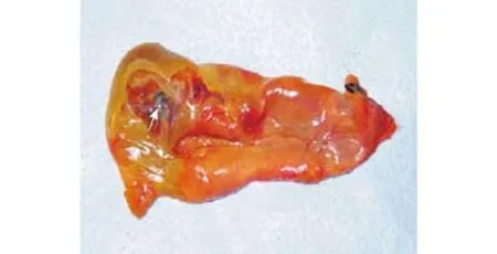
Fig. 1. General view of the excised gallbladder with venous thrombus on the wall. The whole gallbladder was excised and the serous membrane was cut open to expose the thrombus (arrow) in the veins below the serous membrane.

Fig. 2. Microscopic view of the gallbladder wall with venous thrombus (arrow) below the serous membrane.
During the operation, about 1000 ml of light yellow liquid was induced. The gallbladder size was normal,but the gallbladder wall and the upper peritoneum behind the duodenal bulb showed severe edema. In the front wall of the gallbladder there was a 4 cm2fanshaped region in green-brown color, but no dilation of the common bile duct was seen. The gallbladder was resected and cut open; examination showed thicker bottom. In the front wall, immediately below the serous membrane, there were several vessels running vertically,inside which a thrombus was identi fi ed (Fig. 1). The fi nal diagnosis was "thrombosis of gallbladder vein",which was con fi rmed by permanent section examination(Fig. 2). The patient then received anticoagulant therapy and was hospitalized for 8 days.
Discussion
Venous thrombosis of the gallbladder is very rare and we have not found a similar report. Usually the combination of ascites, thickening gallbladder wall and reversal of fl ow in the portal vein suggest vascular occlusive disease within the portal venous system.[1,2]But in our case, the patient only had ascites and a thickened gallbladder wall. Doppler ultrasound did not show any reversal of fl ow in the portal vein. Why a large amount of ascites appeared within 9 hours remains equivocal. Ascites is usually caused by liver disease but may also result from many other disorders, including heart failure, hepatic in fi ltration by tumor,[3]hepatic vein thrombosis,[4]veno-occlusive disease,[5,6]peritoneal carcinoma[7]or in fl ammation. Massive ascites caused by thrombosis of the renal or ovarian veins has also been reported.[5]In trauma, acute-onset ascites due to aggressive fl uid resuscitation and elevated intrathoracic pressure has also been reviewed.[8]However, in our case,why massive ascites appeared in the short term remains elusive. During the operation, we found severe edema of the gallbladder wall and the upper peritoneum behind the duodenal bulb, which seemed to be associated with the fan-shaped necrosis of the front wall of the gallbladder. It might be sensible to consider the severe edema as a source of ascites, which, combined with the occlusion of gallbladder veins, appears to account for the acute onset of ascites in the short term. The formation of ascites from edematous bowels has been demonstrated in animal studies.[9,10]No ascites recurred after excision of the gallbladder.
Deep vein thrombosis occurs frequently, but for venous thrombosis at unusual sites the hereditary thrombophilias[11]are usually considered and generally present as recurrent thrombosis, which is not the case for the present patient. Tuberculosis and antituberculosis drugs may cause venous thrombosis.[12-14]Sarode et al[15]found signi fi cant hyper-aggregation in 88% of patients with intestinal tuberculosis,and the hyper-coagulable state was related to the increased platelet aggregability. The present patient was diagnosed with pulmonary tuberculosis one year ago and then underwent anti-tuberculosis treatment,which is closely related to hypercoagulability.[13]Meantime, the comorbidity of chronic obstructive pulmonary disease is another important cause of the coagulable state.[16]Lastly, although smoking is not an independent risk factor for venous thrombosis, it is reported to exacerbate hemostasis in a predisposed state of hypercoagulation.[17-19]Collectively, in our case the hypercoagulability mechanisms of tuberculosis and chronic obstructive pulmonary disease added to anti-tuberculosis therapy, and smoking increased the blood coagulability and eventually led to the deep vein thrombosis.
Funding: None.
Ethical approval: Not needed.
Contributors: GCL and LXG wrote the fi rst draft of this commentary.All authors contributed to the intellectual context and approved the fi nal version. LYF is the guarantor.
Competing interest: No bene fi ts in any form have been received or will be received from a commercial party related directly or indirectly to the subject of this article.
1 Herbetko J, Grigg AP, Buckley AR, Phillips GL. Venoocclusive liver disease after bone marrow transplantation: fi ndings at duplex sonography. AJR Am J Roentgenol 1992;158:1001-1005.
2 Cerri GG, Habr-Gama A, Paranaguá-Vezozzo D, Machado MC,Pinotti HW, Magalh?es A. Doppler demonstration of cystic vein dilatation secondary to portal vein thrombosis. Surg Endosc 1991;5:92-93.
3 Schwendel A, Siems WG, Grune T, Holzhütter HG. Transitions of hepatic purine metabolism of Ehrlich ascites tumor bearing mice in different phases of tumor growth. Biochem Mol Biol Int 1994;34:457-463.
4 Aucejo F, Winans C, Henderson JM, Vogt D, Eghtesad B, Fung JJ, et al. Isolated right hepatic vein obstruction after piggyback liver transplantation. Liver Transpl 2006;12:808-812.
5 Yamasaki S, Kawabe Y, Nakamura H, Ishikawa H, Kawakami A,Migita K, et al. Long-standing and intractable ascites involved in renal vein thrombosis of a patient with systemic lupus erythematosus. Rheumatology (Oxford) 2000;39:445-447.
6 Tsujikawa T, Ihara T, Sasaki M, Inoue H, Fujiyama Y, Bamba T.Effectiveness of combined anticoagulant therapy for extending portal vein thrombosis in Crohn's disease. Report of a case. Dis Colon Rectum 1996;39:823-825.
7 Madanur MA, Battula N, Azam MO, Heaton N, Rela M. Chylous ascites after pancreatico-duodenectomy cholangiocarcinoma xenografts in nude mice. Hepatobiliary Pancreat Dis Int 2007;6:416-419.
8 Mayberry JC, Welker KJ, Goldman RK, Mullins RJ. Mechanism of acute ascites formation after trauma resuscitation. Arch Surg 2003;138:773-776.
9 Schilling JA, McCoord AB, Clausen SW, Troup SB, McKee FW.Experimental ascites; studies of electrolyte balance in dogs with partial and complete occlusion of the portal vein and of the vena cava above and below the liver. J Clin Invest 1952;31:702-710.
10 Volwiler W, Grindlay JH, Bollman JL. The relation of portal vein pressure to the formation of ascites; an experimental study. Gastroenterology 1950;14:40-55.
11 Pathare A, Al Kindi S, Al Haddabi H, Dennison D, Bayoumi R,Muralitharan S. Hereditary thrombophilia in ethnic Omani patients. Am J Hematol 2006;81:101-106.
12 Gogna A, Pradhan GR, Sinha RS, Gupta B. Tuberculosis presenting as deep vein thrombosis. Postgrad Med J 1999;75:104-105.
13 White NW. Venous thrombosis and rifampicin. Lancet 1989;2:434-435.
14 Robson SC, White NW, Aronson I, Woollgar R, Goodman H,Jacobs P. Acute-phase response and the hypercoagulable state in pulmonary tuberculosis. Br J Haematol 1996;93:943-949.
15 Sarode R, Bhasin D, Marwaha N, Roy P, Singh K, Panigrahi D,et al. Hyperaggregation of platelets in intestinal tuberculosis:role of platelets in chronic in fl ammation. Am J Hematol 1995;48:52-54.
16 Alessandri C, Basili S, Violi F, Ferroni P, Gazzaniga PP,Cordova C. Hypercoagulability state in patients with chronic obstructive pulmonary disease. Chronic Obstructive Bronchitis and Haemostasis Group. Thromb Haemost 1994;72:343-346.
17 Tansavatdi K, McClain B, Herrington DM. The effects of smoking on estradiol metabolism. Minerva Ginecol 2004;56:105-114.
18 Buhl R, Farmer SG. Future directions in the pharmacologic therapy of chronic obstructive pulmonary disease. Proc Am Thorac Soc 2005;2:83-93.
19 Ashitani J, Mukae H, Arimura Y, Matsukura S. Elevated plasma procoagulant and fi brinolytic markers in patients with chronic obstructive pulmonary disease. Intern Med 2002;41:181-185.
BACKGROUND: Thrombosis of the gallbladder vein occurs rarely, and few clinical features have been reported. We report here with a case of gallbladder vein thrombosis presenting as acute peritonitis in a 75-year-old man.
METHODS: The old man with sudden continuous abdominal pain resorted to the emergency room and treated for peritonitis associated with acute cholecystitis. The treatment failed to slow the progress of the disease, and massive ascites appeared with thickening of the gallbladder wall.Laparotomic investigation was conducted later.
RESULT: Pathologically, thrombosis of the gallbladder vein was diagnosed.
CONCLUSIONS: The thrombosis of the gallbladder vein is characterized by thickening of the gallbladder wall, ascites, and sudden continuous abdominal pain. The causes of deep vein thrombosis at this unusual site vary.
Author Af fi liations: Department of General Surgery, First Af fi liated Hospital, China Medical University, Shenyang 110001, China (Ge CL, Luo XG and Liu YF)
Yong-Feng Liu, Professor, Department of General Surgery, First Af fi liated Hospital, China Medical University, Shenyang 110001, China (Tel: 86-24-83283299; Email: chunlinge@yahoo.com.cn)
© 2010, Hepatobiliary Pancreat Dis Int. All rights reserved.
June 25, 2009
Accepted after revision December 2, 2009
杂志排行
Hepatobiliary & Pancreatic Diseases International的其它文章
- Synchronous gastric adenocarcinoma and pancreatic ductal adenocarcinoma
- Liver transplantation for acute intermittent porphyria: a viable treatment?
- Contrast-free air cholangiography-assisted unilateral plastic stenting in malignant hilar biliary obstruction
- Growth inhibition induced by short hairpin RNA to silence survivin gene in human pancreatic cancer cells
- Outpatient versus inpatient laparoscopic cholecystectomy: a single center clinical analysis
- Primary hepatic neuroendocrine carcinoma:clinical analysis of 11 cases