Letters to the Editor
2010-06-29
Letters to the Editor
The Editor welcomes submissions for possible publication in the Letters to the Editor section.
Letters commenting on an article published in the Journal or other interesting pieces will be considered if they are received within 6 weeks of the time the article was published. Authors of the article being commented on will be given an opportunity to offer a timely response to the letter. Authors of letters will be notified that the letter has been received. Unpublished letters cannot be returned.
Gallbladder and cystic duct agenesis diagnosed laparoscopically
To the Editor:
Anatomical variations of the biliary tree are not uncommon but isolated agenesis of the gallbladder is rare,[1]with a reported incidence of 0.013%-0.075%.[2]This variation remains undiagnosed since the patient is often asymptomatic. In spite of available diagnostic modalities, preoperative diagnosis is sometimes difficult.
A 28-year-old woman presented with symptoms of dyspepsia and upper abdominal discomfort. Routine biochemical and hematological investigations demonstrated no abnormalities. With a shrunken gallbladder with an acoustic shadow shown by sonography, the patient was hospitalized for laparoscopic cholecystectomy. Intraoperatively, no gallbladder was observed in its normal position. The whole supraduodenal part of the common bile duct was explored but no evidence of the gallbladder or cystic duct was found. Other abnormal locations, including the retrohepatic on the left side, falciform ligament, and lesser omentum were also excluded. The procedure was terminated at this stage. In the postoperative period, magnetic resonance cholangiopancreatography was carried out to confirm the diagnosis of gallbladder agenesis (Fig.). Associated congenital anomalies like annular pancreas and lumbar hernia were also re-examined intra- and post-operatively, but nothing was found. The patient was put on proton pump inhibitors and responded well.

Fig. Magnetic resonance cholangiopancreatography showing absence of the gallbladder.
In the 3rd week of fetal life, ventral thickening of the endoderm at the distal end of the foregut forms the liver. The caudal end of this endoderm proliferates to form the gallbladder and cystic duct. Gallbladder or cystic duct agenesis is a rare condition, about 450 cases have been reported.[1-8]It is difficult to diagnose gallbladder agenesis preoperatively. Annular pancreas and lumbar hernia are other anomalies associated with gallbladder agenesis.[9,10]In the present case, there was no other associated anomaly.
If the gallbladder is not found at its normal or abnormal locations on laparoscopy, open exploration of the extrahepatic biliary system can be avoided. Newer imaging modalities are relatively non-invasive and can provide good information on biliary tract anatomy preoperatively. All patients with gallbladder agenesis undergoing abdominal exploration pose a challenge to the surgeon to correctly identify the anomaly and avoid injury to the bile duct.[11]
Nikhil Gupta, Department of Surgery, Maulana Azad Medical College, New Delhi-02, India
(Tel: 91- 011-45526090;
Email: nikhil_ms26@yahoo.co.in); Sandeep K Gupta and Harmeet Singh Kapoor, Department of Surgery, Sri Balaji Action Medical Institute, Paschim Vihar, New Delhi-63, India
1 Singh B, Moodley J, Haffejee AA, Rajaruthnam P. Laparoscopic diagnosis of gallbladder agenesis. Surg Laparosc Endosc 1997;7: 129-132.
2 Singh B, Satyapal KS, Moodley J, Haffejee AA. Congenital absence of the gall bladder. Surg Radiol Anat 1999;21:221-224.
3 Gotohda N, Itano S, Horiki S, Endo A, Nakao A, Terada N, et al. Gallbladder agenesis with no other biliary tract abnormality: report of a case and review of the literature. J Hepatobiliary Pancreat Surg 2000;7:327-330.
4 Sarli L, Violi V, Gobbi S. Laparoscopic diagnosis of gallbladder agenesis. Surg Endosc 2000;14:373.
5 Galli A, della Porta M, Rebuffat C. Laparoscopic diagnosis of gallbladder agenesis: clinical case. Chir Ital 2003;55:295-298.
6 Kabiri H, Domingo OH, Tzarnas CD. Agenesis of the gallbladder. Curr Surg 2006;63:104-106.
7 Balakrishnan S, Singhal T, Grandy-Smith S, El-Hasani S. Agenesisof the gallbladder: lessons to learn. JSLS 2006;10:517-519.
8 Nardello O, Muggianu M, Cagetti M. Agenesis of the gallbladder. Ann Ital Chir 2007;78:45-47.
9 Heij HA, Niessen GJ. Annular pancreas associated with congenital absence of the gallbladder. J Pediatr Surg 1987;22:1033.
10 Nghiem DD, Read RC. Agenesis of the gallbladder and congenital lumbar hernia. South Med J 1979;72:1151-1153.
11 Planjar M, Knezevic A, Maksimovic V. Agenesis of the gallbladder -- case report and a short review of the literature. Acta Chir Iugosl 1979;26:79-83.
Melanoma of the gallbladder misdiagnosed as gallbladder cancer
To the Editor:
Metastatic melanoma of the gallbladder is extremely rare, and despite appropriate therapy its prognosis is poor with few patients surviving more than 2 years. We present a case of gallbladder involvement by malignant melanoma in a 63-year-old man initially diagnosed with gallbladder cancer. The man with a history of malignant melanoma resected from his back 18 years ago was admitted to our hospital because of severe right upper abdominal pain two days before admission.
Ultrasonography and magnetic resonance showed an 8 cm mass in the gallbladder lumen (Fig. 1). Chest X-ray, tumoral markers (CEA and CA19-9), and blood analyses showed nothing abnormal. Gallbladder cancer was suspected for immediate surgical treatment. A cholecystectomy with resection of liver segments Ⅳb and Ⅴ and a lymphadenectomy of the hepatic hilus were performed for the gallbladder cancer. The mass inside the gallbladder lumen measured 8 cm and had a single attachment point to the mucosal surface, darkly colored (Fig. 2). Histological evaluation showed a malignant melanoma with no signs of lamina propria or muscularis invasion. None of the 12 dissected lymph nodes showed signs of metastasis. The postoperative course was uneventful and the patient was discharged on postoperative day 5.
Melanoma can metastasize to any organ, and gastrointestinal metastases occur in 2%-4% of the patients.[1]Isolated metastasis to the gallbladder is rare and most patients have widespread disease by the time of diagnosis.[2]Over 50% of metastatic gallbladder lesions are from melanomas and this can be explained by the hematogenous spread of melanoma to the abdomen.[3]

Fig. 1. Magnetic resonance image showing a solid mass in the gallbladder lumen.
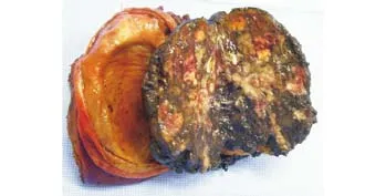
Fig. 2. Opened gallbladder and the 8-cm tumoral mass with dark coloration.
Most patients with melanoma metastatic to the gallbladder are asymptomatic, and the most common symptom is acute cholecystitis due to cystic duct obstruction.[3]Right upper quadrant or epigastric pain, weight loss, nausea, and vomiting may be present.[3]Radiographic examination is useful to determine gallbladder masses. Ultrasonography is the most useful tool for assessing gallbladder lesions and can reveal an intracholecystic nodule or mass. CT scan has an important role in the detection of metastatic disease.
In the present case, the mass inside the gallbladder was misdiagnosed as a gallbladder cancer, and was treated according to this hypothesis.
Since gallbladder melanoma is a rare entity, there is no optimal therapy for this kind of tumor. Its prognosis is very poor, with a survival period of 8.54 months for metastatic lesions.[4]Apparently, aggressive surgical therapy tends to prolong the survival rate, and to improve the quality of life of patients.[2]The treatment is dependent on the extension of the disease and the status of the patient. In the present case the tumor was limited to the gallbladder, and surgery, misindicated for a gallbladder cancer, aimed to prevent symptoms or tumor complications and to improve the prognosis. The patient remained well without symptoms, and 2 years latter showed a metastasis to the tonsil that was resected. Ten months later, the patient presented 2 pulmonary metastases and was referred to reavaliation by the clinical oncologist.
The role of adjuvant chemotherapy and immunotherapy is still controversial for metastatic gastrointestinal melanoma.[1]In patients with a gallbladder mass and a history of cutaneous melanoma, isolated metastatic melanoma has to be diagnosed despite its rarity. In spite of its poor prognosis, a surgical approach seems to be the best treatment.
Renata Potonyacz Colaneri and Bárbara dos Santos Nunes, University of São Paulo Medical School, São Paulo/SP, Brazil,
(Tel: +55-11-8126-6852;
Fax: +55-11-2942-8956;
Email: pcol_renata@yahoo.com.br);
Paulo Herman, Associate professor,
Department of Gastroenterology, University of São Paulo Medical School, São Paulo/SP, Brazil
References
1 Seelig MH, Schonleben K. Laparoscopic cholecystectomy for a metastasis of a malignant melanoma in the gallbladder. Z Gastroenterol 1997;35:673-675.
2 Velez AF, Penetrante RB, Spellman JE Jr, Orozco A, Karakousis CP. Malignant melanoma of the gallbladder: report of a case and review of the literature. Am Surg 1995;61:1095-1098.
3 Dong XD, DeMatos P, Prieto VG, Seigler HF. Melanoma of the gallbladder: a review of cases seen at Duke University Medical Center. Cancer 1999;85:32-39.
4 Guida M, Cramarossa A, Gentile A, Benvestito S, De Fazio M, Sanbiasi D, et al. Metastatic malignant melanoma of the gallbladder: a case report and review of the literature. Melanoma Res 2002;12:619-625.
杂志排行

Hepatobiliary & Pancreatic Diseases International的其它文章
- Thrombosis of gallbladder vein presenting as acute peritonitis
- Synchronous gastric adenocarcinoma and pancreatic ductal adenocarcinoma
- Liver transplantation for acute intermittent porphyria: a viable treatment?
- Contrast-free air cholangiography-assisted unilateral plastic stenting in malignant hilar biliary obstruction
- Growth inhibition induced by short hairpin RNA to silence survivin gene in human pancreatic cancer cells
- Outpatient versus inpatient laparoscopic cholecystectomy: a single center clinical analysis