Seeing beneath the surface: Harnessing point-of-care ultrasound for internal jugular vein evaluation
2024-05-08VichayutChayapinunAbhilashKoratalaTaweevatAssavapokee
Vichayut Chayapinun,Abhilash Koratala,Taweevat Assavapokee
Abstract Point-of-care ultrasound (POCUS) of the internal jugular vein (IJV) offers a noninvasive means of estimating right atrial pressure (RAP),especially in cases where the inferior vena cava is inaccessible or unreliable due to conditions such as liver disease or abdominal surgery.While many clinicians are familiar with visually assessing jugular venous pressure through the internal jugular vein,this method lacks sensitivity.The utilization of POCUS significantly enhances the visualization of the vein,leading to a more accurate identification.It has been demonstrated that combining IJV POCUS with physical examination enhances the specificity of RAP estimation.This review aims to provide a comprehensive summary of the various sonographic techniques available for estimating RAP from the internal jugular vein,drawing upon existing data.
Key Words: Point-of-care ultrasound;Bedside ultrasound;Internal jugular vein;Right atrial pressure;Central venous pressure
INTRODUCTION
An accurate assessment of hemodynamic status at the bedside is of utmost importance in guiding proper management through altering blood volume and cardiac output,especially in critically ill patients.Central venous pressure (CVP) is a physiologic parameter that is used to assess cardiac preload within this evaluation.CVP is identical to right atrial pressure (RAP) in the absence of vena caval obstruction,and these terms are often used interchangeably.Assessing CVP clinically by performing a neck veins examination can be challenging at times and yield low sensitivity,especially in the elderly,obese,and short-necked patients[1-4].The gold standard for measuring CVP isviainserting a central venous catheter (CVC) into the superior vena cava,however,this method is invasive,time-consuming,and can cause serious complications[4-6].With the help of point-of-care ultrasound (POCUS),RAP can be non-invasively measured through various methods including using compression ultrasound on the forearm vein[7],evaluation of the inferior vena cava(IVC),and evaluation of the internal jugular vein (IJV).The most common method is the evaluation of IVC diameter and collapsibility[8,9] (Figure 1).However,the association between IVC parameters and RAP is moderate and lacks reliability in mechanically ventilated individuals or those with abdominal compartment syndrome[10].Moreover,imaging the IVC can be challenging in situations like morbidly obese individuals,those unable to recline,patients with cirrhosis,ascites,or those who have undergone recent abdominal surgery[11].Owing to its location and convenient accessibility,IJV POCUS presents a viable alternative in such instances.Unlike visual inspection,where identifying the vein can be difficult in many cases,POCUS makes the IJV easily visible,saving time for physicians and improving the accuracy of the examination[12,13].
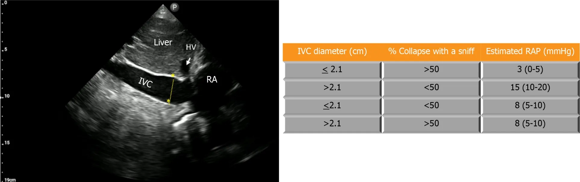
Figure 1 Estimation of right atrial pressure by inferior vena cava ultrasound in spontaneously breathing patients,based on current guidelines[8]. IVC: Inferior vena cava;RA: Right atrium;HV: Hepatic vein;RAP: Rright atrial pressure.
SONOGRAPHIC METHODS OF IJV ASSESSMENT
Multiple techniques have been described to assess CVP by IJV ultrasound.Based on available data,these methods encompass measuring the vertical height of the blood column in the IJV[13-16],utilizing the IJV height-to-width ratio(aspect ratio)[17,18],IJV collapsibility index (IJV-CI)[13,17,19-22],IJV distensibility index[23],measuring the maximal anteroposterior diameter of the IJV (AP-IJV Dmax)[18,24],calculating the difference in the percentage of the IJV crosssectional area (IJV-CSA) or diameter before and after the Valsalva maneuver[25,26],and combining IJV vertical height with right atrial depth measured with echocardiography[27,28].
The first technique,pioneered by Lipton[14],uses POCUS to visualize the height of the blood column in the IJV above the sternal angle of Louis where a venous collapse is seen (the ‘wine bottle’ or ‘paint brush’ appearance),thereby being able to measure the height of the IJV more accurately than compared to only performing neck veins examination(Figure 2).The height is then added to the hypothesized right atrial depth below the sternum of 5 cm to calculate the RAP,abiding by the doctrine of Sir Thomas Lewis,who first proposed the use of neck veins examination in 1930.This method’s accuracy is questionable,owing to the presumed right atrial depth of 5 cm,which is proven to be inaccurate[27,29].In a study by Deolet al[30] ultrasound-assisted assessment of the column height underestimated CVP by 4.7 cm H2O,whereas visual inspection underestimated it by 6.1 cm H2O.Moreover,this approach can be time consuming as it requires two tools to assess the height of the column from the sternal angle (e.g.,two rulers,a pen and a ruler,or a tongue depressor and a ruler or tape,commonly employed by physicians at the bedside),further compromising measurement accuracy[31].
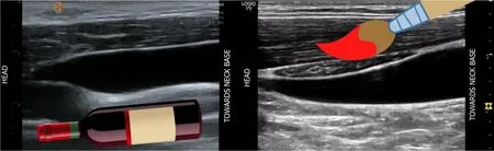
Figure 2 Internal jugular vein collapse point compared to a wine bottle and paint brush. Figures adapted from NephroPOCUS.com with permission.
The second technique uses the height-to-width ratio (aspect ratio) to estimate the CVP as a binary variable (e.g.,10 mmHg),without precisely quantifying it.The transducer is positioned at the level of the cricoid cartilage in the transverse plane,then the height (anteroposterior diameter) and the width (transverse diameter) are measured to obtain the aspect ratio.A ratio of <0.75 has shown to correlate with RAP of <10 mmHg with a sensitivity and specificity of 62% and 67%respectively in one study[17].This method yields mixed results,and its utility is currently not well-demonstrated[18].
The third technique uses the collapsibility index of IJV (IJV-CI) to estimate RAP.The IJV-CI is calculated by its respiratory variation,(IJVmax– IJVmin)/IJVmax expressed in percentage (Figure 3).The actual RAP cannot be quantified,but studies[13,21,22] have shown that an IJV-CI of approximately ≤ 30% signifies an elevated CVP of ≥ 8 mmHg,demonstrating good sensitivity and specificity.Worthy of particular mention,in a study by Leal-villarrealet al[21] in patients with cirrhosis,IJV-CI (anteroposterior diameter 2 cm above the sternoclavicular joint) ≤ 24.8% was better at predicting a CVP ≥ 8 mmHg with 100% sensitivity and 97.1% specificity outperforming IVC POCUS.In this cohort,IVC POCUS was unattainable in 18% of the cases correlating with our real-life experience in cirrhotic patients.
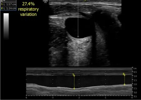
Figure 3 Anteroposterior diameter of the internal jugular vein. M-mode tracing depicts respiratory variation in the diameter.
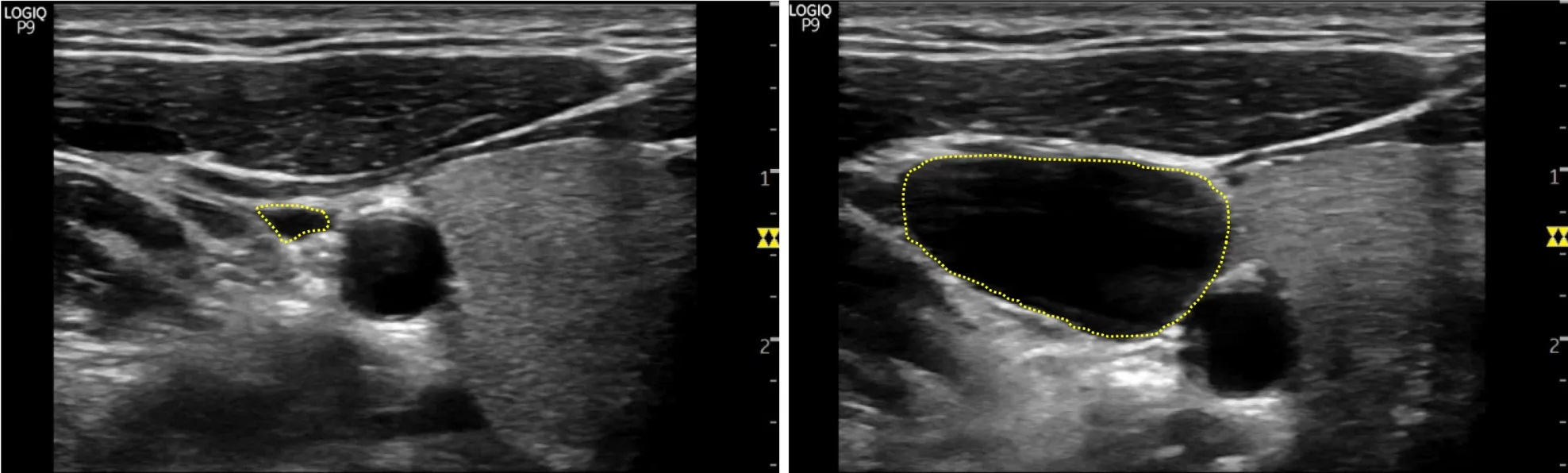
Figure 4 Increase in the size of internal jugular vein with Valsalva maneuver by several folds in a spontaneously breathing person with normal right atrial pressure.
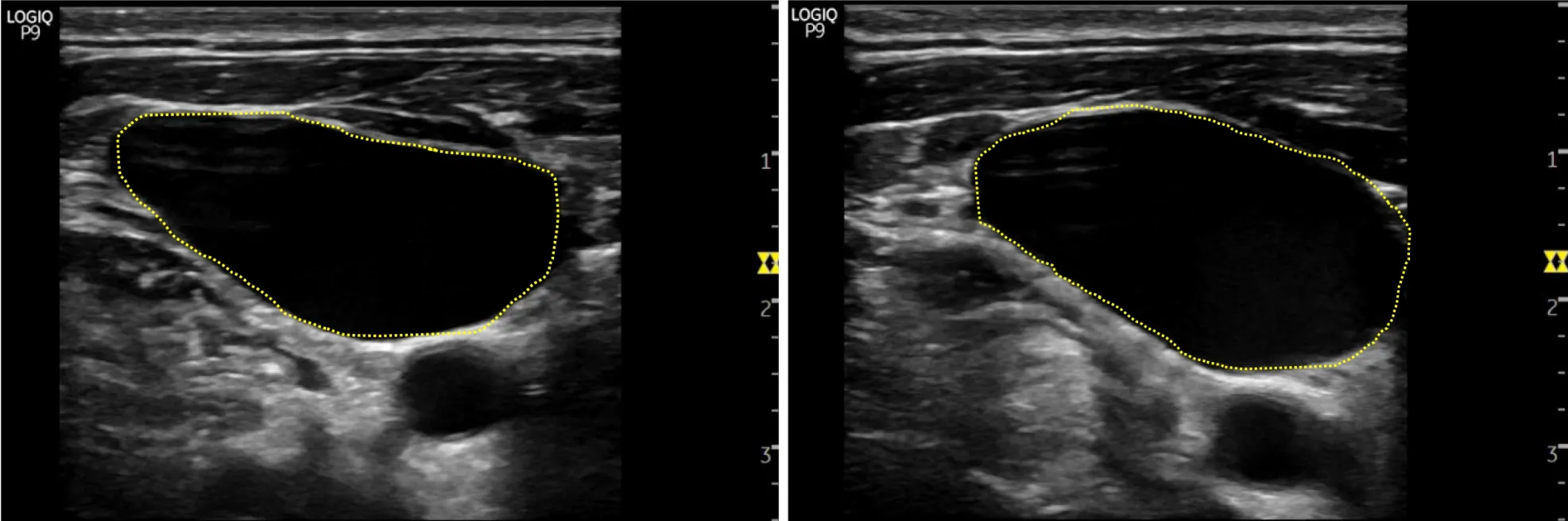
Figure 5 Minimal increase in the size of internal jugular vein with Valsalva maneuver in a spontaneously breathing heart failure patient with elevated right atrial pressure.
The fourth technique uses the IJV distensibility index to predict the fluid responsiveness in patients undergoing mechanical ventilation.IJV is imaged at the cricoid cartilage level in the transverse plane and the distensibility index is calculated by (IJVmax– IJVmin)/IJVmin,expressed in percentage.In one study,an IJV distensibility index of >18%before volume challenge (7 mL/kg crystalloid) had an 80% sensitivity and 85% specificity to predict fluid responsiveness[23].While this technique does not quantify the RAP,it could be a valuable adjunct to other hemodynamic parameters in assessing fluid status in mechanically ventilated patients.
The fifth technique uses the IJV maximal anteroposterior diameter (AP-IJV Dmax) to predict the RAP whether it is low(<8 mmHg) or high.Similar to the second technique of acquiring the aspect ratio,the transducer is positioned transversely 2 cm above the clavicle,at end-expiration and the diameter is collected to correlate with RAP.It has been shown that AP-IJV Dmax has the best correlation with RAP,the best validity in predicting its values,and a very good inter-rater reliability[24] along with its accuracy in predicting low RAP of <8 mmHg[18].
The sixth technique uses the difference in the percentage of the IJV cross-sectional area (IJV-CSA) before and after the Valsalva maneuver to assess for RAP.The Valsalva maneuver increases the IJV-CSA by about 20%-30% in patients with normal RAP,and it is assumed that in volume-overloaded patients with a decrease in venous compliance,the increase in IJV-CSA will be blunted.This technique measures the patient’s IJV-CSA at the vertical column height where a venous collapse is seen and re-measures that same parameter while the patient performs the Valsalva maneuver,then calculates the difference in the percentage of the IJV-CSA (Figures 4 and 5).One study[25] found that a <17% increase in IJV-CSA with Valsalva predicted elevated RAP (≥ 12 mmHg) with 90% sensitivity,74% specificity,and a 94% negative predictive value.Alternatively,the ratio between maximum IJV diameter during Valsalva maneuver and diameter at rest (JVD ratio)<4 has also shown to predict elevated RAP[26].
Another recently described technique uses the IJV vertical height combined with right atrial depth measured with echocardiography to quantify the RAP.While similar to the first technique in principle,this approach enhances accuracy by directly measuring the right atrial depth instead of relying on the assumed 5 cm.It involves marking the highest point of the venous collapse (wine bottle sign) and then performing a focused cardiac ultrasound to visualize the heart in long axis (parasternal long axis view).Right atrial depth is measured from the surface to the location of the non-coronary cusp of the aortic valve attachment to the posterior left ventricular outflow tract (Figure 6).The two parameters,the column height,and the right atrial depth,are then added together for an estimation of the RAP in cm H2O,later converted to mmHg.This method predicted actual RAP within 3 mmHg 74% of the time when compared to cardiac catheterizationderived value[27].
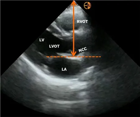
Figure 6 Measurement of right atrial depth using parasternal long axis view on focused cardiac ultrasound. RVOT: Right ventricular outflow tract;LV: Left ventricle;LVOT: LV outflow tract;LA: Left atrium;NCC: Non-coronary cusp of aortic valve.
In an alternative technique described by Xinget al[28],the initial step of measuring the height of collapse point is the same but the right atrial depth is calculated using an adjusted apical 4-chamber view with the right atrium in the center of the view,where distance between the transducer contact point and the center of the right atrium is recorded.A pencil and a ruler are then used in relation to the transducer to derive the surface projection of the center of the right atrium.The RAP is then estimated by adding the IJV column height to the right atrial depth.This technique demonstrates a substantial correlation and high accuracy,with a mean difference of 0.22 mmHg in RAP compared to CVC.Nevertheless,it lacks practicality in a clinical setting where swift assessment is essential,as it involves multiple operators and the use of various measuring instruments.
With respect to estimation of RAP using IJV POCUS in mechanically ventilated patients,there is limited data.One noteworthy study is by Hilbertet al[32] in a cohort of 47 patients,where they evaluated the ratio between IJV diameter in the 30° and 0° head position (30/0 ratio).A 30/0 ratio of <0.45 indicated a low CVP,whereas a cutoff of >0.65 predicted a CVP ≥ 10 mmHg with reasonable accuracy.
CONCLUSION
In essence,IJV POCUS serves as a quick,non-invasive bedside tool for assessing RAP.However,the diversity in techniques requires cautious interpretation,avoiding overestimation of accuracy.Interpretation of IJV POCUS in the appropriate clinical context is crucial.Additionally,CVP is not a surrogate for volume status but just one component in bedside hemodynamic assessment,to be considered alongside other variables.For instance,Figure 7 presents an algorithm for hemodynamic assessment in cirrhotic patients with acute kidney injury,utilizing IJV for fluid tolerance assessment[33].Future research should compare various techniques in different clinical settings to establish a standardized,practical method for routine use.
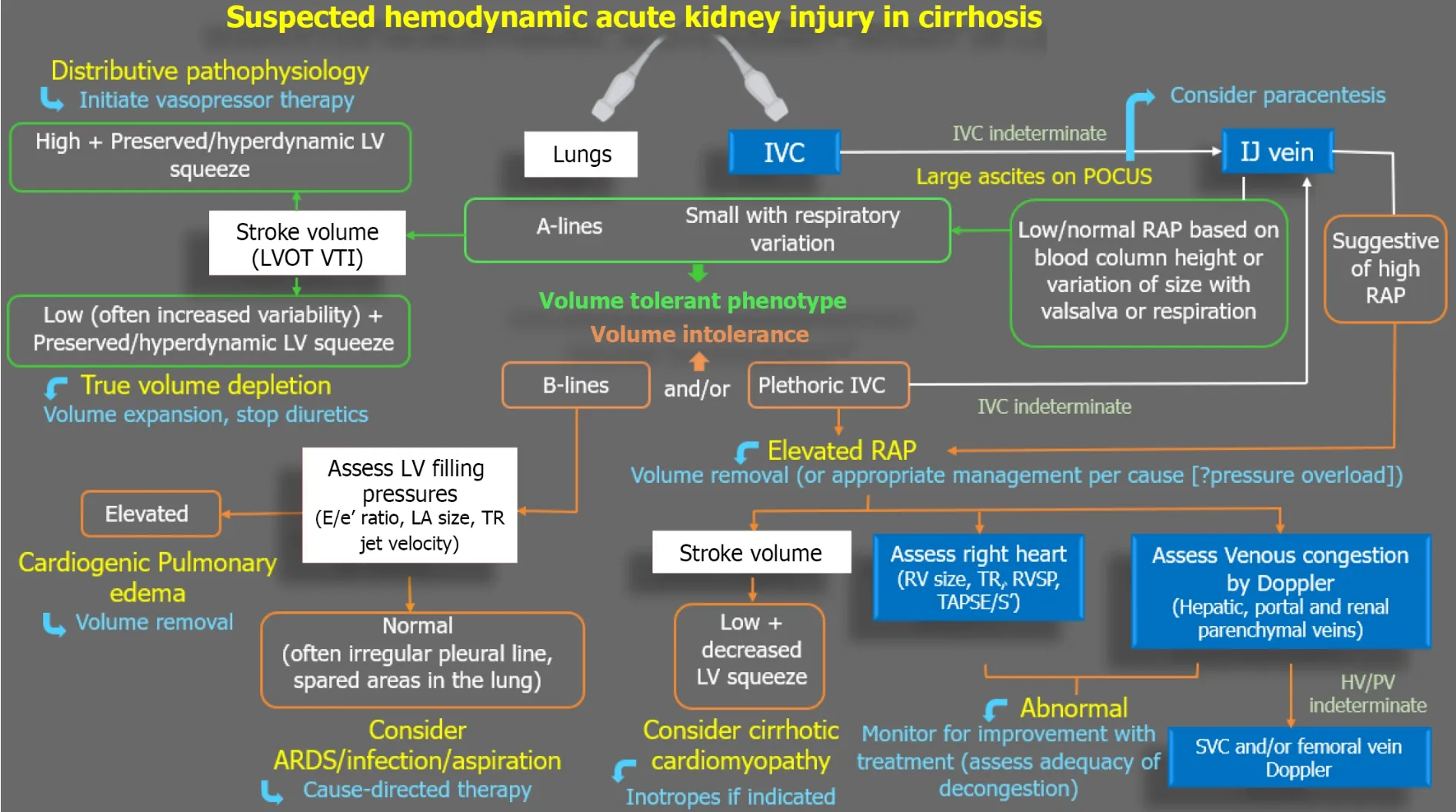
Figure 7 Diagnostic algorithm in a case of cirrhosis and suspected hemodynamic acute kidney injury. Incorrect angle of insonation is a frequent source of error when assessing LVOT VTI (surrogate for stroke volume) and other Doppler measurements listed.Adapted from Ref.33 with kind permission of the publisher (corresponding author’s prior open access publication).Blue boxes: Right heart;Red boxes: Left heart-related sonographic parameters;Green outlines:Volume tolerance phenotype;Orange outlines: Volume intolerance.POCUS: Point-of-care ultrasonography;VTI: Velocity time integral;E/e′: Ratio of the early diastolic waves of the mitral inflow Doppler and mitral annular tissue Doppler;LA: Left atrium;RV: Right ventricle;RVSP: Right ventricular systolic pressure;TAPSE:Tricuspid annular plane systolic excursion;S′: Tricuspid annular systolic velocity;SVC: Superior vena cava;ARDS: Acute respiratory distress syndrome;HV: Hepatic vein;PV: Portal vein.
FOOTNOTES
Author contributions:Chayapinun V drafted the initial version of the manuscript;Assavapokee T and Koratala A have designed the manuscript;Assavapokee T and Koratala A have revised the manuscript for critical intellectual content.
Conflict-of-interest statement:All authors have no conflicts of interest to declare.
Open-Access:This article is an open-access article that was selected by an in-house editor and fully peer-reviewed by external reviewers.It is distributed in accordance with the Creative Commons Attribution NonCommercial (CC BY-NC 4.0) license,which permits others to distribute,remix,adapt,build upon this work non-commercially,and license their derivative works on different terms,provided the original work is properly cited and the use is non-commercial.See: https://creativecommons.org/Licenses/by-nc/4.0/
Country/Territory of origin:United States
ORCID number:Abhilash Koratala 0000-0001-5801-3574.
S-Editor:Gong ZM
L-Editor:A
P-Editor:Zhao S
杂志排行

World Journal of Cardiology的其它文章
- Facing ethical concerns in the age of precise gene therapy: Outlook on inherited arrhythmias
- Spontaneous coronary artery rupture after lung cancer surgery: A case report and review of literature
- Development and validation of a nomogram model for predicting the risk of pre-hospital delay in patients with acute myocardial infarction
- Risk of permanent pacemaker implantation following transcatheter aortic valve replacement: Which factors are most relevant?
- Cardiac rehabilitation after cardiac surgery: An important underutilized treatment strategy
- Inflammation as a cause of acute myocardial infarction in patients with myeloproliferative neoplasm