Effects of simulated zero gravity on adhesion,cell structure,proliferation,and growth behavior,in glioblastoma multiforme
2024-01-08SaifaldeenAltaieandAmeraAlrawi
Saifaldeen Altaie and Amera Alrawi
AFFILIATIONS
1 School of Precision Instrument and Optoelectronic Engineering,Tianjin University,Tianjin,China
2College of Medicine,Ninevah University,Mosul,Iraq
3College of Science,University of Mosul,Mosul,Iraq
ABSTRACT All life on Earth has evolved under the influenc of continuous gravity,and methods have been developed to balance this influenc with the biological evolution of organisms at the cellular and system levels.However,when exposed to zero gravity in space,the balance between cell structure and external forces is destroyed,resulting in changes at the cellular level(e.g.,cell morphology,adhesion,viability,apoptosis,etc.),and understanding the molecular mechanism of cell response to zero gravity will help to cope with diseases that rely on mechanical response.Therefore,biological research in space and zero gravity is a unique step in developing the best anti-cancer treatments,which is a great challenge to humanity.In this study,multicellular glioma cancer cells from a brain tumor in a 72-year-old Iraqi patient were subjected to simulated zero gravity for 24 h,and the results showed that most of the cells lost their adhesion,which is considered to be the firs step toward cell apoptosis.In addition to the formation of multicellular spheroids,the results also showed that the inhibition rate for cell death was 32%in comparison to the control cells.Moreover,the cells showed a clear change in their cellular morphology and growth behavior.These results give new hope for fightin cancer distinctively,and such a treatment method has no side effects in comparison to traditional chemical and radiological ones.
KEYWORDS 3D cell culture,Space biology,Glioblastoma,Simulated microgravity,Cancer biology
I.INTRODUCTION
Gravity is an external force that affects all life and biological evolution on Earth.However,the development of space science has led to new discoveries about how zero gravity(ZG)affects life and biological processes,and it has been shown that ZG can alter important cellular properties including external morphology,growth,and migration.1In particular,how gravity affects cancer cells is now receiving considerable attention,with studies showing physiological changes in the morphology,growth behavior,adhesion,proliferation,migration,and viability of cancer cells in ZG,2,3and these results have attracted research attention in the search for new strategies to treat this disease.For example,a study of colorectal cancer cells showed increased apoptosis in ZG.4Furthermore,in another study of prostate cancer-derived cells cultured in simulated ZG for three and fiv days,most of the cells lost their adhesion,with the adherent cells growing in a monolayer;however,after fiv days,the detached cells aggregated to form multicellular spheroids that grew significantl with variations in size and shape.2Recently,Chenet al.5studied how ZG affects thyroid cancer cells6and found increased apoptosis after 24 h in simulated microgravity,while another study of breast cancer cells showed increased apoptosis after 72 h.7
ZG causes changes in both the innate and adaptive immune systems,resulting in changed immune responses.The effect of ZG on the immune system was obtained through several studies in the Soyuz,Skylab,Salyut,and Space Shuttle programs,which showed that the function of immune cells decreases in efficienc as a result of different fligh times.8In addition,in Russian studies conducted onboard the Mir space station,a 50% decrease in the lymphatic response was observed after the mission compared to the prefligh response.9Other studies in mice have shown suppression of lymphocyte division and cytotoxic T-cell activity as a result of the effect of ZG on immune cells;also shown was that T-cell percentages,numbers,response to a strong mitogen,and secretion of cytokines—which are essential for optimal immune defense and homeostasis—were significantl affected.10
One of the challenges that scientists face in the fiel of cancer treatment is glioblastoma multiforme (also known as glioma),which is one of the most dangerous type of brain tumor because of its high growth rate and usually arises in the astrocytes of the central nervous system.11Glioma tumors account for 17% of all types of brain tumors that affect adults,with patients living on average for 16–21 months after diagnosis and then dying.12There are currently many obstacles preventing the success of treatments for this type of cancerous tumor,the most significan one being the inaccessible site of tumor origination within the brain,as well as the rate of spread of the tumor to neighboring tissues within the brain13and the lines of defense used by the cancerous tumor against conventional therapies.14
The aim of the present work was to grow a cell line from brain tumor cells of the glioma type under standard conditions and then expose the cells to ZG in order to study their external morphology,growth behavior,and adhesion.
II.EXPERIMENTAL WORK
A.Materials and methods
The practical side of this study was conducted in the laboratories of the Iraqi Center for Cancer Research and Medical Genetics at Al-Mustansiriya University in Iraq.
1. Cell line
The brain tumor cell line with type identifiabl glioblastoma multiform(ANGM5)as established already by Al-Shammariet al.15was adopted at the Iraqi Center for Cancer Research and Medical Genetics at Al-Mustansiriya University in Baghdad in Iraq.The patient from whom the tumor was derived was a 72-yearold man admitted to a neurosurgery hospital in Baghdad in 2005;the patient lived in Baghdad for the last 30 years of his life,suffered from migraine on the left side for one month with blurred vision,and did not receive chemotherapy or radiation therapy before the operation.
2. Cell cultures
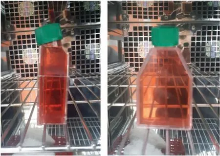
FIG.1.Front and side views of flas full of medium in incubator containing 5%CO2 at 37○C.
Glioblastoma cells were prepared and grown using the following method adopted by Freshney.16(1) The cells were transferred from frozen at-80○C and then thawed in a water bath at 37○C.(2)The cells were placed in a flas (T25)with a volume of 25 cm2and containing the culture medium RPMI-1640 and 10%bovine serum.(3)The flas was placed in an incubator containing 5%CO2at 37○C.(4)After incubation,when it was confirme that there was growth in the flas and that it was free from contamination,a secondary cell line was cultured and produced.(5) The cells were examined using an inverted microscope to ensure that they had vitality,were free from contamination,and had grown to the required number(ca.4–5×10⁵cells);they were then placed in an incubator at 37○C until the cell growth reached ∼70%.(6) As shown in Fig.1,the culture flas was fille completely with the medium with no air bubbles therein to reduce the effect of the shear stress of the liquid in the flask
3. Zero gravity simulation
As shown in Fig.2,ZG in outer space was simulated using a newly designed device(known as Gravity Challenge)as used previously by Altaieet al.17The sample inside the device is subjected to motion in a 3D geometric path resulting from the rotation of two perpendicular axes.The rotation rates must be constant and rapid enough to prevent gravitropic responses but slow enough to avoid significan acceleration forces,particularly centrifugal forces greater than that of the sensitivity threshold for the system being studied.18Therefore,the angular velocity adopted for the main axis was 10 rpm,while that for the secondary axis was 70%of that for the main axis.Because increasing distance from the center of rotation at the same velocity results in increased accelerations at the outer edges of the sample,the distance from the center of rotation was 80 mm,while the time for flippin the direction of rotation of the axis was 60 s.The LogStart feature was chosen,which allows sample data to be recorded in tabular form,and the program used displays the mean gravity for the coordinates,and the total mean gravity as a function of time.
4. Creating biological conditions
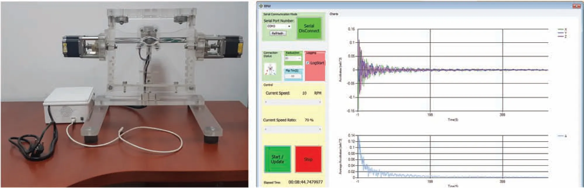
FIG.2.Gravity Challenge device(left)and screenshot of program accompanying this device(right).Zero gravity(ZG)is simulated by means of continuous change of the object’s orientation relative to the gravity vector(gravity always acts normal to the ground),which can generate effects comparable to those of true ZG when the changes are faster than the object’s response time to gravity.

FIG.3.Schematic of hood containing ZG device.The sample inside the device is subjected to motion in a 3D geometric path resulting from the rotation of two perpendicular axes.One flas is put in the ZG device,the second one is a control,and both are fille completely with the medium so that there are no air bubbles.The temperature and humidity sensor,the heat source,and the temperature and humidity controller create a stable temperature(37○C±1○C)and humidity(24±1 RH%)inside the hood.
To subject the cells to simulated ZG,it was firs necessary to create environmental conditions suitable for natural cell growth as follows.(1) The flask were covered with Parafilm which is a plastic paraffi tape characterized by being flexibl and retractable and used to close and isolate the covered containers against air pollution and prevent the medium from leaking out of the flasks (2) The hood was prepared for conducting the experiment by sterilizing both it and the ZG device using Valera Surgical Spirit Spray containing 68.6% alcohol and 3.7%methanol.(3)To create biological conditions for natural cell growth inside the hood,a board containing a smart electronic circuit was placed in the hood consisting of the following: (a) a current controller (Contactor-3TB41) to control the amount of alternating current entering the smart electronic circuit;(b)a temperature and humidity controller (SHT2000) to sense the temperature and humidity inside the hood and then set them within specifi ranges(temperature: 37○C ± 1○C;humidity: 24 ± 1 RH %);(c) a heater with an adjustable power of 100–400 W.This smart circuit was designed ergonomically to provide a suitable environment for natural cell growth by controlling the heater temperature via the controller,which contains a sensor to read the temperature and humidity inside the hood and display the readings digitally,as shown in Fig.3.(4) Two flask of the same type and quantity of cells were used,one subjected to ZG and the other as a control,in order to compare the results between them.(5) The sensor was placed near the control flas and the device in order to obtain accurate readings of both temperature and humidity.(6) The ZG device was operated remotely via a program on a laptop,and the motion of the device was monitored and the data recorded by the program were followed up for a period of 24 h.19,20
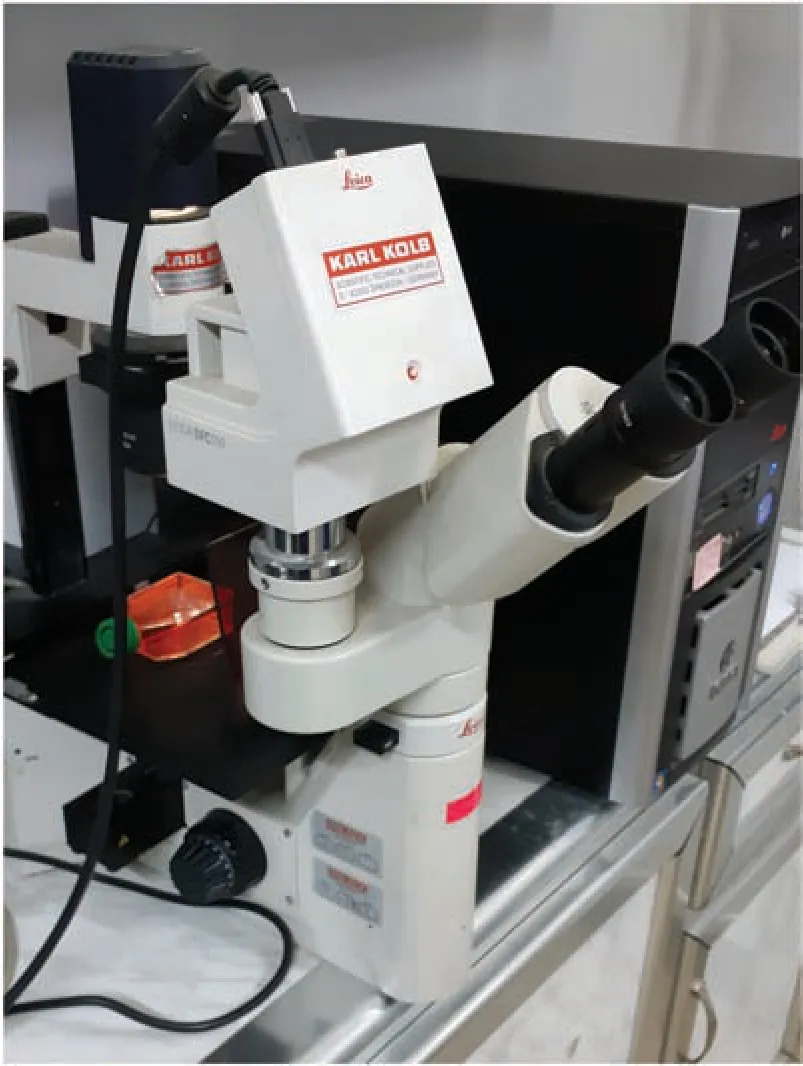
FIG.4.Microscope(Karl Kolb)supported by program that enables images to be modifie and viewed directly on computer screen.
B.MTT cytotoxicity assay
To identify the percentage of dead cells after exposure to ZG,a cytotoxicity test was performed as follows.(1) The flask were placed in the incubator for 24 h in order for the semi-spherical bodies to become stable at the bottom of the flasks (2) The cells were washed using phosphate buffer solution(PBS),and the process was repeated twice for 10 min.(3)A sufficien amount of trypsin enzyme solution was added to the remaining adherent cells,and they were placed in the incubator for 30–60 s at 37○C and monitored until they changed from a single layer of cells to single cells.(4)The cells were placed in a centrifuge at a rate of 3000 rpm for 10 min to expel the trypsin and used medium.(5)MTT[3-(4,5dimethylthiazol-2-yl)2,5-diphenyltetrazolium bromide]solution was used.(6)Cells were seeded in a 96-hole plate at a density of 10 000 cells/hole and MTT was added at a fina concentration of 1 mg/ml per hole and incubated for 3 h under standard culture conditions.(7) Medium was discarded for each hole and 200 μl of dimethyl sulfoxide(DMSO)was added to dissolve the formazan crystal precipitation and incubated for 10 min.(8) The absorbance was measured by using an ELISA device at a wavelength of 570 nm.(9) The results were expressed as a percentage of MTT inhibition compared to the control sample.
III.RESULTS AND DISCUSSION
A.Results
As shown in Fig.4,the T25 flask of glioma tumor cells(ANGM5) were photographed using a microscope (Karl Kolb)before subjecting them to simulated ZG conditions.As shown in Figs.5(a) and 5(b),the cells appeared in elongated multipolar or polygonal form,in addition to their growth in the form of a monolayer.
After 24 h of subjecting the cells to ZG,two different groups of cells appeared inside the T25 flask Some cells grew in the form of adherent cells,while the majority lost their adhesion and began to form 3D multicellular spheroids,as shown in Fig.5(c),where the red arrows indicate these cells and the black arrows indicate adherent cells.Both groups showed a change in cellular morphology to a semi-spherical shape compared with the control cells,which grew naturally with an increase in growth density,maintaining their elongated multipolar shape,as shown in Fig.5(d).The multicellular spheroid cells showed increased size and number as the time for which they were subjected to simulated ZG increased.
After exposing the tumor cells (ANGM5) to ZG for 24 h,they were transferred to an incubator under standard conditions(5% CO2,37○C,1g) and left for 24 h.It was observed that the cells continued to form multicellular spheroid cells,as shown in Fig.5(e),compared with the control cells under natural conditions,which grew naturally in the form of a multicellular layer,as shown in Fig.5(f).
The ZG environment stimulates the formation of 3D multicellular spheroids of cancer cells,given that the study showed semi-spherical structures resulting from the accumulation of cells after losing their adhesion.As shown in Fig.6,after exposing glioma cells (ANGM5) to ZG for 24 h,these structures were distinctively semi-spherical with different dimensions of 215 μm×165 μm.
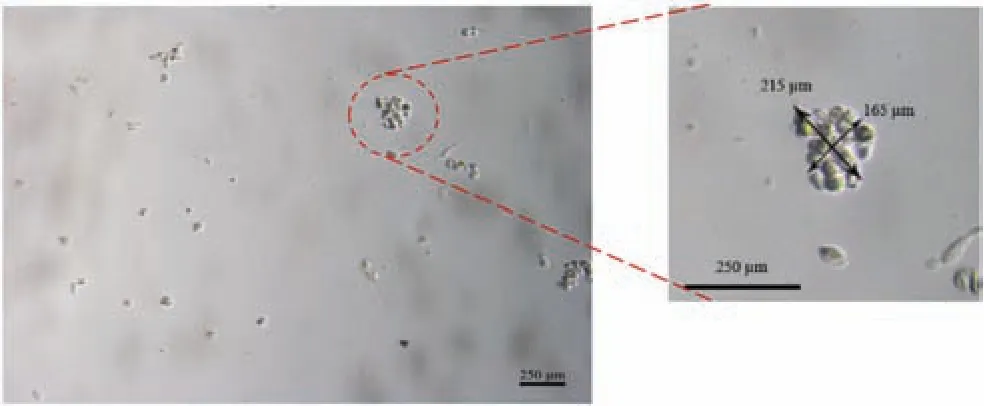
FIG.6. Semi-spherical structure of glioma cells with dimensions of 215 μm×165 μm after exposing cells to ZG for 24 h.Scale:250 μm.

FIG.7.Glioma cells grouped in a complex irregular structure with dimensions of 315 μm×360 μm.Scale:250 μm.
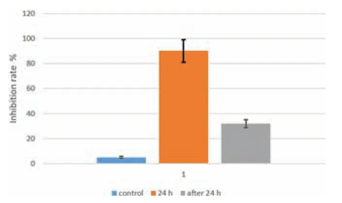
FIG.8.Percentage of dead cells from cytotoxicity test(MTT).First column(blue):percentage of dead control cells;second column (orange): percentage of dead cells directly after ZG;third column (gray): percentage of dead cells after 48 h(24 h under ZG)and 24 h under normal conditions(5%CO2,37○C,1g).
A large number of previous studies have been focused on the behavior of cancer cells using different types of tissues exposed to ZG;all showed that the majority of cells grew in a 3D spherical shape after different exposures times to ZG,and the spherical growth was very similar for all types.2,3When cancer cells are exposed to ZG,they lose their adhesion gradually and form spherical bodies similar to small malignant tumors;when gravity is restored,the spheroids re-adhere to the surface on which they rest.In the present study,after the glioma cells were exposed to gravity for 24 h and then placed in an incubator under normal conditions for another 24 h,the results showed the development of semi-spherical bodies with multiple cells into complex structures with irregular shapes similar to glands and with dimensions of 315 μm×360 μm,as shown in Fig.7.This often happens when the cells are exposed to ZG for long periods of time,and this is consistent with the study by Koppet al.7
The viability of live cells was evaluated by the MTT test and compared with the control cells under normal gravity.The results showed an initial significan increase in the number of dead cells at 24 h,but after transferring the cells to an incubator under standard conditions and natural gravity and leaving them there for 24 h,the number of dead cells decreased,reaching 32%,as shown in Fig.8,while maintaining a semi-spherical shape.A possible explanation for these results is that the glioma cells(ANGM5)became accustomed to the new growth condition as a result of adaptation to the effect of ZG,so they continued to grow and spread after the effect of ZG disappeared,but with an abnormal external appearance compared to the control cells(Fig.9).

FIG.9.Flow chart of experiment.
B.Discussion
Many of the physiological processes of cancer depend on the time required for cell adhesion and the adhesion environment,21this being because adhesion allows cells to transfer forces,sense the surrounding microenvironment,and coordinate their biological behaviors.22It is known that adherent cells are linked to each other through their cytoskeleton,therefore mechanical and chemical stimuli are transferred and control cell proliferation and migration.In turn,adhesion between cancer cells is a very important factor in combating this disease,as studies have shown that adhesion between cells promotes the movement of cancer cells by increasing the polarization of individual cells,which plays an important role in the pathogenesis of tumors.23Intercellular contact critically regulates cell survival,being the main mechanism that controls cell proliferation,and it is regulated by a protein called plakoglobin.24
The changes in cell morphology such as adhesion,viability,apoptosis,etc.may be due to a disruption in the proteins of tumor cells subject to the effect of ZG.Studies have shown that Caveolin-1 controls the stability of focal adhesions and contributes to mechanosensing and adaptation in response to mechanical stimuli including cell detachment.25These studies show that Caveolin-1 regulates yes-associated protein activity,which in turn modulates pathophysiological processes such as 3D multicellular spheroids.It is known that ZG affects gene regulation and even DNA damage.A study was conducted on thyroid cancer cells by exposing them to ZG during the Shenzhou-8 space experiment;the results showed the formation of huge 3D aggregates by FTC-133 cells that showed altered expression ofEGFandCTGFgenes under true ZG,26and the authors suggested that this regulation could determine the onset and progression of tumor development.In another future study,the behavior of proteins and gene regulation in glioma cells under ZG will be studied.
The present study—which was conducted on the local cancer cell line in the Iraqi Center for Cancer Research laboratories—showed that more than 90% of the cells lost the adhesion feature,in addition to changing their appearance and composition as a result of the effect of ZG.Furthermore,the toxicity examination showed that 32%of the cells died after exposure to simulated ZG for 24 h,as well as a clear change in the behavior of cell growth.These results herald new science because the ZG simulator provides environmental conditions that living organisms on Earth have not encountered,and it may provide special conditions that help discover changes in proteins that may become new targets for the development of a drug to treat cancer by conducting more studies under ZG conditions to investigate tumor growth,progression mechanisms,and malignancy progression.
AUTHOR DECLARATIONS
Conflict of Interest
The authors have no conflict to disclose.
DATA AVAILABILITY
The data that support the finding of this study are available from the corresponding author upon reasonable request.
杂志排行
纳米技术与精密工程的其它文章
- Stress distribution variations during nanoindentation failure of hard coatings on silicon substrates
- Electrode design for multimode suppression of aluminum nitride tuning fork resonators
- Design and analysis of longitudinal–flexuralhybrid transducer for ultrasonic peen forming
- An advanced cost-efficient IoT method for stroke rehabilitation using smart gloves
- Electrical characterization of an individual nanowire using flexible nanoprobes fabricated by atomic force microscopy-based manipulation
- An image mosaic technique with non-overlapping regions based on microscopic vision in precision assembly