Reduced Imaging Time and Improved Image Quality of 3D Isotropic T2-Weighted Magnetic Resonance Imaging with Compressed Sensing for the Female Pelvis
2023-11-14HaoMeiFengXiaoMingDeng
Hao Mei, Feng Xiao, Ming Deng
Abstract: This study is to compare three-dimensional (3D) isotropic T2-weighted magnetic resonance imaging (MRI) with compressed sensing-sampling perfection with application optimized contrast (CS-SPACE) and the conventional image (3D-SPACE) sequence in terms of image quality,estimated signal-to-noise ratio (SNR), relative contrast-to-noise ratio (CNR), and the lesions’ conspicuous of the female pelvis.Thirty-six females (age: 51, 28–73) with cervical carcinoma (n=20),rectal carcinoma (n=7), or uterine fibroid (n=9) were included.Patients underwent magnetic resonance (MR) imaging at a 3T scanner with the sequences of 3D-SPACE, CS-SPACE, and twodimensional (2D) T2-weighted turbo-spin echo (TSE).Quantitative analyses of estimated SNR and relative CNR between tumors and other tissues, image quality, and tissue conspicuity were performed.Two radiologists assessed the difference in diagnostic findings for carcinoma.Quantitative values and qualitative scores were analyzed, respectively.The estimated SNR and the relative CNR of tumor-to-muscle obturator internus, tumor-to-myometrium, and myometrium-to-muscle obturator internus was comparable between 3D-SPACE and CS-SPACE.The overall image quality and the conspicuity of the lesion scores of the CS-SPACE were higher than that of the 3D-SPACE (P <0.01).The CS-SPACE sequence offers shorter scan time, fewer artifacts, and comparable SNR and CNR to conventional 3D-SPACE, and has the potential to improve the performance of T2-weighted images.
Keywords: compressed sensing; sampling perfection with application-oriented contrasts (SPACE)using variable flip angle evolutions; three-dimensional (3D) imaging; magnetic resonance imaging(MRI); pelvis
1 Introduction
Magnetic resonance imaging (MRI) has become an increasingly important tool in the diagnosis and noninvasive assessment of female pelvic disease.In MRI, the two-dimensional (2D) T2-weighted turbo-spin echo (TSE) sequence plays a crucial role in the standardized protocols for the female pelvis, which provides excellent tissue contrast and high in-plane spatial resolution [1–5].However, the 2D TSE sequence has relatively thick image sections, so it needs to acquire multiple planes, resulting in a long total acquisition time.More than that, if 2D imaging is off-plane or sub-optimal, it may need to be repeated.Based on these obvious flaws, three-dimensional (3D)isotropic imaging is proposed.Previous studies demonstrated that a 3D T2-weighted TSE sequence with high spatial resolution and multiplanar reconstruction capability showed certain advantages over the 2D T2-weighted TSE sequence in uterine tumors [6–8].
In recent years, the 3D sampling perfection with application-oriented contrasts using variable flip angle evolutions (3D SPACE; Siemens Healthcare, Erlangen, Germany) sequence has been applied to the brain, bone, rectum, prostate,and female pelvis [6–12].Compared to the conventional 2D TSE image, 3D SPACE has the potential to improve the delineation of anatomical structures with many positive results [7, 8,12–17].The refocusing pulse train of the 3D SPACE sequence consists of variable flip-angle pulses.With flip angles less than 180°, the radiofrequency energy deposition decreases, which allows for longer echo trains, shorter echo spacing, and higher turbo factors [18].In clinical practice, T2-weighted image (T2WI) with 3D SPACE yields a sequence with high signal to noise ratio (SNR) and contrast-to-noise ratio(CNR), excellent contrast, and superior anatomic definition, as well as the ability to facilitate retrospective alignments of MRI in any arbitrary plane according to the anatomical and pathological structures [19].However, despite its promising added value, the T2WI with 3D SPACE is rarely used clinically, because of its long acquisition time.Moreover, physiological movements such as intestinal gurgling and breathing induce motion artifacts in pelvic imaging.
Compressed sensing (CS) technique is the one with the most potent method to accelerate magnetic resonance (MR) acquisition by reconstructing interactively incoherently under sampled data using a regularization term [20, 21].CS has been introduced to MRI, including time-offlight MR angiography [22, 23], and dynamic contrast-enhanced MRI [24].Recently, it has been applied to sampling perfection with application optimized contrast (SPACE) imaging to reduce the scan time, showing varied effects on image quality and diagnostic value [25–29].While CS technology is of great value to clinical institutions, to date no systematic evaluation of the clinical feasibility of a 3D T2WI sequence using CS-SPACE for pelvic imaging accelerated has been conducted.The purpose of our retrospective study was to qualitatively and quantitatively compare a CS-SPACE sequence with the routine SPACE for 3D T2WI of the female pelvis.
This study is to compare CS-SPACE and the conventional 3D-SPACE sequence in terms of image quality, estimated SNR, relative CNR, and conspicuity of female pelvis, aimed to improve the performance of T2-weighted image for the evaluation of tumor extent and improve the diagnostic confidence of radiologists.
2 Methodology
2.1 Patients
This retrospective study was approved by the Medical Ethics Committee of Zhongnan Hospital of Wuhan University (Approval number: 2017021)with waiver of informed consent.Between August 2020 and November 2020, 36 patients (age range 28–73 years, mean age 51 years) were identified.Out of 36 patients, 27 patients (age range 35–73 years, mean age 52.3 years) underwent radical hysterectomy, in 1 to 14 days (mean 4.3 days)after MR examination (tumor size range 1.6–8 cm, mean 3.54 cm).The pathological findings revealed cervical cancer in 20 patients, and rectal cancer in 7 patients.The remaining 9 patients(age range 28–57 years, mean age 45.7 years)underwent endocrinologic therapy following their MR examination.
2.2 MR Imaging
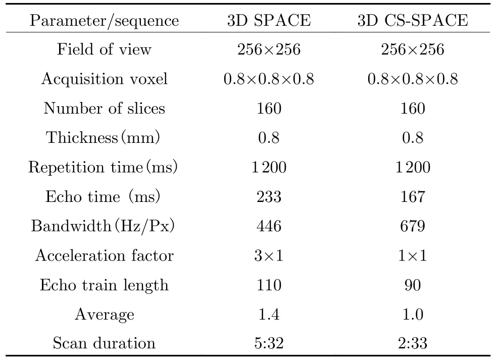
MRI was performed at 3T (Prisma, Siemens Healthcare, Germany) scanner using a 24-element pelvic phased-array coil and 32-element spine coil.In each patient, a sandbag was placed after the pelvic coil was covered before examination, to maximize coil fit and reduce motion artifacts.2D TSE T2WI images were obtained in sagital, coronal, and axial planes, while axial 3D T2WI with SPACE and 3D T2WI with CSSPACE were obtained in the axial plane.The sampling factor of axial 3D T2WI with CSSPACE is 0.14; regularization parameter was 0.000 5; and iteration was 40.The detailed imaging parameters of the 2D and 3D T2-weighted sequences are shown in Tab.1.After acquisition of the T2WI, axial T1-weighted 2D TSE, axial contrast-enhanced dynamic T1-weighted sequence, and sagital contrast-enhanced T1-weighted sequence were obtained for all patients, but those images were not used in this study.

Tab.1 MR parameters of 3D SPACE, and 3D CS-SPACE sequences
2.3 Imaging Analysis
2.3.1 Quantitative Imaging Analysis
Quantitative analyses were performed in all 36 patients.All images were transferred to picture archiving and communication system (PACS).The analysis was performed by one radiologist(Y.N.Hu, with more than 6 years’ experience in genitourinary imaging).Conventional 3DSPACE and CS-SPACE were quantitatively analyzed.Mean signal intensity (SI) was measured by placement of circular regions of interest(ROIs) over the tumor foci, myometrium and internal obturator muscle in identical locations on both axial 3D T2WI images.For the tumors,an ROI was drawn to encompass as much of the lesion as possible on a representative optimal panel image.For cases with multiple uterine leiomyomas, only the largest lesion was analyzed.Care was taken to size and place ROIs consistently for the same patient.For the internal obturator muscle, a 1 cm2circular ROI was used,placed over areas of relatively homogeneous region.Because CS and parallel imaging technique was used, the usual SNR or CNR measurements could not be performed [30, 31].Instead,estimated SNR was calculated as the ratio between the mean SI and standard deviation of the SI in the same tumor ROI as previously reported [6–8].Similarly, the relative CNR between tissues was estimated by using different ratios in SI.The relative CNR between the paired tissue types was calculated by using the following equation: the relative CNR = (A–B)/(A+B), whereAandBare the mean SIs of the two different tissue types.
2.3.2 Qualitative Analysis
Two radiologists reviewed both axial 3D SPACE and 3D CS-SPACE images.They performed sideby-side comparisons for the conspicuity of the lesion and the sharpness of the lesion edges using a 3-point scale as follows [6, 7]: score 1, conspicuity or sharpness of the lesion edge was bad; score 2, conspicuity or sharpness of the lesion edge was general; and score 3, conspicuity or sharpness of the lesion edge was better.The overall image quality was also assessed based on the artifact degree concerning motion, susceptibility, and dielectric artifacts using a 5-point scale as follows: score 1, poor quality with severe artifacts;score 2, below-average quality with diagnostically relevant artifacts; score 3, fair quality with moderate but not diagnostically relevant artifacts; score 4, good quality with minimal artifacts; and score 5, excellent quality without artifacts.These standards have been widely used in previous research and have been widely accepted.
2.3.3 Statistical Analysis
The statistical analyses were performed by using software (Statistical Product and Service Solutions, SPSS 25.0 for Windows; SPSS, International Business Machines Corporation).A pairedt-test was used to compare the estimated SNRs and the relative CNRs, and the Wilcoxon signedrank test for comparison of five- and three-point scales used for qualitative evaluation.
2.4 Results
2.4.1 Quantitative Analysis
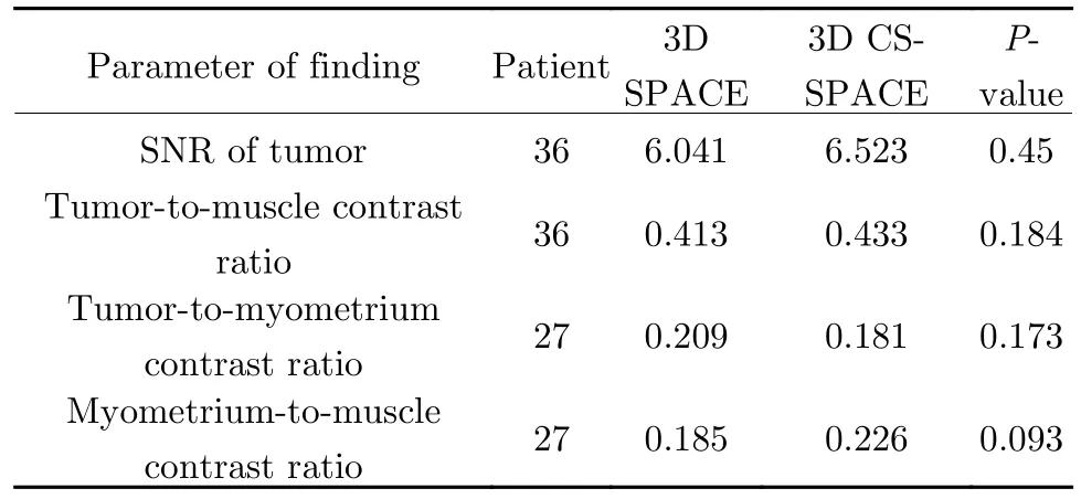
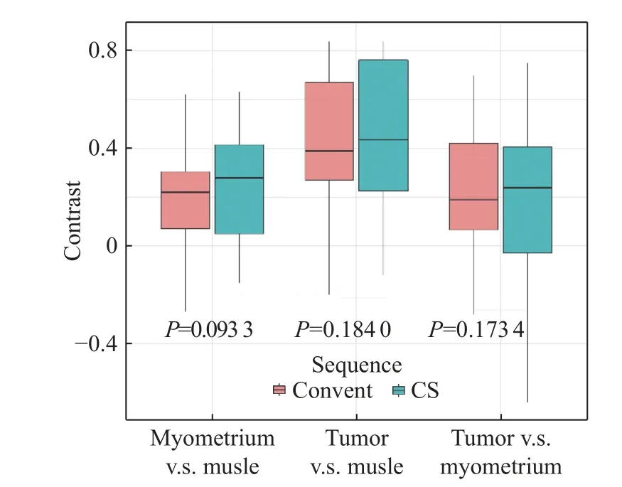
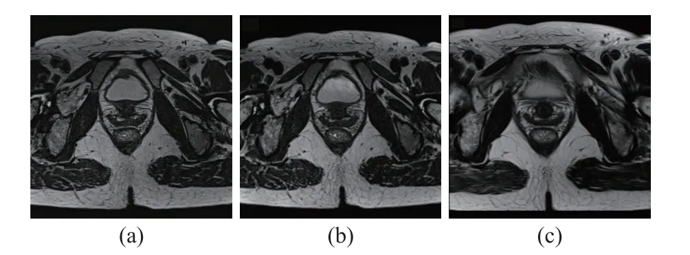
There was no significant difference in estimated SNRs (6.523 vs 6.041,P=0.450) between the two sequences for the lesion.And no significant difference is found in relative CNR tumor-to-musculi obturator internus (0.434 vs 0.413,P=0.178),tumor-to-myometrium (0.181 vs 0.209,P=0.169),and myometrium-to-musculi obturator internus(0.227 vs 0.209,P=0.091).The detailed results are shown in Tab.2, Fig.1, Fig 2, and Fig.3.The pelvic anatomy can be clearly showed with the 3D CS-SPACE (2:33 min), 3D SPACE (5:32 min) and 2D TSE T2WI (2:33 min) sequence,and there is no significant difference in SNR and CNR.

Tab.2 Comparison of 3D SPACE and 3D CS-SPACE sequences for estimated SNR and relative tumor contrast ratio

Fig.1 Box and whisker plots that show results of the relative CNRs
2.4.2 Qualitative Analysis

Fig.2 T2WI of a 49-year-old female with irregular menstruation for more than 2 months: (a) axial 3D CS-SPACE T2WI; (b) 3D SPACE T2WI; (c) 2D TSE T2WI

Fig.3 T2-weighted MR images of a 31-year-old woman with subserous leiomyoma with pathologically proved endometrioid adenocarcinoma (pT1N0MX): (a) axial 3D CS-SPACE T2WI; (b) sagital reformation of 3D CS SPACE; (c) coronal reformation of 3D CS SPACE; (d)3D SPACE T2WI; (e) sagital reformation of 3D SPACE; (f) coronal reformation of 3D SPACE
The overall image quality score for 3D CSSPACE images was higher than that for 3D SPACE images (Median: 4 vs 3,P<0.001)(Tab.3).Both radiologists agreed that there was a difference between the lesions shown in the 3D CS-SPACE sequence and the conventional 3D SPACE images.The 3D CS-SPACE sequence showed sharpness of the lesion edge and fewer artifacts.The conspicuity of lesion was better on 3D CS-SPACE images than on 3D SPACEimages (Average: 2.39 vs 1.81,P<0.001) (Tab.4).The specific case in Fig.3 also validated our results.3D CS SPACE shows fewer motion artifacts and better distinction between the mucous layer and the smooth muscle layer of the uterus.

Tab.3 Image quality scores for 3D CS-SPACE versus 3D SPACE images

Tab.4 Conspicuity of lesion scores for 3D CS-SPACE versus 3D SPACE images
3 Conclusion
The 3D CS-SPACE sequence offers advantages,such as shorter scan time, greater tumor conspicuity, fewer artifacts, and equal SNR and CNR, so it has the potential to improve the performance of T2WI for the evaluation of tumor extent and improve the diagnostic confidence of radiologists.In particular, we believe that this sequence holds promise to increase the quality of pelvic tumor imaging, especially for the patient with physiological movement.
杂志排行
Journal of Beijing Institute of Technology的其它文章
- Analysis of Dynamic Characteristics of Forced and Free Vibrations of Mill Roll System Based on Fractional Order Theory
- Automated Segmentation of the Brainstem, Cranial Nerves and Vessels for Trigeminal Neuralgia and Hemifacial Spasm
- Nitrogen Content Inversion of Corn Leaf Data Based on Deep Neural Network Model
- Application of Opening and Closing Morphology in Deep Learning-Based Brain Image Registration
- Tissue Microstructure Estimation of SANDI Based on Deep Network
- Spatial-Spectral Joint Network for Cholangiocarcinoma Microscopic Hyperspectral Image Classification
