CO2地质封存中储层岩石润湿性测量研究进展
2023-07-27


王 欣, 李少華, 刘 瑜, 张 毅, 蒋兰兰, 宋永臣
(大连理工大学能源与动力学院,大连116024)
摘要: 多孔介质的润湿性是 CO2 地质封存过程中的重要参数。基于润湿性测量方法和光学成像技 术综述了 CO2 封存条件下不同尺度的多孔介质润湿性测量技术,并分析了相关润湿现象。目前,岩石润湿性的测量主要分为: 实验室尺度的表面润湿性测定、孔隙尺度的内部壁面接触角测定, 以及宏观尺度的岩心整体润湿性评价。孔隙结构、矿物组成成分和表面粗糙度是孔隙尺度接触角 的关键影响因素, 它们会影响多孔介质的混合润湿特性并造成润湿滞后现象。根据不同局部驱替 事件(如排水、渗吸)的接触角分布建立了孔隙尺度与连续尺度的岩石润湿性关系。最新研究发 现, 随着驱替的发展,岩石润湿性在排水和渗吸过程中发生了显著改变,但不同尺度的岩石润湿 性的关系及润湿转变机理仍需要进一步研究。
关键词: 润湿性;CO2 地质封存 ;孔隙尺度 ;润湿转变 ;成像技术
中图分类号: X 701 文献标志码: A
A review of wettability measurement of the reservoir rock in CO2 geological storage
WANG Xin, LI Shaohua, LIU Yu, ZHANG Yi, JIANG Lanlan, SONG Yongchen
(School of Energy and Power Engineering, Dalian University of Technology, Dalian 116024, China)
Abstract: The wettability of porous media is an important parameter in the process of CO2 geological storage. Based on the wettability measurements and optical imaging techniques, the wettability measurement techniques of porous media at different scales under CO2 storage conditions were reviewed, and the related wettability phenomena were analyzed. At present, the rock wettability measurements are mainly described from the measurement of surface wettability at the laboratory scale, the contact angle at the porous inner surface, and the overall wettability evaluation of core samples at the macro scale. It was shown that the pore structure, mineral composition, and surface roughness were critical factors for the pore-scale contact angle, affecting the mixed wetting characteristics of porous media and causing the wetting hysteresis phenomenon. The relationship between pore-scale and continuum-scale rock wettability was established based on the contact angle distribution in different local displacement events (such as drainage and imbibition). A recent study found that the rock wettability changed significantly during the drainage and imbibition process with the development of the displacement process. However, the correlation between the wettability of rocks at different scales and the mechanism of wettability alternation still need to be further studied.
Keywords: wettability ; CO2 geological storage; pore-scale; wettability alternation; imaging technique
CO2地质封存指通过技术手段将 CO2注入深部盐水层、枯竭油气藏、不可开采的煤层等储层中进行长久封存,是实现温室气体减排的有力措施之一[1]。岩石润湿性是评估储层封存能力和安全性的重要指标[2],它是指一种流体黏附到固体表面的能力[3],直接影响 CO2?盐水?岩石多相系统中残余相的饱和度[4]、流体形态分布[5]、界面面积、相对渗透率[6]及毛细管压力[7-8]等渗流参数。润湿性是影响储层物理性质的关键因素[9],储层流体特性(如温度、压力、相态)和组成(如矿物成分和相互作用)会直接影响岩石的润湿性[10-13]。岩石润湿性按均匀程度可以分为均匀润湿和混合润湿体系。均匀润湿体系的接触角分布通常较为集中[14],然而,大部分储层都不是均匀润湿体系。实际储层存在复杂的孔隙结构,非均质性强[15],大多为混合润湿体系,通常表现出不同的流体流动特性[16]。 Akai 等[17]发现,在混合润湿体系下,水的有效渗透率与实验结果更相似。 Zou 等[18]认为混合润湿体系会增加流体连通性和团簇运动,降低系统有效渗透率。因此,准确描述多孔介质的润湿性,有利于探明流体的流动特性和 CO2的封存机制。
目前,已有文献总结了 CO2?流体?岩石体系的润湿性特征[19-20],尤其是岩石表面润湿性,并分析了岩石润湿性的影响因素以及润湿性在流体运移过程中的作用[21-22]。本文针对 CO2封存条件下多孔介质润湿性的最新测量技术,综述了多孔介质在不同尺度下的润湿性测量方法,主要包括:实验室尺度的多孔介质表面润湿性测定、孔隙尺度多孔介质局部壁面接触角测定,以及宏观尺度岩心样品的整体润湿性评价。同时,本文指出了测量过程中润湿性表现出的新现象,如润湿滞后、混合润湿现象,以及不同驱替事件(即排水和渗吸)下的润湿性变化。这将有助于设置不同的润湿性条件,以获得最佳的 CO2封存能力,进一步促进 CO2地质封存的实施。
1 多孔介质表面润湿性
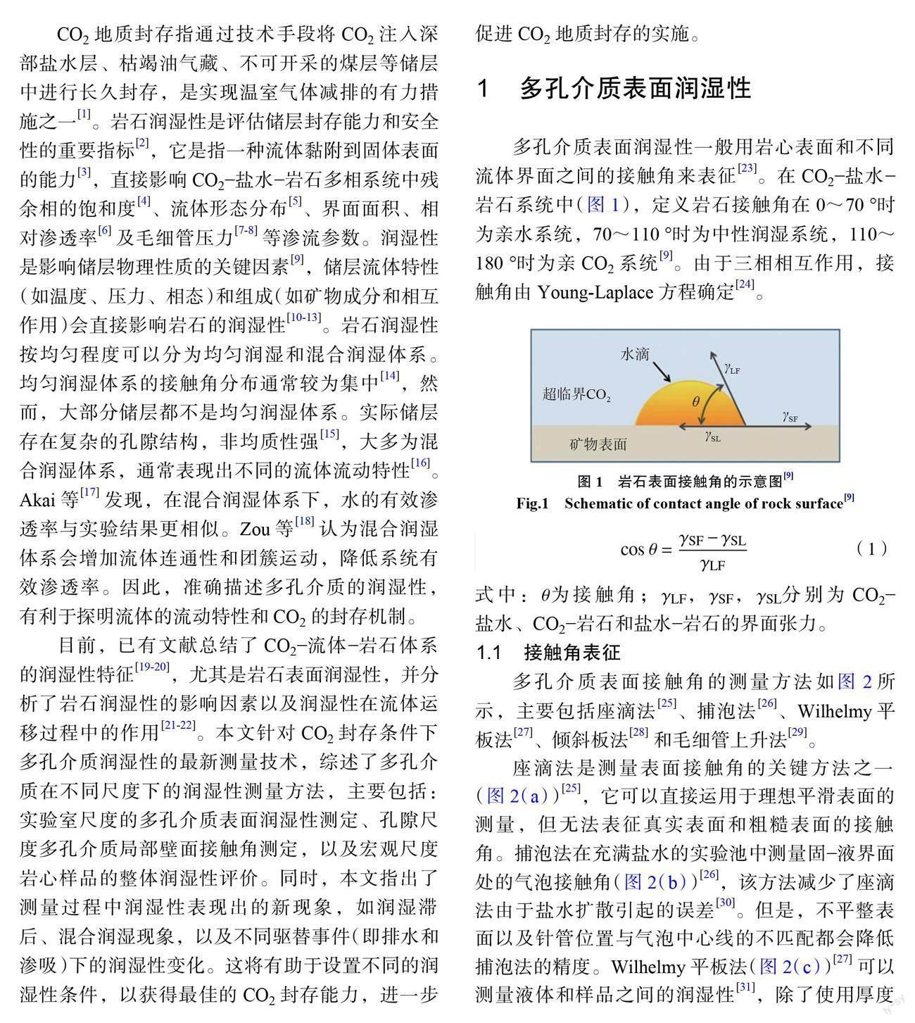
多孔介质表面润湿性一般用岩心表面和不同流体界面之间的接触角来表征[23]。在 CO2?盐水?岩石系统中(图1),定义岩石接触角在0~70°时为亲水系统,70~110°时为中性润湿系统,110~180°时为亲 CO2系统[9]。由于三相相互作用,接触角由 Young-Laplace 方程确定[24]。
式中:θ为接触角; LF , SF , SL分别为 CO2?盐水、 CO2?岩石和盐水?岩石的界面张力。
1.1 接触角表征
多孔介质表面接触角的测量方法如图2所示,主要包括座滴法[25]、捕泡法[26]、Wilhelmy 平板法[27]、倾斜板法[28]和毛细管上升法[29]。
座滴法是测量表面接触角的关键方法之一(图2(a))[25],它可以直接运用于理想平滑表面的测量,但无法表征真实表面和粗糙表面的接触角。捕泡法在充满盐水的实验池中测量固?液界面处的气泡接触角(图2(b))[26],该方法减少了座滴法由于盐水扩散引起的误差[30]。但是,不平整表面以及针管位置与气泡中心线的不匹配都会降低捕泡法的精度。Wilhelmy 平板法(图2(c))[27]可以测量液体和样品之间的润湿性[31],除了使用厚度均匀的矩形板外,也可以在三角形和不规则形状的平板中计算接触角[32]。倾斜板法主要用于测量动态接触角(图2(d))[28],其操作简单,但接触角的测量强烈依赖于板的倾斜、液滴的大小和形状。毛细管上升法根据毛细效应可以有效地获得毛细管的动态润湿过程(图2(e))[29],但缺乏实用性。
1.2 接触角滞后
接触角滞后(contact angle hysteresis, CAH)由表面粗糙度、表面变形、表面不均匀等因素引起,通常表现为前进接触角( advancing contact angle, ACA)和后退接触角(receding contact angle, RCA)之差[33]。近年来提出了一系列动态接触角的测量方法[26, 34-37]。Lander 等[38]发现, Wilhelmy 平板法是测量 CAH 的最佳方法,可以减少操作者的主观性,其次是倾斜板法,而座滴法实施难度最大。当 CO2注入储层后,排水过程中 CO2驱替水相时,水接触角为后退接触角;渗吸过程中水驱替 CO2时,水接触角为前进接触角[39]。排水过程的后退接触角一般小于渗吸过程的前进接触角[40]。岩石的非均质性也会影响接触角滞后, ACA 对非均质表面中疏水的成分更敏感,而 RCA 对亲水的成分更敏感[19]。目前,虽然有接触角滞后的定性分析,但复杂的拓扑结构和非均质性对接触角滞后的影响仍需进行大量的研究。
多孔介质表面接触角可以表征岩石表面润湿性和动态滞后过程,但表面粗糙度和岩石结构可能会影响测量精度。渗吸方法揭示了岩石内部的孔隙连通特征[20],可以测量岩石的润湿性[41-42],但受流体界面相互作用的影响较大。多孔介质表面润湿性只能衡量实验室尺度的岩心表面特性[43]。未来应准确测量具有复杂结构的岩石润湿性,完善多孔介质的物性表征,为 CO2封存选址提供有力的理论支撑。
2 多孔介质局部壁面接触角
岩石的表面粗糙度、孔隙几何形状和化学成分直接影响流体分布,进而影响接触角测量的准确性[44]。目前,对孔隙尺度下岩石的润湿特征仍缺乏基本的认识[45],尚未完全了解流体注入后的壁面润湿行为。 Li 等[46]发现平板表面测量的静态接触角小于孔隙内接触角,用表面接触角来预测孔接触角是不合适的。多孔介质局部壁面的润湿性测量至关重要,需要通过一些先进的技术(如微模型和 X 射线计算机断层扫描)来实现。
2.1 微模型
微模型由两块薄玻璃板组成,玻璃板表面通过化学手段和几何设计进行处理[47],模拟真实的岩石状况(图3(a))[48],该模型有助于观察储层中复杂的多相、多组分的相互作用[49]。高压微模型可以用于研究 CO2?盐水?岩石系统中的流体运动现象(图3(b),3(c))[50-51],测量多孔介质壁面与液面之间的接触角[52-53],也可以獲得流体运动过程中的动态接触角[54]、渗吸过程中的前进接触角和排水过程中的后退接触角,如图3(d)所示[53]。除了常规的角度测量方法外,还可以用图像拟合流体界面来测量微模型的接触角[55]。在微模型中,可以直接测量静态、动态和平衡接触角,方法简单,易于操作。但润湿性主要通过测量微模型上的局部接触角获得,即使在高度光滑和均匀的微模型中,不同的位置会有不同的接触角分布,因此,该方法测得的结果并不能代表整体的润湿性。
目前已有学者研究了不同润湿性对微模型中流动、驱替的影响[54-55]。与亲水状态相比,中性润湿的微模型降低了 CO2?盐水驱替前缘速度,促进了非润湿相的孔隙填充,提高了驱替效率[56]。 Avenda?o 等[57]发现亲水多孔介质中的驱替前缘不均匀,残余油饱和度较大。与通过控制几何形状来改变微模型的方法相比, Lee 等[58]通过改变亲水性和亲油性组分的比例来定量控制微模型的混合润湿特性,减少了制造不同微模型的时间,实现了润湿性的合理调控。然而,运用润湿性量化流体驱替过程中渗流参数的研究仍不全面,未来还需研究微模型中不同润湿性的内部机理。
微模型的一个独特优势是多孔介质之间没有接触[59],研究者可以清楚地观察润湿性变化并描述其动态特性。然而,大多数微模型的改性是通过改变原油组分来模拟亲油状态[52, 60],亲 CO2的 微模型研究较少。同时,均匀蚀刻的二维孔隙网络不能代表真实的多孔介质特性,制备具有真实三维多孔结构的微模型仍然是现有技术的难点。最近有研究提出采用不同刻蚀深度的2.5-D 微模型来模拟真实多孔介质[61],如 Xu 等[62]利用2.5-D 微模型观察到夹断现象, Peng 等[63]发现了能表征多孔介质三维特征的临界刻蚀深度标准,从而有助于从二维尺度研究真实多孔介质的渗吸动力学行为。2.5-D 微模型作为新发展的微流控技术,比2- D 微模型更容易观察到复杂的孔隙动力学行为,但是,这种刻蚀技术目前在国内应用不多,是未来微模型研究的难点和焦点。虽然3D 打印技术已经出现[64],但高分辨率的打印技术还未完全成 熟,基于3D 打印的刻蚀技术还有待研究。孔隙尺度下微模型中的多相流动机制仍不明晰,因此,还需要大量的微模型实验来验证微模型技术在 CO2封存中的实际应用能力。
2.2 X 射线计算机断层扫描
X射线计算机断层扫描是一种无损成像技术,可以表征岩石样品的三维孔隙结构,重建样品的三维几何形状,获得孔隙尺度下的样品成像[65]。X 射线成像系统主要由 X 射线源、样品及旋转系统、X 射线检测系统、计算机系统(硬件和软件)组成。由射线源产生的 X 射线照射在被扫描的样品上得到图像信号,并利用图像重建算法和三维图像处理软件进行储层岩石样品的内部可视化分析[66]。
Andrew 等[67]首次提出孔隙尺度的局部接触角的测量方法(图4),将获得的 CT 图像进行图像预处理和相位分割以测量局部接触角,近年来该方法已大量应用于原位润湿性的研究。在孔隙尺度研究中,二维接触角定义为流体?流体和流体?固体界面之间的角度,而三维接触角由平面向量的点积计算。准确确定平面是计算局部接触角的关键,表1是孔隙尺度下平面的确定方法。
手动测量方法一般是随机选取接触线上的300个点进行接触角测量,无法获得足够多的接触角值,计算较慢,且主观性较大。因此,AlRatrout 等[69]提出了自动测量方法描述局部接触角,该方法与手动方法相比,在成像不良处的测量准确度更高,得到的接触角分布范围更大[74]。这种孔隙尺度的研究相比于多孔介质表面润湿性的测量更准确,也有助于进一步研究润湿性的影响因素。 Xie 等[75]发现,随着盐度增加,润湿性向水湿性降低的方向转变。对比低盐度注水和高盐度注水实验,低盐度注水期间润湿性从弱亲油向弱亲水转变,而高盐度注水的润湿性几乎恒定[76]。在 CO2?盐水?玻璃珠/石英砂系统中,润湿性随着离子强度的增加和 CO2从气态向超临界态的转变而缓慢减弱[77]。Alhammadi等[70]认为与原油接触的有机层会改变方解石的润湿性。 Qin 等[78]发现超临界 CO2注入会引起亲油碳酸盐的润湿性反转。大部分孔隙尺度局部接触角是静态接触角,为了衡量动态润湿性, Mascini 等[79]基于 Young-Laplace 方程提出了一种与驱替事件有关的后退接触角的概念(图5),发现动态接触角的分布范围比静态接触角小,这主要是由于静态接触角未考虑孔隙内流体界面行为。因此,动态接触角在驱替过程中更有助于描述实际动态过程,并可应用于孔隙尺度建模研究。 R*为某一时刻的曲率半径, Rht 为发生驱替事件的曲率半径,θ*为对应 R*的接触角,θht 为对应于 Rht 的接触角。
X 射线计算机断层扫描可以表示储层的真实特征,该技术提供了数十万次测量,可以有效地减少测量误差,已被用于识别地质封存中流体的分布特性。这种三维成像技术不仅可以观察孔隙的几何特征,也可以观察一些复杂的微观现象,如驱替前缘的演化[80]、海恩斯跳跃[81]、毛细流动中的活塞式驱替和夹断[82-83]及旁路流动等[64, 84]。然而,润湿性引起的微观现象的机理仍不清楚,有关孔隙尺度观测和孔隙事件的分析较少,仍然需要开发一些更先进的方法来研究多孔介质内部的复杂特性,如使用同步加速器 X 射线断层扫描提高实时成像能力[65]、运用环境扫描显微镜研究孔隙微观特性等[85]。随着各种成像技术的发展,未来应考虑综合多種技术来获得流动过程中更准确的原位表征,进而分析动态润湿特性。孔隙尺度下的原位接触角大多在油?盐水?岩石系统下进行测量,含 CO2的多相体系的原位润湿分析是未来的一大研究热点。
2.3 孔隙尺度润湿滞后现象
多孔介质的表面杂质、吸附作用和表面粗糙度会引起润湿滞后现象的出现[67]。Jafari 等[53]证明了 RCA 比 ACA更有可重复性,发现在较低的压力下动态滞后更明显。然而, Lv 等[86]在中性润湿的玻璃珠实验中没有发现明显的润湿滞后现象。 Khishvand 等[87]发现油?盐水、气?油和气?盐水的接触角滞后几乎相同(约10°)。孔隙尺度下不同体系的润湿滞后结果仍需更多的研究来证明。
表面粗糙度对润湿性的研究至关重要,会导致储层条件下排水和渗吸的接触角分布存在较大偏差[86]。表面粗糙度的作用如图6(a)所示,粗糙度会降低亲水表面的接触角,增加疏水表面的接触角[22, 88]。Sari 等[89]发现:接触角随着表面粗糙度的增加而减小,在低盐度情况下接触角减小得更少,表面粗糙度的作用较低。 AlRatrout 等[90]发现:在润湿性变化不大的亲水条件下,界面曲率与粗糙度无关,非润湿相被很好地捕获,以阻止气体运移,有利于 CO2地质封存;而当体系中的润湿性发生改变,接触角和界面曲率会随着粗糙度的增加而增加(图6(b))。nz2为油?盐水界面的法向向量, nz3为盐水?岩石界面的法向向量,θi为该点的接触角, i 为三相接触线 M 上的某一点,κ为曲率。在储层条件下, CO2与地层盐水相互作用, CO2会发生溶解、沉淀和地质特性转变等复合作用。由于矿物溶解和表面粗糙度作用[13],矿物表面变得更亲水,这会影响实验测量和现场测试结果,因此,孔隙尺度润湿滞后的分析结果对封存机制的确立至关重要。
2.4 混合润湿性
混合润湿性是指不同大小的孔隙其润湿性不同,孔隙结构特性(如孔径、形状、体积和连通性)会使润湿性更加复杂,孔隙中的亲油和亲水组分会对润湿性造成不同影响[91]。在中性润湿条件下,孔隙几何形状也会显著影响润湿性[92]。然而,也有相反的观点认为孔隙结构对封存过程影响较小[14],孔隙结构和润湿性机理还需进一步分析。 Chang 等[93]应用十八烷基三氯硅烷(OTS)制备混合润湿体系,分析不稳定驱替过程(图7),获得的微观接触角分布比其他文献中的接触角分布范围大,这可能是由表面粗糙度和接触线钉扎作用引起的。
为了分析孔隙尺度不同润湿条件下的多相流动分布, Scanziani[94]比较了亲水条件和混合润湿条件下静态和动态实验的结果,发现相比于均质润湿体系,混合润湿体系可以提高驱替效率[95]。与亲水体系相比,混合润湿的碳酸盐有利于 CO2形成大且连通的神经节(图8(a))[96]。在混合润湿的石灰岩中,大孔隙中的盐水驱油,油位于角落处;而在亲水体系下,油存在于孔隙中心,盐水则留在角落里(图8(b))[97]。S 层为油层和盐水层的夹层。 Gao 等[98]发现亲水体系砂岩的平均接触角小于混合润湿体系,混合润湿体系的相对渗透率低于亲水体系(图8(c))。Lin 等[99]发现混合润湿砂岩具有较低的毛细管压力,仅为亲水系统的十分之一。相比于亲水体系,孔隙填充事件更倾向于发生在混合润湿体系中[100]。界面钉扎作用会导致接触角滞后,进而抑制界面衰退,并阻止夹断现象的出现[101]。在亲水条件下,可以增强夹断,提高非润湿相的残余饱和度,进而增强 CO2捕获能力[102]。
润湿性的空间分布会影响流体占有率和连通性, Armstrong 等[103]通过形态学方法构建混合润湿体系,分析空间润湿分布对相对渗透率的影响。随着多孔介质亲水程度的增加,驱替过程的图像变得更加紧凑[104]。然而,针对空间润湿性分布的研究较少,多孔介质表面实验无法准确表征润湿性的空间分布以及内部孔隙空间复杂的相互作用。如何准确刻画多孔介质物理性质并分析空间非均质润湿性的影响,也是未来研究中的一个重点方向。
3 岩心整体润湿性
目前,已有研究将孔隙尺度润湿性与岩心尺度特性联系起来[18, 105]。Deglint 等[106]测得的微观接触角的分布范围比宏观接触角大,微观接触角不能表征宏观尺度的润湿性。这种润湿性在不同尺度上的不一致会导致流体驱替、流体饱和度、毛细管曲线等结果的误差。 Rucker 等[107]也发现了孔隙尺度接触角所测结果和岩心尺度实验的结果有不同的润湿性响应。岩心的整体润湿性减少了因矿物成分变化产生的局部润湿性不足,可以作为多孔介质整体特性的评价指标。目前在石油工程中,可以通过毛细管压力曲线间接推断润湿性,如排水和渗吸过程中的 Amott 指数[23]或 USBM 指数[108]均可以表征润湿性。这两种方法均可以测量岩心样品的平均润湿性[23],但它们不能解释非均质润湿性,无法预测部分润湿体系中的空间润湿性分布。
3.1 核磁共振成像技术
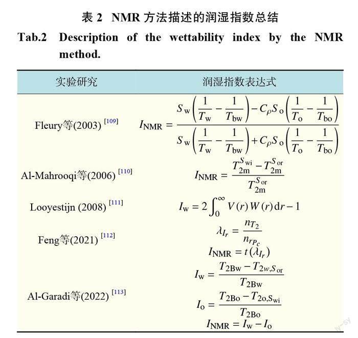
核磁共振成像(NMR)是一种无损成像技术,可以提供孔隙结构和流体分布信息。多孔介质表面可以显著改变核磁共振弛豫时间,亲水表面相对于亲油表面会显著降低弛豫时间[23],中性润湿体系的弛豫时间取决于润湿表面的数量和相互作用强度,因此,可根据弛豫时间测量多孔介质的润湿性。通常采用核磁共振的横向弛豫时间 T2分布获得润湿性指标[109],主要的润湿性描述如表2所示。表中: INMR为无量纲 NMR 润湿指数, S w为水饱和度, S o为油饱和度, Tw 为水主要弛豫时间, To 为油主要弛豫时间, C 为水?油表面弛豫比, Tbw为水体积弛豫时间, Tbo为油体积弛豫时间; T2m 为T2的平均值, Swi为不可还原水饱和度, S or 为残余油饱和度; Iw 为水的润湿性指数, Io 为油的润湿性指数, V (r)dr 为半径 r 和 r+dr 之间的孔隙体积分数, W (r)dr 为半径 r 和 r+dr 之间的孔隙表面积分数;λIr为平均饱和度指数的比, nT2为 T2分布根据 Ir-Sw 关系计算出的平均饱和指数, nrPc 为 MICP(注汞毛细管压力)曲线根据 Ir-Sw 关系计算出的平均饱和指数; Ir 为电阻率指数; T2Bw, T2Bo , T2o;Swi , T2w;S or分别为散装水、散装油、不可还原水和剩余油的主导 T2峰的模态值。
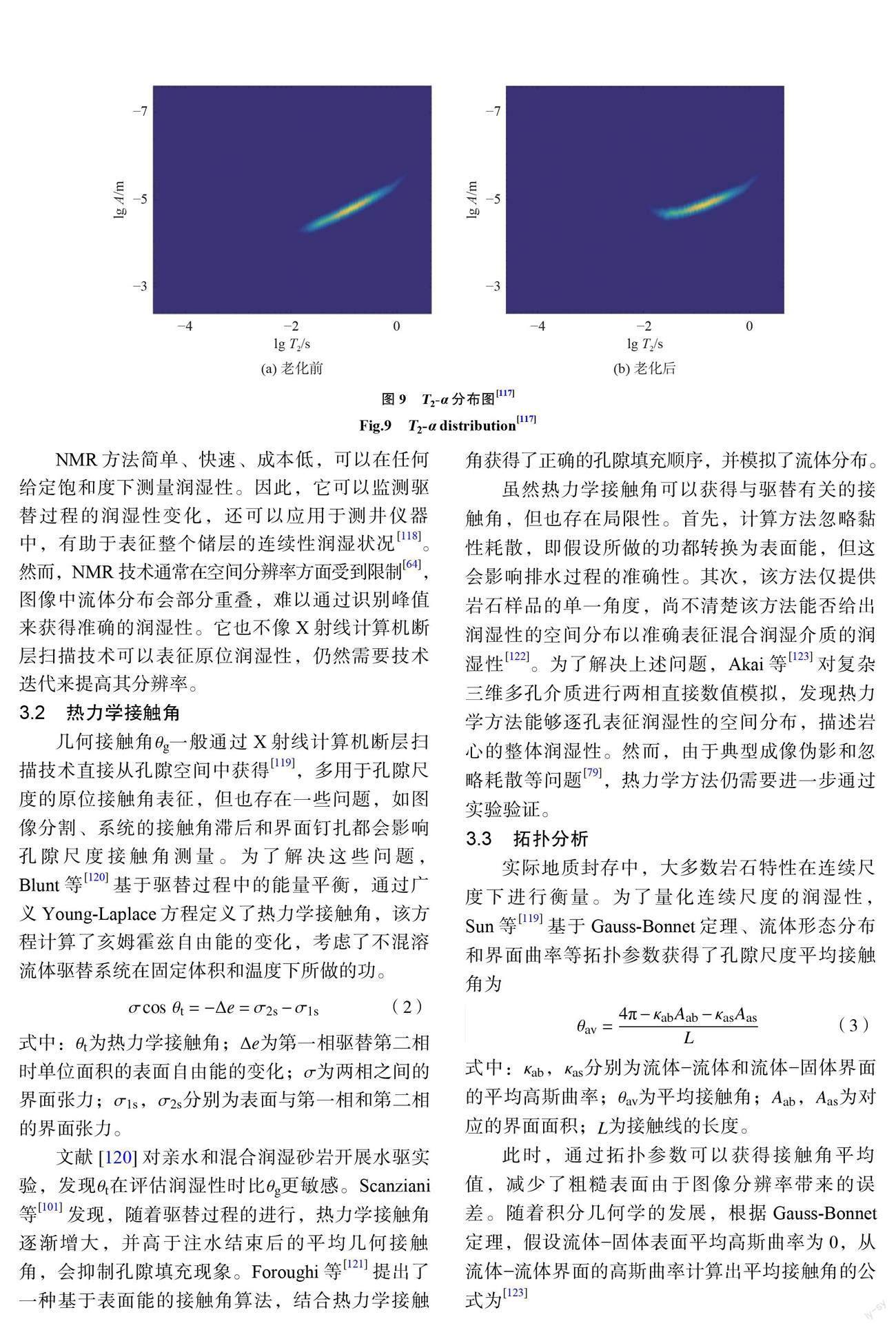
Liang 等[114]采用扩散?横向弛豫时间(D-T2)结合 Amott 实验提供了更完整的致密砂岩润湿性评估。用核磁共振可以研究非均质润湿性的3个主要形成机制:原始矿物组分的不同润湿特性、极性化合物的吸附和有机物在原油中的沉积[115]。这些机制会导致多孔介质的某些部分变成亲油的,而另一些部分变成亲水的。除了表面弛豫的作用,表面覆盖率[116]、电阻率[112]都可以预测润湿性。为了分析非均质润湿性, Wang 等[115]提出了由润湿表面覆盖率和局部润湿性确定的表观接触角,还可以通过分析表面弛豫分布和孔徑分布(T2-α)来研究混合润湿多孔介质的润湿性分布[117],老化前 T2-α分布呈线性分布,老化后表面弛豫率分布随孔径分布的变化而变化(图9)。图中,A 为孔隙长度。老化过程只改变了原油与孔表面接触的大孔的润湿性,小孔表面仍然是水湿的,因此,大孔中的表面弛豫率小于小孔中的表面弛豫率。
NMR 方法简单、快速、成本低,可以在任何给定饱和度下测量润湿性。因此,它可以监测驱替过程的润湿性变化,还可以应用于测井仪器中,有助于表征整个储层的连续性润湿状况[118]。然而,NMR 技术通常在空间分辨率方面受到限制[64],图像中流体分布会部分重叠,难以通过识别峰值来获得准确的润湿性。它也不像 X 射线计算机断层扫描技术可以表征原位润湿性,仍然需要技术迭代来提高其分辨率。
3.2 热力学接触角
几何接触角θg一般通过 X 射线计算机断层扫描技术直接从孔隙空间中获得[119],多用于孔隙尺度的原位接触角表征,但也存在一些问题,如图像分割、系统的接触角滞后和界面钉扎都会影响孔隙尺度接触角测量。为了解决这些问题, Blunt 等[120]基于驱替过程中的能量平衡,通过广义 Young-Laplace 方程定义了热力学接触角,该方程计算了亥姆霍兹自由能的变化,考虑了不混溶流体驱替系统在固定体积和温度下所做的功。
式中:θt 为热力学接触角;Δe为第一相驱替第二相时单位面积的表面自由能的变化;σ为两相之间的界面张力;σ1s ,σ2s 分别为表面与第一相和第二相的界面张力。
文献[120]对亲水和混合润湿砂岩开展水驱实验,发现θt 在评估润湿性时比θg更敏感。 Scanziani 等[101]发现,随着驱替过程的进行,热力学接触角逐渐增大,并高于注水结束后的平均几何接触角,会抑制孔隙填充现象。Foroughi 等[121]提出了一种基于表面能的接触角算法,结合热力学接触角获得了正确的孔隙填充顺序,并模拟了流体分布。
虽然热力学接触角可以获得与驱替有关的接触角,但也存在局限性。首先,计算方法忽略黏性耗散,即假设所做的功都转换为表面能,但这会影响排水过程的准确性。其次,该方法仅提供岩石样品的单一角度,尚不清楚该方法能否给出润湿性的空间分布以准确表征混合润湿介质的润湿性[122]。为了解决上述问题, Akai 等[123]对复杂三维多孔介质进行两相直接数值模拟,发现热力学方法能够逐孔表征润湿性的空间分布,描述岩心的整体润湿性。然而,由于典型成像伪影和忽略耗散等问题[79],热力学方法仍需要进一步通过实验验证。
3.3 拓扑分析
实际地质封存中,大多数岩石特性在连续尺度下进行衡量。为了量化连续尺度的润湿性, Sun 等[119]基于 Gauss-Bonnet 定理、流体形态分布和界面曲率等拓扑参数获得了孔隙尺度平均接触角为
式中:κab ,κas 分别为流体?流体和流体?固体界面的平均高斯曲率;θav为平均接触角;Aab ,Aas 为对应的界面面积; L为接触线的长度。
此时,通过拓扑参数可以获得接触角平均值,减少了粗糙表面由于图像分辨率带来的误差。随着积分几何学的发展,根据 Gauss-Bonnet 定理,假设流体?固体表面平均高斯曲率为0,从流体?流体界面的高斯曲率计算出平均接触角的公式为[123]
式中: n为第1相和第2相接触表面的三相接触线的闭合回路数;κG12为界面 S 12高斯曲率的积分值。
该方法提供了拓扑结构和润湿性的简单关系,适合评估孔隙空间的接触角。 Sun 等[124]进一步提出了缺陷曲率,建立接触角和流体拓扑结构的直接联系(图10(a),10(b)),定义宏观接触角为
式中:κd为缺陷曲率; Nc为固体表面上形成的闭合接触线环的数量。
θm考虑了由于表面非均质性和流体动力学引起的接触角滞后的平均影响,同时,该方法不需要平衡条件。对于给定的流体团簇,较大的缺陷曲率决定了较大的接触角。将拓扑性质与热力学接触角相联系,宏观接触角表征了多相系统的润湿性[125]。另外,不需要三相接触线就可以获得空间分布的表观接触角θa ,减轻了接触线的钉扎效应(图10(c),10(d)),在低分辨率情况下可以得到更准确的结果,并可以获得不同尺度润湿性之间的相互关系。图10中: Mce 为团簇和流体的表面; Mcs为团簇和固体的表面。
综上,热力学方法以及拓扑结构方法是表征整体润湿性的2个关键方法,不仅可以描述几何特征,还考虑了多孔介质的空间分布。相比于孔隙局部接触角,宏观接触角方法减小了由于图像分辨率和成像不清产生的测量误差,是目前新提出的有效的整体润湿性评估方法。未来更应该关注热力学方法和几何方法的结合,包括耗散事件和拓扑变化,进一步优化宏观接触角的计算过程。同时,可以结合不同尺度的润湿性分析,如通过岩石表面润湿性分析润湿性影响因素和相关作用,以及运用孔隙尺度的成像技术来观察局部润湿现象,并采用整体润湿性指标来评估岩石的非均质特征和整体拓扑结构。
4 结 论
多孔介质润湿性对 CO2?流体?岩石体系渗流特性具有重要影响,本文综述了目前 CO2地质封存领域岩石润湿性研究的最新进展,主要包括:实验室尺度多孔介质表面润湿性测定、孔隙尺度多孔介质局部壁面接触角测定,以及宏观尺度岩心样品整体润湿性评价。接触角是多孔介质表面润湿性最常见的表征指标,但常规表面接触角测量实验忽略了岩石粗糙度和多孔介质复杂的内部结构,影响了润湿性测量准确性。随着光学成像技术的发展,微模型和 X 射线计算机断层扫描可用于获得孔隙尺度的岩石润湿性。微模型制备简单,可以获得接触角动态变化过程, X 射线计算机断层扫描技术可以获得三维真实岩心局部润湿性及其迟滞特性。通过微观接触角测量技术分析了多孔介质粗糙度和孔隙结构对孔隙尺度润湿性的影响,尤其是一些重要的润湿现象如接触角滞后、混合润湿。这种微观现象对分析渗流特性和封存效率也极为重要。关于岩心整体润湿性表征,基于 NMR 技術的润湿指数可以评估岩心整体润湿性,但容易受到内部磁场的影响。热力学接触角可以表征与驱替过程相关的岩心整体润湿性,拓扑学方法也可以有效地获得连续尺度润湿特性。岩心整体润湿性表征方法极具新颖性,但还缺乏系统的研究。
目前,虽然已进行了大量的实验室润湿性测量,然而很少有将多种尺度联系起来以分析内部机理的实验研究。岩心表面的润湿性测量常用于定量表征并分析润湿性的影响因素,孔隙壁面的局部接触角测量可以有效地分析空间非均质特征,宏观尺度的整体润湿性评估是衡量真实多孔介质结构和物性的最新方法。未来需进一步研究不同尺度下岩石润湿性的内在联系,如用孔隙尺度方法观察孔隙空间的内部现象,结合宏观的整体评价,探明不同储层条件下润湿性的演变规律和转变机理,进一步解析岩石润湿性对封存储量和效率的影响。
參考文献:
[1]蔡博峰 , 李琦 , 张贤.中国二氧化碳捕集利用与封存(CCUS)年度报告(2021)——中国 CCUS 路径研究[R].武汉:生态环境部环境规划院, 中国科学院武汉岩土力学研究所, 2021.
[2] ALI M, SAHITO M F, JHA N K, et al. Effect of nanofluid on CO2-wettability reversal of sandstone formation; implications for CO2 geo-storage[J]. Journal of Colloid and Interface Science, 2020, 559:304–312.
[3] BOBEK J E, MATTAX C C, DENEKAS M O. Reser- voir rock wettability —its significance and evaluation[J]. Transactions of the AIME, 1958, 213(1):155–160.
[4] TENG Y, JIANG L L, LIU Y, et al. MRI study on CO2 capillary trap and drainage behavior in sandstone cores under geological storage temperature and pressure[J]. International Journal of Heat and Mass Transfer, 2018, 119:678–687.
[5] TOKUNAGA T K, WAN J. Capillary pressure and mineral wettability influences on reservoir CO2 capacity[J]. Reviews in Mineralogy and Geochemistry, 2013, 77(1):481–503.
[6] GHARBI O, BLUNT M J. The impact of wettability and connectivity on relative permeability in carbonates: a pore network modeling analysis[J]. Water Resources Research, 2012, 48(12): W12513.
[7] TENG Y, WANG P F, JIANG L L, et al. An experimental study of density-driven convection of fluid pairs with viscosity contrast in porous media[J]. International Journal of Heat and Mass Transfer, 2020, 152:119514.
[8] TENG Y, WANG P F, XIE H P, et al. Capillary trapping characteristics of CO2 sequestration in fractured carbonate rock and sandstone using MRI[J]. Journal of Natural Gas Science and Engineering, 2022, 108:104809.
[9] IGLAUER S, PENTLAND C H, BUSCH A. CO2 wettability of sealand reservoir rocks and the implications for carbon geo-sequestration[J]. Water Resources Research, 2015, 51(1):729–774.
[10] ABBASZADEH M, SHARIATIPOUR S, IFELEBUEGU A. The influence of temperature on wettability alteration during CO2 storage in saline aquifers[J]. International Journal of Greenhouse Gas Control, 2020, 99:103101.
[11] IGLAUER S, SALAMAH A, SARMADIVALEH M, et al. Contamination of silica surfaces: impact on water-CO2- quartz and glass contact angle measurements[J]. International Journal of Greenhouse Gas Control, 2014, 22:325–328.
[12] SARAJI S, GOUAL L, PIRI M, et al. Wettability of supercritical carbon dioxide/water/quartz systems: simultaneous measurement of contact angle and interfacial tension at reservoir conditions[J]. Langmuir, 2013, 29(23):6856–6866.
[13] ZHANG X, WEI B, SHANG J, et al. Alterations of geochemical properties of a tight sandstone reservoir caused by supercritical CO2-brine-rock interactions in CO2-EOR and geosequestration[J]. Journal ofCO2 Utilization, 2018, 28:408–418.
[14] ZHAO X C, BLUNT M J, YAO J. Pore-scale modeling: effects of wettability on waterflood oil recovery[J]. Journal of Petroleum Science and Engineering, 2010, 71(3/4):169–178.
[15] KOVSCEK A R, WONG H, RADKE C J. A pore-level scenario for the development of mixed wettability in oil reservoirs[J]. AIChE Journal, 1993, 39(6):1072–1085.
[16] JAHANBAKHSH A, SHAHROKHI O, MAROTO- VALER M M. Understanding the role of wettability distribution on pore-filling and displacement patterns in a homogeneous structure via quasi 3D pore-scale modelling[J]. Scientific Reports, 2021, 11(1):17847.
[17] AKAI T, ALHAMMADI A M, BLUNT M J, et al. Modeling oil recovery in mixed-wet rocks: pore-scale comparison between experiment and simulation[J].Transport in Porous Media, 2019, 127(2):393–414.
[18] ZOU S M, ARMSTRONG R T, ARNS J Y, et al.Experimental and theoretical evidence for increased ganglion dynamics during fractional flow in mixed-wet porous media[J]. Water Resources Research, 2018, 54(5):3277–3289.
[19] DRELICH J W, BOINOVICH L, CHIBOWSKI E, et al. Contact angles: history of over 200 years of open questions[J]. Surface Innovations, 2020, 8(1/2):3–27.
[20] SHARIFIGALIUK H, MAHMOOD S M, REZAEE R, et al. Conventional methods for wettability determination of shales: a comprehensive review of challenges, lessons learned, and way forward[J]. Marine and Petroleum Geology, 2021, 133:105288.
[21] YEKEEN N, PADMANABHAN E, ABDULELAH H, et al. CO2/brine interfacial tension and rock wettability at reservoir conditions: a critical review of previous studies and case study of black shale from Malaysian formation[J]. Journal of Petroleum Science and Engineering, 2021, 196:107673.
[22] STEVAR M S P, B?HM C, NOTARKI K T, et al. Wettability of calcite under carbon storage conditions[J]. International Journal of Greenhouse Gas Control, 2019, 84:180–189.
[23] ANDERSON W. Wettability literature survey-part 2: wettability measurement[J]. Journal of Petroleum Technology, 1986, 38(11):1246–1262.
[24] KWOK D Y, NEUMANN A W. Contact angle measurement and contact angle interpretation[J]. Advances in Colloid and Interface Science, 1999, 81(3):167–249.
[25] HUHTAM?KI T, TIAN X L, KORHONEN J T, et al. Surface-wetting characterization using contact-angle measurements[J]. Nature Protocols, 2018, 13(7):1521–1538.
[26] XUE J, SHI P, ZHU L, et al. A modified captive bubble method for determining advancing and receding contact angles[J]. Applied Surface Science, 2014, 296:133–139.
[27] HUBBE M A, GARDNER D J, SHEN W. Contact angles and wettability of cellulosic surfaces: a review of pro- posed mechanisms and test strategies[J]. BioResources , 2015, 10(4):8657–8749.
[28] AL-ANSSARI S, ARIF M, WANG S B, et al. Wettabilityof nano-treated calcite/CO2/brine systems: implication for enhanced CO2 storage potential[J]. International Journal of Greenhouse Gas Control, 2017, 66:97–105.
[29] HEBBAR R S, ISLOOR A M, ISMAIL A F. Contact angle measurements[M]//HILAL N, ISMAIL A F, MATSUURA T, et al. Membrane Characterization. Amsterdam: Elsevier, 2017:219–255.
[30] HASHEMI L, GLERUM W, FARAJZADEH R, et al. Contact angle measurement for hydrogen/brine/sandstone system using captive-bubble method relevant for underground hydrogen storage[J]. Advances in Water Resources, 2021, 154:103964.
[31] RAM? E. The interpretation of dynamic contact angles measured by the Wilhelmy plate method[J]. Journal of Colloid and Interface Science, 1997, 185(1):245–251.
[32] PARK J, PASAOGULLARI U, BONVILLE L. Wettability measurements of irregular shapes with Wilhelmy plate method[J]. Applied Surface Science, 2018, 427:273–280.
[33] ERAL H B, T MANNETJE D J C M, OH J M. Contact angle hysteresis: a review of fundamentals and applications[J]. Colloid and Polymer Science, 2013, 291(2):247–260.
[34] SHANG J Y, FLURY M, HARSH J B, et al. Comparison of different methods to measure contact angles of soil colloids[J]. Journal of Colloid and Interface Science, 2008, 328(2):299–307.
[35] DICKSON J L, GUPTA G, HOROZOV T S, et al. Wetting phenomena at the CO2/water/glass interface[J]. Langmuir, 2006, 22(5):2161–2170.
[36] MENNELLA A, MORROW N R. Point-by-point method of determining contact angles from dynamic Wilhelmy plate data for oil/brine/solid systems[J]. Journal of Colloid and Interface Science, 1995, 172(1):48–55.
[37] RUIZ-CABELLO F J M, RODR?GUEZ-VALVERDE M A, CABRERIZO-V?LCHEZ M. A new method for evaluating the most stable contact angle using tilting plate experiments[J]. Soft Matter, 2011, 7(21):10457–10461.
[38] LANDER L M, SIEWIERSKI L M, BRITTAIN W J, etal. A systematic comparison of contact angle methods[J]. Langmuir, 1993, 9(8):2237–2239.
[39] ZHANG L J, KIM Y, JUNG H, et al. Effects of salinity- induced chemical reactions on biotite wettability changes under geologic CO2 sequestration conditions[J]. Environmental Science & Technology Letters, 2016, 3(3):92–97.
[40] WANG S B, TOKUNAGA T K. Capillary pressure- saturation relations for supercritical CO2 and brine in limestone/dolomite sands: implications for geologic carbon sequestration in carbonate reservoirs[J]. Environmental Science & Technology, 2015, 49(12):7208–7217.
[41] ABD A S, ELHAFYAN E, SIDDIQUI A R, et al. A review of the phenomenon of counter-current spontaneous imbibition: analysis and data interpretation[J]. Journal of Petroleum Science and Engineering, 2019, 180:456–470.
[42] LI C X, SINGH H, CAI J C. Spontaneous imbibition in shale: a review of recent advances[J]. Capillarity, 2019, 2(2):17–32.
[43] DONALDSON E C, ALAM W. Wettability[M].Amsterdam: Elsevier, 2013.
[44] ALHAMMADI A M, ALRATROUT A, BIJELJIC B, et al. Pore-scale imaging and characterization of hydrocarbon reservoir rock wettability at subsurface conditions using X-ray microtomography[J]. Journal of Visualized Experiments, 2018(140):57915.
[45] COLE D R, CHIALVO A A, ROTHER G, et al. Supercritical fluid behavior at nanoscale interfaces: implications for CO2 sequestration in geologic formations[J]. Philosophical Magazine, 2010, 90(17/18):2339–2363.
[46] LI X X, FAN X F, BRANDANI S. Difference in pore contact angle and the contact angle measured on a flat surface and in an open space[J]. Chemical Engineering Science, 2014, 117:137–145.
[47] SHE Y, AOKI H, WANG W C, et al. Spontaneous deformation of oil clusters induced by dual surfactants for oil recovery: dynamic study from Hele-Shaw cell to wettability-altered micromodel[J]. Energy & Fuels, 2022, 36(11):5762–5774.
[48] LU H W, HUANG F, JIANG P X, et al. Exsolution effects in CO2 huff-n-puff enhanced oil recovery: water- oil-CO2 three phase flow visualization and measurements by micro-PIV in micromodel[J]. International Journal of Greenhouse Gas Control, 2021, 111:103445.
[49] SONG W, DE HAAS T W, FADAEI H, et al. Chip-off- the-old-rock: the study of reservoir-relevant geological processes with real-rock micromodels[J]. Lab on A Chip, 2014, 14(22):4382–4390.
[50] CHEN Y, LI Y F, VALOCCHI A J, et al. Lattice Boltzmann simulations of liquid CO2 displacing water in a 2D heterogeneous micromodel at reservoir pressure conditions[J]. Journal of Contaminant Hydrology, 2018, 212:14–27.
[51] CAO S C, DAI S, JUNG J. Supercritical CO2 and brine displacement in geological carbon sequestration: micromodel and pore network simulation studies[J]. International Journal of Greenhouse Gas Control, 2016, 44:104–114.
[52] SAADAT M, YANG J Y, DUDEK M, et al. Microfluidic investigation of enhanced oil recovery: the effect of aqueous floods and network wettability[J]. Journal ofPetroleum Science and Engineering, 2021, 203:108647.
[53] JAFARI M, JUNG J. Direct measurement of static anddynamic contact angles using a random micromodel considering geological CO2 sequestration[J]. Sustainability, 2017, 9(12):2352.
[54] CASTRO E R, TARN M D, GINTEROV? P, et al. Determination of dynamic contact angles within microfluidic devices[J]. Microfluidics and Nanofluidics, 2018, 22:1–11.
[55] VAN ROOIJEN W, HASHEMI L, BOON M, et al. Microfluidics-based analysis of dynamic contact angles relevant for underground hydrogen storage[J]. Advances in Water Resources, 2022, 164:104221.
[56] HU R, WAN J M, KIM Y, et al. Wettability effects on supercritical CO2–brine immiscible displacement during drainage: pore-scale observation and 3D simulation[J]. International Journal of Greenhouse Gas Control, 2017, 60:129–139.
[57] AVENDA?O J, LIMA N, QUEVEDO A, et al. Effect of surface wettability on immiscible displacement in a microfluidic porous media[J]. Energies, 2019, 12(4):664.
[58] LEE H, LEE S G, DOYLE P S. Photopatterned oil- reservoir micromodels with tailored wetting properties[J]. Lab on A Chip, 2015, 15(14):3047–3055.
[59] KIM Y, WAN J M, KNEAFSEY T J, et al. Dewetting of silica surfaces upon reactions with supercritical CO2 and brine: pore-scale studies in micromodels[J]. Environmental Science & Technology, 2012, 46(7):4228–4235.
[60] SAADAT M, TSAI P A, HO T H, et al. Development of a microfluidic method to study enhanced oil recovery by low salinity water flooding[J]. ACS Omega, 2020, 5(28):17521–17530.
[61] YANG W P, BROWNLOW J W, WALKER D L, et al. Effect of surfactant - assisted wettability alteration on immiscible displacement: a microfluidic study[J]. Water Resources Research, 2021, 57(8): e2020WR029522.
[62] XU K, LIANG T B, ZHU P, et al. A 2.5-D glass micromodel for investigation of multi-phase flow in porous media[J]. Lab on A Chip, 2017, 17(4):640–646.
[63] PENG X L, WANG X Z, ZHANG Y Z, et al. Experimental study of strong imbibition in microcapillaries representing pore/throat characteristics of tight rocks[J]. Fuel, 2023, 342:127775.
[64] ANBARI A, CHIEN H T, DATTA S S, et al. Microfluidic model porous media: fabrication and applications[J]. Small, 2018, 14(18):1703575.
[65] GAN M G, ZHANG L W, MIAO X X, et al. Application of computed tomography (CT) in geologic CO2 utilization and storage research: a critical review[J]. Journal of Natural Gas Science and Engineering, 2020, 83:103591.
[66] SHEPPARD A, LATHAM S, MIDDLETON J, et al. Techniques in helical scanning, dynamic imaging and image segmentation for improved quantitative analysis with X-ray micro-CT[J]. Nuclear Instruments and Methods in Physics Research Section B:Beam Interactions with Materials and Atoms, 2014, 324:49–56.
[67] ANDREW M, BIJELJIC B, BLUNT M J. Pore-scale contact angle measurements at reservoir conditions using X-ray microtomography[J]. Advances in Water Resources, 2014, 68:24–31.
[68] KLISE K A, MORIARTY D, YOON H, et al. Automated contact angle estimation for three-dimensional X-ray microtomography data[J]. Advances in Water Resources, 2016, 95:152–160.
[69] ALRATROUT A, RAEINI A Q, BIJELJIC B, et al. Automatic measurement of contact angle in pore-space images[J]. Advances in Water Resources, 2017, 109:158–169.
[70] ALHAMMADI A M, ALRATROUT A, SINGH K, et al. In situ characterization of mixed-wettability in a reservoir rock at subsurface conditions[J]. Scientific Reports, 2017, 7(1):10753.
[71] SCANZIANI A, SINGH K, BLUNT M J, et al. Automatic method for estimation of in situ effective contact angle from X-ray microtomography images of two-phase flow in porous media[J]. Journal of Colloid and Interface Science, 2017, 496:51–59.
[72] IBEKWE A, POKRAJAC D, TANINO Y. Automated extraction of in situ contact angles from micro-computed tomography images of porous media[J]. Computers & Geosciences, 2020, 137:104425.
[73] YANG J H, ZHOU Y F. An automatic in situ contact angle determination based on level set method[J]. Water Resources Research, 2020, 56(7): e2020WR027107.
[74] ZANKOOR A, KHISHVAND M, MOHAMED A, et al. In-situ capillary pressure and wettability in natural porous media: multi-scale experimentation and automated characterization using X-ray images[J]. Journal of Colloid and Interface Science, 2021, 603:356–369.
[75] XIE Y, KHISHVAND M, PIRI M. Impact of connate brine chemistry on in situ wettability and oil recovery: pore-scale experimental investigation[J]. Energy & Fuels, 2020, 34(4):4031–4045.
[76] KHISHVAND M, ALIZADEH A H, ORAKI KOHSHOUR I, et al. In situ characterization of wettability alteration and displacement mechanisms governing recovery enhancement due to low-salinity waterflooding[J]. Water Resources Research, 2017, 53(5):4427–4443.
[77] LV P F, LIU Y, WANG Z, et al. In situ local contact angle measurement in a CO2-brine-sand system usingmicrofocused X-ray CT[J]. Langmuir, 2017, 33(14):3358–3366.
[78] QIN Z Q, ARSHADI M, PIRI M. Near-miscible supercritical CO2 injection in oil-wet carbonate: a pore- scale experimental investigation of wettability state and three-phase flow behavior[J]. Advances in Water Resources, 2021, 158:104057.
[79] MASCINI A, CNUDDE V, BULTREYS T. Event-based contact angle measurements inside porous media using time-resolved micro-computed tomography[J]. Journal of Colloid and Interface Science, 2020, 572:354–363.
[80] CIEPLAK M, ROBBINS M O. Dynamical transition in quasistatic fluid invasion in porous media[J]. Physical Review Letters, 1988, 60(20):2042–2045.
[81] BERG S, OTT H, KLAPP S A, et al. Real-time 3D imaging of Haines jumps in porous media flow[J].Proceedings of the National Academy of Sciences of the United States of America, 2013, 110(10):3755–3759.
[82] VALVATNE P H, BLUNT M J. Predictive pore-scale modeling of two-phase flow in mixed wet media[J]. Water Resources Research, 2004, 40(7): W07406.
[83] BLUNT M J. Multiphase flow in permeable media: a pore-scale perspective[M]. Cambridge: Cambridge University Press, 2017.
[84] CHATZIS I, MORROW N R, LIM H T. Magnitude and detailed structure of residual oil saturation[J]. Society of Petroleum Engineers Journal, 1983, 23(2):311–326.
[85] IVANOVA A, MITIUREV N, CHEREMISIN A, et al. Characterization of organic layer in oil carbonate reservoir rocks and its effect on microscale wetting properties[J]. Scientific Reports, 2019, 9(1):10667.
[86] LV P F, LIU Y, JIANG L L, et al. Pore -scale contact angle measurements of CO2-brine-glass beads system using micro-focused X-ray computed tomography[J]. Micro & Nano Letters, 2016, 11(9):524–527.
[87] KHISHVAND M, ALIZADEH A H, PIRI M. In-situ characterization of wettability and pore-scale displacements during two- and three-phase flow in natural porous media[J]. Advances in Water Resources, 2016, 97:279–298.
[88] DE GENNES P G, BROCHARD-WYART F, QU?R? D. Capillarity and wetting phenomena: drops, bubbles, pearls, waves[M]. New York: Springer, 2004.
[89] SARI A, AL MASKARI N S, SAEEDI A, et al. Impact of surface roughness on wettability of oil-brine-calcite system at sub-pore scale[J]. Journal of Molecular Liquids, 2020, 299:112107.
[90] ALRATROUT A, BLUNT M J, BIJELJIC B. Wettability in complex porous materials, the mixed-wet state, and its relationship to surface roughness[J]. Proceedings of the National Academy of Sciences of the United States ofAmerica, 2018, 115(36):8901–8906.
[91] GAO Z Y, FAN Y P, HU Q H, et al. A review of shale wettability characterization using spontaneous imbibition experiments[J]. Marine and Petroleum Geology, 2019, 109:330–338.
[92] RABBANI H S, ZHAO B Z, JUANES R, et al. Pore geometry control of apparent wetting in porous media[J]. Scientific Reports, 2018, 8(1):15729.
[93] CHANG C, KNEAFSEY T J, WAN J M, et al. Impacts of mixed-wettability on brine drainage and supercritical CO2 storage efficiency in a 2.5-D heterogeneous micromodel[J]. Water Resources Research, 2020, 56(7): e2019WR026789.
[94] SCANZIANI A. Immiscible three-phase flow in porous media: dynamics and wettability effects at the pore scale[D]. London: Imperial College London, 2020.
[95] CHANG C, KNEAFSEY T J, TOKUNAGA T K, et al. Impacts of pore network-scale wettability heterogeneity on immiscible fluid displacement: a micromodel study[J]. Water Resources Research, 2021, 57(9): e2021WR030302.
[96] AL-MENHALI A S, MENKE H P, BLUNT M J, et al. Pore scale observations of trapped CO2 in mixed-wet carbonate rock: applications to storage in oil fields[J]. Environmental Science & Technology, 2016, 50(18):10282–10290.
[97] SINGH K, BIJELJIC B, BLUNT M J. Imaging of oil layers, curvature and contact angle in a mixed-wet and a water-wet carbonate rock[J]. Water Resources Research, 2016, 52(3):1716–1728.
[98] GAO Y, RAEINI A Q, SELEM A M, et al. Pore-scale imaging with measurement of relative permeability and capillary pressure on the same reservoir sandstone sample under water-wet and mixed-wet conditions[J]. Advances in Water Resources, 2020, 146:103786.
[99] LIN Q Y, BIJELJIC B, BERG S, et al. Minimal surfaces in porous media: pore-scale imaging of multiphase flow in an altered-wettability Bentheimer sandstone[J]. Physical Review E, 2019, 99(6):063105.
[100] MASCINI A, BOONE M, VAN OFFENWERT S, et al. Fluid invasion dynamics in porous media with complex wettability and connectivity[J]. Geophysical Research Letters, 2021, 48(22): e2021GL095185.
[101] SCANZIANI A, LIN Q Y, ALHOSANI A, et al. Dynamics of fluid displacement in mixed-wet porous media[J]. Proceedings of the Royal Society A: Mathematical, Physical and Engineering Sciences, 2020, 476(2240):20200040.
[102] KREVOR S, BLUNT M J, BENSON S M, et al. Capillary trapping for geologic carbon dioxide storage – from pore scale physics to field scale implications[J]. InternationalJournal of Greenhouse Gas Control, 2015, 40:221–237.
[103] ARMSTRONG R T, SUN C H, MOSTAGHIMI P, et al.Multiscale characterization of wettability in porous media[J]. Transport in Porous Media, 2021, 140(1):215–240.
[104] ZHAO B, MACMINN C W, JUANES R. Wettability control on multiphase flow in patterned microfluidics[J].Proceedings of the National Academy of Sciences of the United States of America, 2016, 113(37):10251–10256.
[105] LIN Q Y, BIJELJIC B, PINI R, et al. Imaging and measurement of pore-scale interfacial curvature to determine capillary pressure simultaneously with relative permeability[J]. Water Resources Research, 2018, 54(9):7046–7060.
[106] DEGLINT H J, CLARKSON C R, GHANIZADEH A, et al. Comparison of micro- and macro-wettability measurements and evaluation of micro-scale imbibition rates for unconventional reservoirs: Implications for modeling multi-phase flow at the micro-scale[J]. Journal of Natural Gas Science and Engineering, 2019, 62:38–67.
[107] RUCKER M, BARTELS W B, BULTREYS T, et al. Workflow for upscaling wettability from the nanoscale to core scale[J]. Petrophysics, 2020, 61(2):189–205.
[108] DONALDSON E C, THOMAS R D, LORENZ P B. Wettability determination and its effect on recovery efficiency[J]. Society of Petroleum Engineers Journal, 1969, 9(1):13–20.
[109] FLEURY M, DEFLANDRE F. Quantitative evaluation of porous media wettability using NMR relaxometry[J]. Magnetic Resonance Imaging, 2003, 21(3/4):385–387.
[110] AL-MAHROOQI SH, GRATTONI C A, MUGGERIDGE A H, et al. Pore-scale modelling of NMR relaxation for the characterization of wettability[J]. Journal of Petroleum Science and Engineering, 2006, 52(1-4):172–186.
[111] LOOYESTIJN W. Wettability index determination from NMR logs[J]. Petrophysics, 2008, 49(2):130–145.
[112] FENG C, FENG J G, FENG Z Y, et al. Determination of reservoir wettability based on resistivity index prediction from core and log data[J]. Journal of Petroleum Science and Engineering, 2021, 205:108842.
[113] AL-GARADI K, EL-HUSSEINY A, ELSAYED M, et al. A rock core wettability index using NMR T2 measurements[J]. Journal of Petroleum Science and Engineering, 2022, 208:109386.
[114] LIANG C, XIAO L Z, ZHOU C C, et al. Two- dimensional nuclear magnetic resonance method for wettability determination of tight sand[J]. Magnetic Resonance Imaging, 2019, 56:144–150.
[115] WANG J, XIAO L Z, LIAO G Z, et al. Theoretical investigation of heterogeneous wettability in porous media using NMR[J]. Scientific Reports, 2018, 8(1): 13450.
[116] CHEN J, HIRASAKI G J, FLAUM M. NMR wettability indices: effect of OBM on wettability and NMR responses[J]. Journal of Petroleum Science and Engineering, 2006, 52(1/4): 161–171.
[117] WANG J, XIAO L Z, LIAO G Z, et al. NMR characterizing mixed wettability under intermediate-wet condition[J]. Magnetic Resonance Imaging, 2019, 56:156–160.
[118] LOOYESTIJN W, HOFMAN J. Wettability-index determination by nuclear magnetic resonance[J]. SPE Reservoir Evaluation & Engineering, 2006, 9(2):146–153.
[119] SUN C H, MCCLURE J E, MOSTAGHIMI P, et al.Linking continuum-scale state of wetting to pore-scale contact angles in porous media[J]. Journal of Colloid and Interface Science, 2020, 561: 173–180.
[120]BLUNT M J, LIN Q Y, AKAI T, et al. A thermodynamically consistent characterization of wettability in porous media using high-resolution imaging[J]. Journal of Colloid and Interface Science,2019, 552: 59–65.
[121]FOROUGHI S, BIJELJIC B, LIN Q Y, et al. Pore-by-poremodeling, analysis, and prediction of two-phase flow inmixed-wet rocks[J]. Physical Review E, 2020, 102(2):023302.
[122]BLUNT M J, AKAI T, BIJELJIC B. Evaluation of methods using topology and integral geometry to assess wettability[J]. Journal of Colloid and Interface Science,2020, 576: 99–108.
[124]SUN C H, MCCLURE J E, MOSTAGHIMI P, et al. Probing effective wetting in subsurface systems[J].Geophysical Research Letters, 2020, 47(5):e2019GL086151.
[125]SUN C H, MCCLURE J E, MOSTAGHIMI P, et al.Characterization of wetting using topological principles[J]. Journal of Colloid and Interface Science,2020, 578: 106–115.
