4-Hydroxycinnamic acid attenuates neuronal cell death by inducing expression of plasma membrane redox enzymes and improving mitochondrial functions
2023-01-03SujinPrkYoonAKimJewngLeeHyunsooSeoSngJipNmDongGyuJoDongHoonHyun
Sujin Prk, YoonA Kim, Jewng Lee, Hyunsoo Seo, Sng-Jip Nm, Dong-Gyu Jo, Dong-Hoon Hyun,*
a Department of Life Science, Ewha Womans University, Seoul 03760, South Korea
b Department of Chemistry and Nano Science, Ewha Womans University, Seoul 03760, South Korea
c School of Pharmacy, Sungkyunkwan University, Suwon 16419, South Korea
Keywords:NADH-quinone oxidoreductase 1 (NQO1)Cytochrome b5 reductase 4-Hydroxycinnamic acid Neuroprotection Improved mitochondrial functions
A B S T R A C T Many approaches to neurodegenerative diseases that focus on amyloid-β clearance and gene therapy have not been successful. Some therapeutic applications focus on enhancing neuronal cell survival during the pathogenesis of neurodegenerative diseases, including mitochondrial dysfunction. Plasma membrane (PM)redox enzymes are crucial in maintaining cellular physiology and redox homeostasis in response to mitochondrial dysfunction. Neurohormetic phytochemicals are known to induce the expression of detoxifying enzymes under stress conditions. In this study, mechanisms of neuroprotective effects of 4-hydroxycinnamic acid (HCA) were examined by analyzing cell survival, levels of abnormal proteins, and mitochondrial functions in two different neuronal cells. HCA protected two neuronal cells exhibited high expression of PM redox enzymes and the consequent increase in the NAD+/NADH ratio. Cells cultured with HCA showed delayed apoptosis and decreased oxidative/nitrative damage accompanied by decreased ROS production in the mitochondria. HCA increased the mitochondrial complexes I and II activities and ATP production. Also,HCA increased mitochondrial fusion and decreased mitochondrial fission. Overall, HCA maintains redox homeostasis and energy metabolism under oxidative/metabolic stress conditions. These f indings suggest that HCA could be a promising therapeutic approach for neurodegenerative diseases.
1. Introduction
Neurodegeneration is a progressive deterioration of neuronal structures and functions associated with aging, leading to a wide range of symptoms in the brain, such as cognitive impairment in Alzheimer’s disease (AD) [1], movement alteration in Parkinson’s disease (PD) [2] and amyotrophic lateral sclerosis (ALS) [3].Genetic and environmental factors are involved in the progression of these neurodegenerative diseases. These age-related disorders are commonly represented by oxidative stress, altered energy metabolism,accumulation of protein aggregates, and apoptotic cell death [4-6].
Approaches to identifying AD, PD, and ALS therapies based on the common mechanisms have been developed. Firstly, acetylcholine esterase inhibitors (donepezil, galantamine, and rivastigmine) and anN-methyl-D-aspartate receptor antagonist (memantine) are the Food and Drug Administration (FDA)-approved drugs for AD. In clinical trials, AD therapies focused on energy metabolism, oxidative stress, inflammation, amyloid-β (Aβ) peptide, protein tau, and synaptic plasticity have been performed [7]. Secondly, dopamine precursors (L-dopa,L-dopa coupled with carbidopa), dopamine agonists (amantadine, apomorphine, bromocriptine, cabergoline),monoamine oxidase inhibitors (selegiline, rasagiline), and catechol-O-methyltransferase inhibitors (entacapone, tolcapone) have been used to attenuate symptoms of PD and slow its progression [8,9].Non-dopaminergic therapies, selective serotonin reuptake inhibitors for psychiatric symptoms, and cholinesterase inhibitors for cognition have been applied. Thirdly, a glutamate receptor antagonist (riluzole)is the only ALS drug approved by the FDA. A new free radical scavenger (edaravone) has been developed and tested in clinical trials for ALS [10].
Recently, many studies on therapies for neurodegenerative diseases have focused on maintaining redox homeostasis because the generation of protein aggregates can be enhanced by oxidative/nitrative stress as well as a genetic mutation [11,12]. In addition, dysfunctional mitochondria and the formation of protein aggregates can be induced by external stressors (e.g., pollution, sunlight, smoking, etc.) [13,14]and/or internal factors (e.g., metabolic production of free radicals) [15].Mitochondrial dysfunction is commonly found at the early stage of neurodegenerative diseases and contributes to the progression of these degeneration processes by causing a shortage of ATP and altering ATP-dependent biochemical cascades [16,17]. Damaged mitochondria exhibit decreased glutathione peroxidase activity and lower levels of glutathione [18-20]. In addition, mutations in mitochondrial DNA [21,22]and deficits in mitochondrial complexes I, II, III, and IV have been identified in a variety of neurodegenerative diseases and during the aging process [23-26]. Defects in mitochondrial functions are known to be related to the impairment of the mitochondrial fusion/fission cycle in AD model mice [27], neuronal cells overexpressingα-synuclein [28]. A decrease in levels of mitochondrial fusion proteins(Opa1 and Mfn1) and an increase in the levels of fission proteins(Drp1 and Fis1) have been identified in mice with mutant APP and Aβ peptide [29] and in neuronal cells overexpressing Swedish mutant form amyloid precursor protein (APPswe) [30]. Drp1-dependent mitochondrial fragmentation can be induced by 6-hydroxydopamine,which causes neuronal apoptosis [31].
In response to mitochondrial dysfunction, cells can survive by stimulating glycolysis coupled to lactate fermentation (e.g., strenuous muscle activity, traumatic brain injury, etc.) [32] or by enhancing the plasma membrane redox system (PMRS) (e.g., ρ0cells without functional mitochondria, calorie restriction) [33,34]. Redox enzymes in the PM such as NADH-quinone oxidoreductase 1 (NQO1, Enzyme Commission (EC) 1.6.99.2) and cytochrome b5 reductase (b5R,EC 1.6.5.5) transfer 1 or 2 electron(s) from NAD(P)H to oxidize coenzyme Q (CoQ), resulting in the production of reduced CoQ [35].A reduced form of CoQ, alone or in association withα-tocopherol(vitamin E), scavenges free radicalsin the PM, inhibiting the formation of semiquinone radicals (CoO-·) and propagation of lipid peroxidation [36,37]. These PM redox enzymes are attenuated in aged tissues [33,38] and in the cortex and hippocampus of the triple transgenic mouse AD model (3xTgAD) [39]. But, they can be stimulated by calorie restriction [33,38], suggesting that PM redox enzymes are potential therapeutic targets for neurodegenerative diseases [35].
Current treatments for neurodegenerative diseases are not focused on curing/preventing neurodegeneration but rather on relieving or delaying its symptoms [40,41]. Recently, alternative therapies based on natural compounds have been developed for neurodegenerative diseases. Epidemiological studies of various human populations and animal experiments have proved that phytochemicals (e.g. sulforaphane, curcumin, panaxydol, panaxynol,panaxytol, etc.) presented in fruits and vegetables can protect neurons during the progression of neurodegenerative diseases [35,42,43].Ginkgo biloba[44,45] and many natural compounds such asα-tocopherol [46,47], lycopene [48,49], and resveratrol [50,51]are known to be neuroprotective because they decrease oxidative stress and increase cell survival signaling. Interestingly, NQO1 expression can also be induced by several phytochemicals, including sulforaphane [52,53], catechin [54,55], polyacetylenes [56], the bioactive materials of the fruit ofSchisandra chinensis[57], which have protective effects in a variety of cells. In fact, NQO1 is an inducible phase II enzyme, which is activated by the activated nuclear factor erythroid 2-related factor-2 (Nrf2) associated with the antioxidant response element (ARE) in response to oxidative and metabolic stresses [58,59].
4-Hydroxycinnamic acid (HCA) (Fig. 1A), also calledp-coumaric acid, is a phytochemical synthesized from cinnamic acid (Fig. 1B)by 4-cinnamic acid hydroxylase or fromL-tyrosine (Fig. 1C) by tyrosine ammonia lyase as found in a variety of edible plants,including peanuts, tomatoes, basil and carrots [60]. Studies have shown that HCA protects mice against inflammation through MAPK pathways [61,62], has protective effects against a mutant form of Cu/Zn superoxide dismutase (G85R) through activating autophagy N2a cells [63], and improves oxidative and osmotic stress responses inCaenorhabditis elegans[64]. Interestingly, HCA can protect mice against cerebral ischemia reperfusion injuries by increasing catalase and superoxide dismutase activities and decreasing malondialdehyde levels [65]. Co-treatment of HCA and gallic acid may attenuate neurodegeneration induced by type 2 diabetes through enhancing antioxidant, anti-inflammatory, and anti-apoptotic processes [66].Similarly,o-coumaric acid is known to attenuate obesity induced by a high-fat diet through declining GSSG levels, and increasing levels of GSH and activities of glutathione peroxidase and glutathione reductase [67]. Previous studies showed that HCA had antioxidant effects, which might be more powerful than those caused by catechin and quercetin [68]. This suggests that HCA could be involved in the expression of antioxidant enzymes, including NQO1.

Fig. 1 Chemical structure of HCA and its precursors: (A) HCA,(B) cinnamic acid and (C) L-tyrosine.

Fig. 2 Characterization of SH-SY5Y and HT22 cells with induced PM redox enzymes. Cells were pre-incubated in the absence or presence of the indicated concentrations (micromolar) of HCA for 24 h. The lysates from SH-SY5Y (A, C) and HT22 cells (B, D) were examined by immunoblot analysis using NQO1 and b5R monoclonal antibodies. Three independent experiments for the immunoblot analysis were performed. NQO1 activity (A, B) was determined in the presence or absence of dicoumarol using whole cell lysates. b5R activity (C, D), and NAD+/NADH ratio (E, F) were assessed using these lysates. Six independent experiments for the activity assays were carried out. Values are the mean ± SEM (n = 6). *P < 0.01 compared with the value for cells cultured without HCA. #P < 0.01 compared with the value between cells cultured without or with dicoumarol under the same concentration of HCA.
Although protective effects of HCA were reported in some studies, no one has investigated whether HCA can protect neuronal cells associated with enhanced mitochondrial function. Our previous reports showed that overexpressed NQO1 or b5R could induce resistance to various toxic insults and increase mitochondrial functions [69-71]. To elucidate the involvement of HCA in the regulation of the PMRS, the neuroprotective effects of HCA on the expression of PM redox enzymes and mitochondrial function in the presence of toxic insults were examined and analyzed.
2. Materials and methods
2.1 Cell culture, HCA treatment with HCA, and RNA interference
Two different cell types, SH-SY5Y human neuroblastoma cell line (ATCC) with dopaminergic characteristics [72-74] and HT22 mouse hippocampal neuronal cells (Sigma-Aldrich, St. Louis,MO, USA) with cholinergic features [75,76] were used to examine whether the effects of HCA (Sigma-Aldrich, St. Louis, MO, USA)were similar in the different types of cells. Both neuronal cells were cultured in Dulbecco’s modified Eagle’s medium supplemented with 10% fetal bovine serum (Invitrogen, Carlsbad, CA, USA), 100 IU/mL penicillin (Invitrogen) and 100 µg/mL streptomycin (Invitrogen) in a humidified 5% CO2/95% air atmosphere. Both cell lines were cultured in the absence or presence of HCA (0, 250 or 500 µmol/L) for 24 h.
SH-SY5Y and HT22 cells were seeded for gene silencing.Cells were transfected 24 h later with 100 nmol/L small-interfering RNA (siRNA) targeting NQO1 or scrambled control siRNA (Bioneer,Daejeon, Korea) using Lipofectamine 3000 reagent (L3000001;Thermo Fisher Scientific).
2.2 Assessment of induced PM redox enzymes
Cells cultured in the absence or presence of HCA were lysed.Protein extracted from whole cells were subjected to immunoblot analysis using anti-NQO1 antibody (1:1 000, Genetex, Irvine, CA,USA), anti-β-actin antibody (1:5 000, Genetex, Irvine, CA, USA),anti-b5R antibody (1:1 000, Sigma-Aldrich, St. Louis, MO, USA),anti-Keap1 antibody (1:1 000, Proteintech, Chicago, IL, USA),anti-Nrf-2 antibody (1:1 000, Abcam), or anti-Lamin B1 antibody(1:1 000, Abcam), respectively, as described previously [69,70].These complexes were incubated with horseradish peroxidase(HRP)-conjugated secondary antibody (1:5 000-1:20 000, Abcam)and detected by an image reader (ImageQuant LAS 4 000, GE Healthcare Life Sciences) following exposure to EZ-Western Lumi-picochemiluminiscense reagents.
Following exposure of neuronal cells to HCA for 24 h, cells were lysed, and NQO1 activity was assessed was measured at 550 nm after addition of NADPH (final 0.2 mmol/L) to the buffer A (50 mmol/L Tris, 0.08 & Triton X-100, 0.2 mmol/L NADPH,10 µmol/L menadione and 76 µmol/L cytochrome c, pH 7.4).Absorbance and NQO1 activity determined by calculating the dicoumarol (10 µmol/L)-sensitive coupled reduction of menadione-cytochrome c following the addition of NADPH using a molar extinction coefficient of 29.5 L/(mmol·cm), as described previously [33,38]. NADH-ascorbate free radical reductase activity(mainly b5R activity) was measured at 340 nm after addition of fresh ascorbate oxidase (66 × 103) to the buffer B (50 mmol/L Tris,0.2 mmol/L NADH, and 0.4 mmol/L fresh ascorbate, pH 7.4) using a molar extinction coefficient of 6.22 L/(mmol·cm).
2.3 Levels of metabolites
Levels of NAD+and NADH were determined separately using a NAD/NADH quantification kit (BioVision, Mountain View, CA,USA). Briefly, to measure total levels of NAD+and NADH, cell lysates were transferred into a 96-well plate. Then, 100 µL of NAD+cycling buffer and 2 µL of NAD+cycling enzyme mix were added.The mixtures were incubated at room temperature for 5 min to convert NAD+to NADH, and the NADH developer was then added to each well. The plate was further incubated for 2 h, and the absorbance was read at 450 nm by using a SpectraMax M3 microplate reader(Molecular Devices, Sunnyvale, CA, USA). To measure NADH,NAD+was decomposed by heating the cell lysates at 60 °C for 30 min, followed by employing the same procedures described above.A standard curve was generated by the serial dilution of a standard NADH solution, and the NAD+/NADH ratio was calculated as[(NADtotal-NADH)/NADH].
2.4 Cell viability assay and evaluation of apoptotic features
When cells reached 70% confluence, they were pre-incubated in the absence or presence of HCA for 24 h and then exposed to a normal culture medium containing rotenone (25, 50, 75, 100 µmol/L) or Aβ1-42(1, 2, 5, 10 µmol/L) or 2-deoxyglucose (2-DG, 5, 10, 15, 20 mmol/L)for 24 h. Cell viability was assessed by trypan blue exclusion based on the membrane integrity and by a 3-(4,5-dimethyl-thiazol-2-yl)-2,5-diphenyltetrazolium bromide (MTT) (Sigma, St. Louis, MO, USA)assay based on metabolic activity [77]. Briefly, cells were trypsinized and washed twice with phosphate buffered saline (PBS) (Invitrogen).Then, trypan blue dye solution was added, and the number of dye-excluding cells was counted on a hemocytometer in triplicate dishes. Cell suspensions with equal numbers were transferred into a 96-well plate for 1 day. MTT solution (10 µL) was then added to each well. The absorbance was read using a SpectraMax M3 microplate reader at 450 nm following incubation of the mixture for 1 h at 37 °C.
Cells cultured in the presence or absence of HCA were lysed after exposure of cells to rotenone for 24 h. Cell membrane damage(propidium iodide-positive cells) and chromatin condensation were also assessed using propidium iodide and Hoechst 33258, as described previously [77,78]. Briefly, trypsinized cells were fixed with 70% (V/V) ethanol for 30 min on ice and washed twice with PBS(pH 7.4). Propidium iodide (100 mg/mL, Sigma) or Hoechst 33258(100 mg/mL, Sigma) were added to cells and incubated for 10 min at room temperature. The cells were examined under the fluorescence microscope (600 nm for propidium iodide and 450 nm for Hoechst 33258) and quantified by counting positive cells and being divided by total cells in each group.
2.5 Levels of oxidative/nitrative damage and assay of scavenging free radicals
Levels of lipid peroxidation were measured using the 8-Isoprostane Assay Kit (Oxis Research, Portland, OR, USA) as described previously [39]. Briefly, cells were lysed after exposure to rotenone, and cell extracts (100 µL) were added to a 96-well plate and incubated with 100 µL HRP-conjugated antibody at room temperature for 1 h. After the incubation, 200 µL of substrate was added to the plate, and it was incubated for 30 min. Absorbance was read at 450 nm after stopping the reaction by adding 50 µL of 3 mol/L sulfuric acid. Protein carbonyl content was determined by method A of Lyras et al. [79], except that the final protein pellets were dissolved in 1 mL of 6 mol/L guanidinium hydrochloride. Carbonyl content was calculated as nmol/mg protein [80]. Measurement of the protein-bound 3-nitrotyrosine content of isolated plasma membranes was performed using a nitrotyrosine assay kit (Oxis Research).
2,2-Diphenyl-1-picrylhydrazyl (DPPH), which is a radical scavenger, was used to assess whether HCA could decrease the levels of free radicals directly [81]. 10 mmol/L DPPH dissolved in ethanol was mixed withα-tocopherol as a control or HCA in 1:3 ratio (final 0.125 mmol/L). Then, the mixtures were loaded into a 96-well plate and incubated for 20 min in the dark. Absorbance was read at 517 nm.
2.6 Isolation of mitochondrial fractions
Mitochondrial fractions were isolated from cells by centrifugation as described previously with minor modifications [82]. Briefly,following culturing neuronal cells in the presence or absence of HCA for 24 h, freshly harvested cells were washed with ice-cold PBS and homogenized in 10 mmol/L Tris buffer (pH 7.6) containing a protease inhibitor cocktail (1.5 mmol/L) (Sigma) using a mortar and pestle homogenizer. The homogenates were centrifuged at 600 r/min for 10 min at 4 °C. The supernatants were centrifuged again at 14 000 r/min for 10 min at 4 °C. The resulting pellets were carefully collected and resuspended in the assay buffer (25 mmol/L potassium phosphate, pH 7.4) for further assays.
2.7 Examination of levels of reactive oxygen species (ROS)
Levels of hydrogen peroxide (H2O2) released from isolated mitochondrial fractions were determined using Amplex Red(Molecular Probes, Eugene, OR, USA), which is a fluorescent dye that reacts with H2O2at a 1:1 stoichiometry in the presence of HRP,producing highly fluorescent resorufin [83,84]. Briefly, the reaction buffer (50 mmol/L Tris, pH 7.4) was supplemented with 5 µmol/L Amplex Red, 0.5 U/mL HRP and 20 U/mL Cu,Zn-superoxide dismutase, which prevents the auto-oxidation of Amplex Red and interference allowing for quantitative assessment of low rates of H2O2production. The supplemented buffers were pre-incubated at 37oC, and mitochondrial fractions and electron donors (5 mmol/L glutamate/malate in the presence of rotenone or antimycin A) were added to the reaction mixture. Levels of H2O2produced in mitochondrial suspensions(0.1 mg pro/mL) were detected as an increase in Amplex Red fluorescence using excitation and emission wavelengths of 560 and 590 nm, respectively. The response of Amplex Red to H2O2was calibrated by sequential additions of known amounts of H2O2solution at 100-1 000 pmol. The concentration of a commercial 30% H2O2solution was calculated from light absorbance at 240 nm using E240mmol/L = 43.6 cm-1, and the stock solution was diluted to 100 µmol/L with water and used for calibration immediately.
Generation of mitochondrial superoxide was examined by measuring mitoSOX levels (Thermo Fisher Scientific) in live cells treated with the indicated drugs for 24 h. 5 µmol/L mitoSOX was used to stain cells. After 1 h, the stained cells were observed on a fluorescent microscope at 625 nm. The mean fluorescent intensity of each group was analyzed using ImageJ software and was normalized to that of the control group.
2.8 Activities of mitochondrial complexes I and II
Activities of mitochondrial complex I and II were assessed using decylubiquinone and dichloroindolphenol (DCIP), as described earlier [70,71,82], with minor modifications. Briefly, the isolated mitochondrial fractions (10 µg) were pre-incubated in the reaction buffer in the complex I assay (70 µmol/L decylubiquinone,60 µmol/L DCIP, 1 µmol/L antimycin A, 0.35% BSA, 80 mmol/L potassium phosphate, pH 7.4) or the complex II assay (70 µmol/L decylubiquinone, 60 µmol/L DCIP, 1 µmol/L antimycin A, 50 µmol/L rotenone, 500 µmol/L EDTA, 200 µmol/L ATP, 0.1% BSA, 80 mmol/L potassium phosphate, pH 7.4) at 37oC. The reactions were initiated by the addition of electron donors (5 mmol/L glutamate/malate for the complex I assay, or 5 mmol/L succinate and 300 µmol/L potassium cyanide for the complex II assay) to the mixtures, and absorbance was read at 600 nm for 5 min at 20 s intervals. Rotenone (1 µmol/L)was further added to the reaction mixture for measuring complex I activity, and absorbance was read at 15 s intervals for 5 min.
2.9 Assessment of ATP production rate
An assay of the ATP production rate (APR) was conducted as previously described with minor modifications [70,71]. Isolated mitochondria (10 µg) were suspended with reaction buffer A (0.1%BSA, 150 mmol/L KCl, 0.1 mmol/L MgCl2, 25 mmol/L Tris-HCl,10 mmol/L potassium phosphate, pH 7.4) containing 160 µmol/L diadenosine pentaphosphate, 1 mmol/L pyruvate, 100 µmol/L ADP,and either 5 mmol/L glutamate/malate or 5 mmol/L succinate. Then,a luciferase assay was carried out following addition of buffer B(50 mmol/L Tris-acetate, pH 7.75) containing 20 µg/mL luciferase(Invitrogen) and 0.8 mmol/L D-luciferin (Invitrogen). The light emission was monitored using a luminometer (20/20n, Turner Biosystems, Sunnyvale, CA) for 5 min at 10 s intervals. A standard curve for luminescence was made using an ATP solution with a
different concentration.
2.10 Determination of mitochondrial fission and fusion
Levels of proteins regulating the fusion/fission process were assessed by immunoblot analysis using antibodies against Opa1(1:1 000, Sigma-Aldrich, St. Louis, MO, USA), Mfn1 (1:1 000,Sigma-Aldrich, St. Louis, MO, USA) and Drp1 (1:1 000, Sigma-Aldrich, St. Louis, MO, USA), respectively.
2.11 Statistical analysis
Statistical differences were determined by one-way analysis of variance (ANOVA). Multiple comparisons were performed with the post-hoc Bonferronit-test. All experiments excluding immunoblot analysis (triplicates,n= 3) were carried out with six replicates (n= 6).Statistical significance was considered when thePvalue was less than 0.01. The statistical tests were performed using IBM SPSS Statistics version 22.0 (IBM, Armonk, NY, USA).
3. Results
3.1 HCA induces the expression of PM redox enzymes and changes cellular redox potential
In order to examine whether HCA could induce expression of the PM redox enzymes, two different neuronal cell lines (SH-SY5Y and HT22) were cultured in the absence or presence of HCA, and their expression levels were assessed by immunoblot analysis. HCA increased NQO1 expression in both cell lines in a dose-dependent manner (Figs. 2A and B). Also, when cells were cultured with HCA,levels of Nrf-2 protein increased were in the nucleus and cytoplasm in a dose-dependent manner. On the other hand, the Keap1 level decreased in the presence of HCA (Figs. S1E and F). The NQO1 activity was about 2-fold higher in SH-SY5Y cells treated with HCA compared to that in control cells (P< 0.01) (Fig. 2A). A dicoumarol-sensitive NQO1 activity, which belongs to PM-specific NQO1, in SH-SY5Y cells was also increased by HCA (P< 0.01). Inversely, the expression of b5R in SH-SY5Y cells was not significantly increased by HCA(Fig. 2C). A b5R activity in SH-SY5Y cells exposed to HCA tended to be increased, but it was not significant. However, expression of b5R in HT22 cells was induced by HCA in a dose-dependent manner, consistent with a concomitant increase in b5R activity(P< 0.01) (Fig. 2D). HCA did not significantly affect the viability of both cell lines until 500 µmol/L HCA, although the viability of the two cells was decreased by HCA in a dose-dependent manner, as confirmed by membrane integrity (Figs. S1A and B) and metabolic activity (Figs. S1C and D).
In order to assess the impact of induced NQO1 and b5R on cellular energy metabolism, levels of metabolites (NAD+and NADH) in both neuronal cells cultured with or without HCA were measured. The NAD+/NADH ratio was significantly elevated (about 2 to 2.5-fold)in SH-SY5Y cells treated with HCA compared with control cells (P< 0.01) (Fig. 2E). A higher NAD+/NADH ratio (around 2 to 3-fold) was also identified in HT22 cells incubated with HCA(P< 0.01) (Fig. 2F).

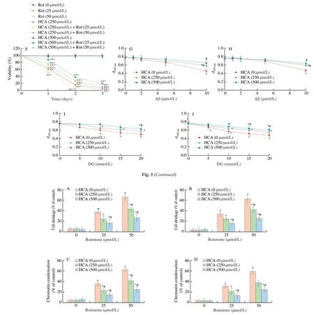
Fig. 3 Effects of HCA on viability of cells following the addition of toxic insults. NQO1 expression level was confirmed by immunoblot after HCA and siRNA treatment for 24 h (A, B). After pre-incubation with HCA, cells were exposed to indicated toxic insults (C-J) for 24 h, and cell viability was determined by trypan blue exclusion (E, F) and the MTT assay (C, D, G, H, I, J). (A, C, E, G, I) SH-SY5Y; (B, D, F, H, J) HT22. Six independent experiments for the cell viability assays were carried out. Values are the mean ± SEM (n = 6). *P < 0.01 compared with the value for the same cells cultured under normal culture conditions. **~P < 0.01 compared with the value between cells cultured without or with indicated toxic insults in the group of cells exposed to the same HCA concentration at the same time point. #P < 0.01 compared with the value between cells cultured in the absence or presence of HCA under the same concentration of indicated toxic insults.).

Fig. 4 Effects of HCA on apoptotic features following treatment with rotenone. Following exposure to rotenone, levels of cell membrane permeabilization(propidium iodide-positive cells) (A, B) and chromatin condensation (C, D) were also measured after the addition of rotenone for 24 h. (A, C) SH-SY5Y;(B, D) HT22. Six independent experiments for the apoptosis assays were carried out. Values are the mean ± SEM (n = 6). *P < 0.01 compared with the value for the same cells cultured under normal culture conditions. #P < 0.01 compared with the value between cells cultured in the absence or presence of HCA under the same concentration of rotenone.
3.2 HCA protects neuronal cells against toxic insults
It has been shown that overexpressed NQO1 and b5R protected neuronal cells from various toxic insults [69,70]. Considering increased NQO1 levels in two cell lines, we designed various NQO1 expressional models by using HCA and siRNA fragments Figs. 3A and B.In order to investigate whether mitochondrial dysfunction could also be attenuated by induced PM redox enzymes, rotenone (a mitochondrial complex I inhibitor) was used to induce mitochondrial dysfunction. Changes in the cell viability were monitored for 1-3 days following exposure to the insults. Treatment of SH-SY5Y and HT22 cells with rotenone significantly decreased viability in a dosedependent manner (P< 0.01) (Figs. 3C-F). However, two neuronal cells with high levels of NQO1 following incubation with HCA were more resistant to being killed by rotenone at the same concentration than were cells cultured without HCA (P< 0.01) (Figs. 3C-F). This meant that high expression of NQO1 increased cell survival under abnormal mitochondrial function, overcoming defect of energy metabolism. Further, two different toxins affecting energy metabolism in neurons, such as Aβ1-42(a misfolded peptide causing Aβ toxicity) and 2-DG (a glycolysis inhibitor), were applied in the presence or absence of HCA. Exposure of the two cells to Aβ1-42lowered metabolic activity (P< 0.01) (Figs. 3G-J), which was not attenuated by treatment with HCA (Figs. 3G-J). 2-DG also decreased metabolic activity in a dose-dependent manner (P< 0.01)(Figs. 3G-J). HCA also protected both cells from 2-DG (P< 0.01),but its protection became significant when exposed to 500 µmol/L HCA (Figs. 3G-J).
3.3 HCA delays apoptotic cell death
In order to investigate how induced of NQO1 or b5R protect neuronal cells against mitochondrial dysfunction, rotenone was used to cause apoptotic cell death, oxidative/nitrative damage and impaired mitochondrial functions. Under normal culture conditions (i.e. culture medium without rotenone), there was slight cell shrinkage in both SH-SY5Y and HT22 cells, confirmed by a fluorescent microscope(P< 0.01) (Figs. 4A and B). Rotenone accelerated cells shrinkage(P< 0.01). HCA addition made the cells resistant to the morphological change induced by rotenone (P< 0.01) (Figs. 4A and B). Similarly, under normal culture conditions, both cells exhibiting chromatin condensation were very rare (Figs. 4C, D and S4A-D).Exposure to rotenone caused increased chromatin condensation.HCA attenuated the appearance of chromatin condensation (P< 0.01)(Figs. 4C, D and S4A-D). Moreover, rotenone increased propidium iodide positive cells, while HCA treatment reduced propidium stained cell levels (Figs. S4E-H).
3.4 HCA attenuates oxidative/nitrative damage
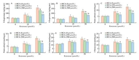
Levels of 8-isoprostane, a lipid peroxidation marker, were not increased in cells pre-incubated with HCA compared to control cells (Figs. 5A and B). 8-Isoprostane levels were greatly elevated in both SH-SY5Y and HT22 cells following exposure to rotenone(P< 0.01), but these levels were attenuated by the addition of HCA in a dose-dependent mannerP< 0.01) (Figs. 5A and B). Levels of protein carbonyls, a hallmark of protein oxidation, were slightly lower in SH-SY5Y cells treated with HCA compared with control cells under normal culture conditions (Figs. 5C and D). Following the addition of rotenone, levels of protein carbonyls in control cells were significantly increased in a dose-dependent manner (P< 0.01),whereas these levels were attenuated by exposure of the cells to HCA(P< 0.01) (Figs. 5C and D). Levels of 3-nitrotyrosine, a biomarker of protein nitration, were also dramatically increased in control cells after the addition of rotenone (P< 0.01) (Figs. 5E and F). However,these levels were attenuated in cells cultured in the presence of HCA(P< 0.01) (Figs. 5E and F).
3.5 HCA is not a direct ROS scavenger
Some studies reported that HCA could be a better antioxidant molecule than catechin and quercetin [68]. In order to examine whether HCA scavenges ROS directly, a DPPH assay was performed using H2O2. HCA itself did not decrease ROS levels in the absence of cells (Fig. S2A), whereasα-tocopherol diminished ROS levels directly (Fig. S2B). Levels of ROS produced in SH-SY5Y cells dramatically increased following treatment with rotenone (P< 0.01)(Fig. S2C). These levels were lower in SH-SY5Y cells supplemented with HCA (P< 0.01) (Fig. S2C). However, these levels were not significantly changed following addition of 2-DG (Fig. S2D).

Fig. 5 Effects of HCA on levels of oxidative/nitrative damage to lipids and proteins after exposure to rotenone. Cell extracts were used to measure levels of 8-isoprostane (A, B), protein carbonyls (C, D) and nitrotyrosine (E, F) following exposure to the indicated concentration of rotenone for 24 h. (A, C, E) SH-SY5Y;(B, D, F) HT22. Six independent experiments for the oxidative/nitrative damage assays were carried out. Values are the mean ± SEM (n = 6). *P < 0.01 compared with the value for the same cells cultured under normal culture conditions. #P < 0.01 compared with the value between cells cultured in the absence or presence of HCA under the same concentration of rotenone.

Fig. 6 Attenuation of mitochondrial ROS production by HCA. Cells were cultured with HCA, and then mitochondrial fractions were isolated by centrifugal fractionation. Production of the mitochondrial ROS was assessed in the presence of glutamate/malate (A, B) or succinate (C, D). (A, C) SH-SY5Y;(B, D) HT22. Six independent experiments for the ROS assays were performed. Values are the mean ± SEM (n = 6). *P < 0.01 compared with the value for the same cells cultured under normal culture conditions. #P < 0.01 compared with the value between cells cultured in the absence or presence of HCA under the same concentration of rotenone.

Fig. 7 Stimulation of mitochondrial functions by HCA. Cells were pre-incubated with HCA, and then mitochondrial fractions were isolated by centrifugal fractionation. Activities of the mitochondrial complexes I and II were measured in the presence of glutamate/malate (A, B) and succinate (C, D). APR was also determined using the same electron donors, glutamate/malate (E) or succinate (F). (A, C, E) SH-SY5Y; (B, D, F) HT22. Six independent experiments for the mitochondrial activity and ROS assays were performed. Values are the mean ± SEM (n = 6). *P < 0.01 compared with the value for the same cells cultured under normal culture conditions. #P < 0.01 compared with the value between cells cultured in the absence or presence of HCA under the same concentration of rotenone.
3.6 HCA-induced PM redox enzymes suppress mitochondrial ROS production
A major portion of ROS in cells is produced in the mitochondria,and our previous studies showed that overexpressed NQO1 and b5R diminished levels of ROS production in response to addition of mitochondrial inhibitors (rotenone and antimycin A) [70,71]. In order to verify whether, and to what extent, production of mitochondrial ROS was weakened in cells cultured in the presence of HCA, ROS generation following addition of rotenone was measured using inhibitors such as rotenone (for the complex I) and antimycin A (for the complex III). In the absence of the mitochondrial inhibitors, ROS levels in both SH-SY5Y and HT22 cells were decreased by addition of HCA (Figs. 6A-D). Following treatment with rotenone, levels of ROS production in both cells were significantly elevated (P< 0.01)(Figs. 6A and B). However, the increased ROS levels in two cells were decreased when cells were exposed to HCA compared to those from the cells cultured without HCA (P< 0.01) (Figs. 6A and B).Moreover, in live cells, levels of mitochondrial superoxide were increased in cells treated with rotenone only, but these levels were attenuated by HCA (P< 0.01) (Figs. S2E-H). Levels of ROS were also greatly increased by treatment of both SH-SY5Y and HT22 with antimycin A (P< 0.01) (Figs. 6C and D). But HCA addition led the cells produce lower levels of ROS incubated with antimycin A(P< 0.01) (Figs. 6C and D).
3.7 HCA-induced PM redox enzymes promote mitochondrial functions
It has been reported that overexpressed NQO1 and b5R can stimulate mitochondrial activity without causing a further increase in ROS generation [70,71]. Activities of the mitochondrial complexes I and II and APR were determined to investigate the mechanism by which the PM redox enzymes regulate the mitochondrial functions in neuronal cells. Mitochondrial complex I activity was dramatically increased in SH-SY5Y cells when cultured in the presence of HCA in a dose-dependent manner (P< 0.01) (Fig. 7A). Mitochondrial complex I activity was significantly decreased by exposure to rotenone in a dose-dependent manner (P< 0.01); however, the inhibition of complex I activity was attenuated by culturing SH-SY5Y cell with HCA (P< 0.01) (Fig. 7A). Complex II activity was also higher in SH-SY5Y cells supplemented with HCA than that in the control cells(P< 0.01) (Fig. 7C). Treatment of SH-SY5Y cells with rotenone tended to decrease complex II activity, but it was not significant(Fig. 7C). In association with the enhanced activity of mitochondrial complexes I and II, APR was also significantly increased in SH-SY5Y cells cultured with HCA in a dose-dependent manner (P< 0.01)(Fig. 7E). APR in SH-SY5Y cells was decreased after addition of rotenone, but it was attenuated by incubation in the presence of HCA (P< 0.01) (Fig. 7E). Similarly, HCA made activities of the complexes I and II in HT22 cells higher than those in control cells cultured under normal culture conditions (Figs. 7B and D). Complex I activity in HT22 cells were decreased by rotenone (P< 0.01), but it was diminished by culturing the cells with HCA (P< 0.01) (Fig. 7B).Complex II activity in HT22 cells was also increased by HCA (P< 0.01),but the activity was not significantly changed by rotenone (Fig. 7D).APR decreased after incubating HT22 cells with rotenone was greatly elevated by addition of HCA (Fig. 7F).
3.8 HCA stimulates mitochondrial fusion and inhibits mitochondrial fission
Opa1 and Mfn1 (which are biomarkers of mitochondrial fusion)and Drp1 (which is a marker of mitochondrial fission) were monitored to elucidate the mechanism by which induction of NQO1 or b5R regulates mitochondrial elongation and fragmentation processes.Levels of Opa1 and Mfn1 were significantly elevated in SH-SY5Y cells supplemented with HCA in a dose-dependent manner compared to those in control cells under normal culture conditions (Fig. 8A). In contrast, Drp1 levels in SH-SY5Y cells were dramatically reduced by exposure to HCA (Fig. 8A). Similarly, treatment of HT22 cells with HCA induced higher expression of Opa1 and Mfn1 and lower levels of Drp1 (Fig. 8B).

Fig. 8 Effects of HCA on mitochondrial fusion and fission processes.Cells were cultured with HCA and lysates from SH-SY5Y (A) and HT22 cells (B) were assessed by the immunoblot analysis using antibodies against Opa1, Mfn1 and Drp1, respectively. Three independent experiments for the immunoblot analysis were carried out.
4. Discussion
Mitochondrial dysfunction is a common mechanism found at the early stage of many neurodegenerative disease. Cells can survive in response to mitochondrial dysfunction by stimulating glycolysis or alternative electron transport through activated PM redox enzymes.Previously, we showed that overexpressed PM redox enzymes such as NQO1 and b5R enhanced mitochondrial functions and made neuronal cells more resistant to some toxic insults [69-71].
This study found that treatment of SH-SY5Y cells with HCA induced expression of NQO1 (Fig. 2A), similar to other neurohormetic phytochemicals such as sulforaphane, curcumin, and catechin [85]. HCA-dependent induction of NQO1 expression was consistent with its elevation of NQO1 activity (Fig. 2A). In addition,HCA also caused higher expression and increased activity of another PM redox enzyme, b5R, in HT22 cells (Fig. 2D), suggesting the cell type specific induction of PM redox enzymes by HCA. These induced PM redox enzymes consequently increased the NAD+/NADH ratio, as expected (Figs. 2E and F), and protected both neuronal cells against toxic insults such as rotenone, Aβ and 2-DG, as measured by cell viability assays (Figs. 3C-J). These data were consistent with previous results using SH-SY5Y cells transfected with NQO1 or b5R [69,70].The fact that, following exposure to the toxic insults, decreased metabolic activity in both SH-SY5Y and HT22 cells was ameliorated by HCA (Fig. 7) suggests its protective effects of HCA, especially on mitochondrial functions. Therefore, rotenone was used to investigate how HCA protects neuronal cells under the toxic culture condition of the mitochondria.
HCA attenuated the appearance of apoptotic features (Fig. 4) and markers of oxidative/nitrative damage (Fig. 5) following treatment with rotenone, meaning that HCA can protect neuronal cells from oxidative/nitrative stress-induced cell death. HCA diminished ROS production in the mitochondria by exposure to rotenone (Figs. 6 and S2E-H), consistent with the previous result [68]. However, this decreased ROS was not due to the direct scavenging by HCA because HCA was not a direct antioxidant (Fig. S3A), suggesting that induced PM redox enzymes attenuated ROS generation. In addition, HCA enhanced mitochondrial functions such as activities of complexes I and II and ATP production (Fig. 7). These data suggest that some of these neuroprotective effects of HCA were caused by induction of ARE gene expression through the Nrf2-Keap1 pathway (Figs. 9 and S1E-F), consistent with previous reports [69-71]. This study showed that HCA could be a good Nrf2 inducer likeo-coumaric acid, which can also reduce oxidative stress [67], indicating its structural similarity too-coumaric acid. Other Nrf2 inducers (including sulforaphane,hydrocytyrosol and 3H-1,2-dithiole-3-thione) can also induce the expression of NQO1 and protect cells against toxic insults [86-88].
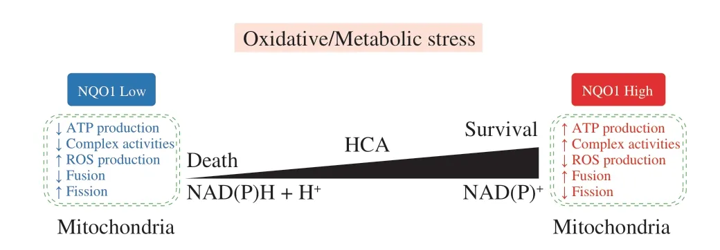
In a previous study, we suggested that overexpressed PM redox enzymes in neuronal cells could protect cells by decreasing the production of O-2· through more efficient electron transport in the mitochondria [70,71]. In fact, mitochondrial fission is a typical process during apoptotic cell death [89,90]. Mitochondrial fission contributes to ROS production under stress conditions and is promoted by Drp1, while mitochondrial fusion can attenuate ROS generation and is modulated by Opa1 and Mfn1 [91], which is consistent with this study (Figs. 6 and 8). In AD, increased ROS can cause excessive mitochondrial fragmentation and enhance defective mitophagy [92]. Mutant APP and Aβ caused abnormal mitochondrial structures, resulting in a loss of dendritic spines and cognitive impairment [29]. In addition, Aβ processed from overexpressed APP down-regulates Drp1 and up-regulates Opa1 and Mfn1 [30]. Our previous study showed that the toxicity of Aβ could be attenuated by enhanced NQO1 [39], consistent with lower levels of Drp1 in neuronal cells cultured with HCA (Fig. 9).

Fig. 9 A schematic diagram of neuroprotective mechanisms induced by phytochemicals. Phytochemicals including HCA activate the up-regulation of neuroprotective gene products such as NQO1, HO1 and GST. Electron flows are marked with red arrows and neuroprotective roles of phytochemicals are shown in orange. Translocated proteins are indicated by green arrows.
5. Conclusion
Taken together, our study found that HCA induces the expression of PM redox enzyme, NQO1 and b5R, protecting neuronal cells from metabolic stress through maintaining redox homeostasis and improving mitochondrial functions. Our finding that HCA-induced PM redox enzymes could protect cells against mitochondrial dysfunction, suggesting that a phytochemical, HCA, could be used as a therapeutic molecule because it can attenuate Aβ toxicity(Figs. 3G and H), and enhance mitochondrial function (Fig. 9).The extent of PM redox enzyme induction by HCA can be various depending on the cell type. Therefore, further research will be required to identify specific PM inducers for therapeutic intervention for neurodegenerative disease.
Conflict of interest
The authors declare that there are no conflicts of interest.
Acknowledgements
This work was supported by the National Research Foundation of Korea (NRF) of the Korean Government (NRF-2021R1F1A1051212),and by Logsynk Co. Ltd. (2-2021-1435-001).
Appendix A. Supplementary data
Supplementary data associated with this article can be found, in the online version, at http://doi.org/10.1016/j.fshw.2022.10.011.
杂志排行
食品科学与人类健康(英文)的其它文章
- Emerging natural hemp seed proteins and their functions for nutraceutical applications
- A narrative review on inhibitory effects of edible mushrooms against malaria and tuberculosis-the world’s deadliest diseases
- Modulatory effects of Lactiplantibacillus plantarum on chronic metabolic diseases
- The role of f lavonoids in mitigating food originated heterocyclic aromatic amines that concerns human wellness
- The hypoglycemic potential of phenolics from functional foods and their mechanisms
- Insights on the molecular mechanism of neuroprotection exerted by edible bird’s nest and its bioactive constituents
