Completed attention convolutional neural network for MRI image segmentation①
2022-10-22ZHANGZhongLVShijieLIUShuangXIAOBaihua
ZHANG Zhong(张 重), LV Shijie, LIU Shuang, XIAO Baihua
(*Tianjin Key Laboratory of Wireless Mobile Communications and Power Transmission,Tianjin Normal University, Tianjin 300387, P.R.China)
(**State Key Laboratory for Management and Control of Complex Systems, Institute of Automation,Chinese Academy of Sciences, Beijing 100190, P.R.China)
Abstract Attention mechanism combined with convolutional neural network (CNN) achieves promising performance for magnetic resonance imaging (MRI) image segmentation,however these methods only learn attention weights from single scale,resulting in incomplete attention learning. A novel method named completed attention convolutional neural network (CACNN) is proposed for MRI image segmentation. Specifically, the channel-wise attention block (CWAB) and the pixel-wise attention block (PWAB) are designed to learn attention weights from the aspects of channel and pixel levels.As a result, completed attention weights are obtained, which is beneficial to discriminative feature learning. The method is verified on two widely used datasets (HVSMR and MRBrainS),and the experimental results demonstrate that the proposed method achieves better results than the state-of-theart methods.
Key words:magnetic resonance imaging (MRI) image segmentation,completed attention convolutional neural network (CACNN)
0 Introduction
Magnetic resonance imaging (MRI) is one of the fundamental technologies to detect different diseases,such as brain tumour,cardiovascular lesions and spinal deformity[1]. This technology is widely used due to its non-invasive characteristic and multi-modality information. As the basis of medical image technologies, MRI medical image segmentation is valuable for research and practical application. For example,it can assist doctors in clinical diagnosis, surgical guidance and so on.
With the development of deep learning, convolutional neural network (CNN) dominates the field of MRI image segmentation. Some methods[2-3]apply the fully convolutional network (FCN) for segmentation,which changes the fully connected layer into the convolution layer and fuses the features of pooling layers and the last convolution layer. Ronneberger et al.[4]designed the U-shape architecture network (U-Net) for biomedical image segmentation, which utilizes the contracting path and the symmetric expanding path along with skip connections to obtain the pixel-wise prediction. Because of the superiority of U-Net, some variants[5-7]were proposed to apply in the field of medical image segmentation.
Recently, the attention mechanism[8], which is prone to pay attention to the important parts of an image rather than the whole one, is introduced into the medical image segmentation. With the attention mechanism, the attention-aware features were generated to adaptively adjust the weights of features[9]. Pei et al.[10]proposed the position attention module and the channel attention module in single scale so as to make the network concentrate on the core location of colorectal tumour. Mou et al.[11]presented CS2-Net which applies the self-attention mechanism to learn rich hierarchical features for medical image segmentation. However, the above-mentioned attention-based methods only learn attention weights from single scale, which is difficult to obtain completed attention information.
In this paper, a novel method named completed attention convolutional neural network (CACNN) is proposed for MRI image segmentation, which learns channel-wise attention weights and pixel-wise attention weights simultaneously. To this end, CACNN is designed as the symmetric structure including the encoder, the decoder, the channel-wise attention block( CWAB ), and the pixel-wise attention block(PWAB). Specifically, CWAB learns the attention weights for each channel so as to adaptively fuse the feature maps of the encoder and the decoder at the same scale. Meanwhile, PWAB learns the attention weights for each pixel in order to fuse the feature maps from different blocks of decoder. In a word, CACNN could learn the attention weights for different aspects,which forces the deep network to focus on extracting the discriminative features for MRI image segmentation.
1 Approach
The framework of the proposed CACNN is shown in Fig.1, which includes the encoder, the decoder,CWAB, and PWAB. In this section, each component of CACNN is introduced in detail.
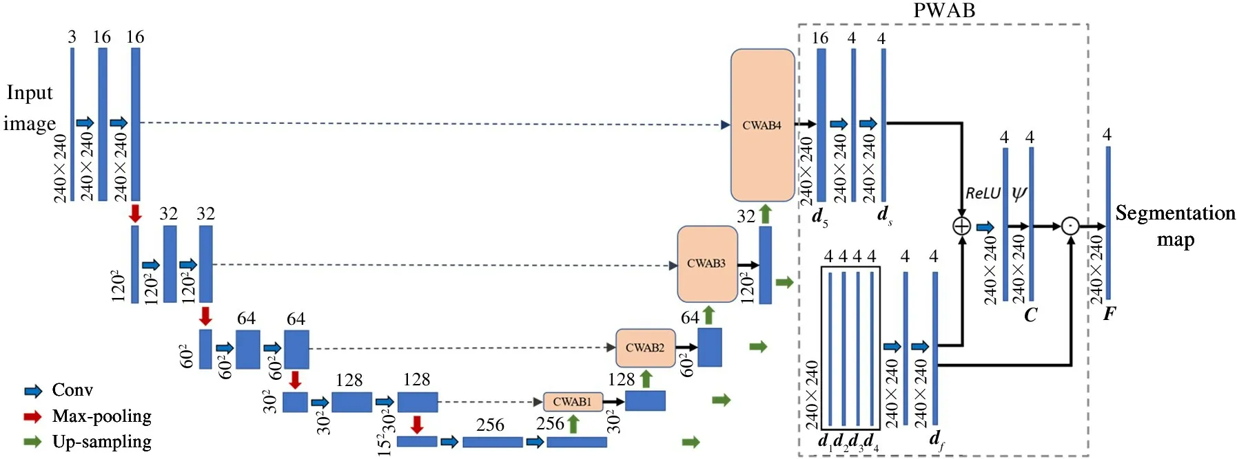
Fig.1 The framework of the proposed CACNN
1.1 The structure of CACNN
CACNN is the symmetric structure where the encoder and the decoder both contain four blocks. As for the encoder, each block consists of two convolution layers and one max-pooling layer, where the kernel size of convolution layer is 3 ×3 with the sliding stride of 1 and the kernel size of max-pooling layer is 2 ×2 with the sliding stride of 2. As for the decoder, each block includes the up-sampling operations to obtain the feature maps with the same size of the corresponding encoder block. Instead of the skip connections, the feature maps of the corresponding blocks from the encoder and the decoder are fed into CWAB. Afterwards,the outputs of CWAB1-3 and the minimum scale feature maps are fed into PWAB after up-sampling operations. Meanwhile, the output of CWAB4 is also as the input of PWAB to generate the final segmentation map.
1.2 Channel-wise attention block
In order to fuse the information from the encoder,some traditional segmentation methods[2-4]adopt the skip connections to directly concatenate the feature maps from the encoder and the decoder. However, the skip connections neglect the importance of different channels of feature maps. Hence, CWAB is proposed to assign different attention weights to each channel.Since there are four blocks of the encoder and the decoder, four CWABs are inserted into CACNN. The structures of four CWABs are similar, and therefore taken CWAB1 is an example. The structure of CWAB1 is shown in Fig.2. The feature maps (IandT) of the corresponding block from the encoder and the decoder are as the input of CWAB1, and they are first conducted by the max-pooling operation to obtainH∈R1×128andQ∈R1×128, respectively. Then, the attention weights can be obtained as
W= softmax(θ(A× [H‖Q])) (1)
where, ‖ denotes the concatenation operation,Ais the learnable transformation vector; × indicates the matrix multiplication operation;θis the non-linear transformation, and it is implemented by the LeakReLU activation function in the experiment. As a result,Wcontains the attention weights for each channel, and the output of CWAB is represented as
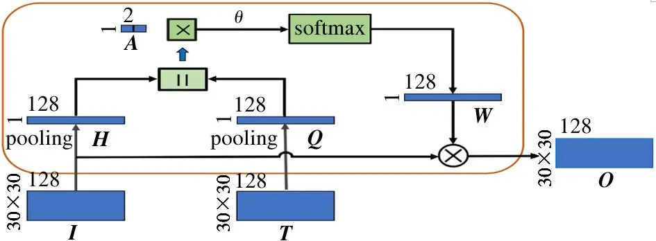
Fig.2 The structure of CWAB

where ⊗indicates the channel-wise multiplication. In a word, the output of CWAB reflects the important of feature maps, and therefore the representation ability is improved.
1.3 Pixel-wise attention block
Most existing segmentation methods[4-5]utilized the feature maps of the last block from the decoder to calculate the final segmentation map, and they only learn the feature maps from single scale. Therefore,PWAB is proposed to fuse feature maps from different scales in order to obtain accurate segmentation map as shown in the right part of Fig.1. Firstly, the feature maps from different blocks are conducted by the upsampling operations to obtain the feature maps with the same size, i. e., 4 × 240 × 240. Then, the feature maps are concatenated in a weighted way and utilize the convolution operation to obtaindf∈R4×240×240:

whereφrepresents the convolution operation. The output of CWAB4 isd5∈R16×240×240, and thends∈R4×240×240is obtained after convolution operations. The attention weights are defined as

whereψrepresents the Sigmoid function. The final segmentation mapFis formulated as

where ⊙indicates the pixel-wise multiplication. As a result, it could fuse the feature maps of multiple scales by assigning attention weights to each pixel. In order to train the network,the cross-entropy function is employed as the loss.
2 Experiments and analysis
2.1 Datasets and implementation details
The experiments is conducted on two challenging datasets: HVSMR[12]dataset and MRBrainS[13]dateset. HVSMR has one modality information, i. e., T2 sequences and it aims to segment blood pool and myocardium in cardiovascular MR images. It includes 1868 training slices and 1473 test slices. The images in HVSMR have different size, and the size of the original images is maintained and fed into the network. MRBrainS contains MR brain scans of three modality information, i. e., T1, T1 inversion recovery and FLAIR sequences. The task of MRBrainS is to segment cerebrospinal fluid, gray matter and white matter. It consists of 104 slices for training and 70 slices for testing,where the size of each slice is 240 ×240. The training images are conducted by a skull stripping pre-processing.
The Adam algorithm is adopted for deep network optimization with the weight decay of 5 ×10⁻⁴and the learning rate of 6 ×10⁻⁴. Furthermore, the batch normalization is utilized in CWABs.
In order to evaluate the performance of MRI image segmentation, two matrices is employed, i. e., pixel accuracy and dice score[1,10]. The pixel accuracy indicates the ratio between the number of correctly classified pixels and the total number of pixels. The dice score reflects the overlap between the prediction results and the ground-truth. The two matrices are defined as
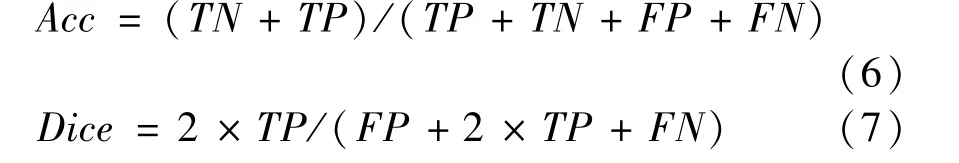
whereTPis the true positive,FPis the false negative,TNis the true negative andFNis the false negative.
2.2 Comparing to the state-of-the-art methods
The proposed CACNN is compared with the stateof-the-art methods, such as FCN[2], SegNet[14]and U-Net[4]where different encoders are utilized, i. e.VGG16, ResNet50 and U-Net structure. The pixel accuracy and the dice score are listed in Table 1, where the following four conclusions can be drawn. Firstly,CACNN gains the best results on the two datasets, because it learns completed attention weights including the channel level and pixel level. The proposed CACNN achieves the pixel accuracy and the dice score of 94.15% and 88.39% on the HVSMR dataset, and 97.13% and 90.48% on the MRBrainS dataset. Secondly, the proposed two attention mechanisms (CWAB and PWAB) both boost the segmentation performance compared with the baseline (U-Net). Note that the backbone of CACNN is designed as U-Net, and therefore it is reasonable to treat U-Net as the baseline.Compared with U-Net, CWAB and PWAB raise the dice score by 0.12% and 1.42% on the HVSMR dataset. It proves that the channel-level attention and the pixel-level attention are both essential for performance improvement. Thirdly, U-Net structure performs better than other network architecture. For example, U-Net with VGG 16 and U-Net with ResNet 50 obtain the best performance for the same encoder (VGG 16 or ResNet 50). Hence, U-Net is chosen as the backbone of CACNN. Finally, MRBrainS contains three kinds of modality information, while HVSMR includes one kind. Comparing their performance, it shows that multiple information is beneficial to the performance improvement.
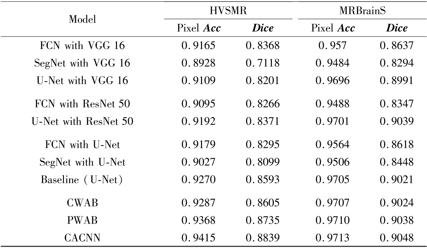
Table 1 Comparison on the HVSMR dataset and the MRBrainS dataset with different methods
Fig.3 illustrates some results of CACNN and other methods on the two datasets. It shows that CACNN is the superiority on dealing with detail information because of the completed attention learning.
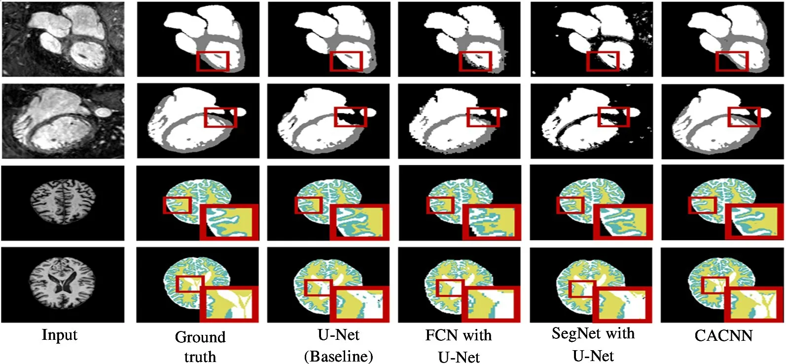
Fig.3 Some results of CACNN and other methods (The first two rows are the samples from HVSMR and the last two rows are samples from MRBrainS)
2.3 Parameter analysis
There are three key parameters in CACNN and therefore a series of experiments are conducted to search the optimal parameter values. Firstly, it tests different number of fused feature maps for PWAB in Eq.(3), and the results are listed in Table 2. It shows the performance improvement with the number of fused feature maps. Hence,the feature maps are fused from four blocks. Then, it study which feature maps to multiply with the attention weights for CWAB and PWAB respectively in Eq.(2) and Eq.(5). From Table 3 and Table 4, it shows thatIanddfare optimal in CACNN.
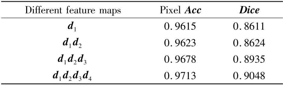
Table 2 Comparison of different numbers of fused feature maps on the MRBrainS dataset
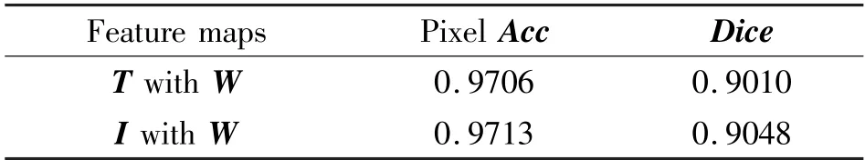
Table 3 Comparison of different feature map combination in CWAB on the MRBrainS dataset
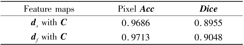
Table 4 Comparison of different feature map combination in PWAB on the MRBrainS dataset
3 Conclusion
CACNN is proposed for MRI image segmentation to learn completed attention weights. CACNN mainly contains CWAB and PWAB which learn attention weights for each channel and each pixel, respectively.With the completed attention weights, the deep network focuses on extracting the discriminative features.CACNN is validated on two datasets HVSMR and MRBrainS, and the experimental results demonstrate that the proposed method outperforms the other methods.
杂志排行

High Technology Letters的其它文章
- Multi-layer dynamic and asymmetric convolutions①
- Feature mapping space and sample determination for person re-identification①
- Energy efficiency optimization of relay-assisted D2D two-way communications with SWIPT①
- Efficient and fair coin mixing for Bitcoin①
- Deep learning based curb detection with Lidar①
- Research on will-dimension SIFT algorithms for multi-attitude face recognition①