Mosquito larva distribution and natural Wolbachia infection in campus areas of Nakhon Ratchasima, Thailand
2022-08-26ThunyaratSurasiangSirilakChumkiewPongsakornMartvisetPathaninChantreeMantanaJamklang
Thunyarat Surasiang, Sirilak Chumkiew, Pongsakorn Martviset, Pathanin Chantree, Mantana Jamklang
1Institute of Molecular Biosciences, Mahidol University, Salaya, Nakhon Pathom, Thailand
2School of Biology, Institute of Science, Suranaree University of Technology, Thailand
3Research Unit in Nutraceuticals and Food Safety, Thammasat University, Pathumthani, Thailand
4Department of Preclinical Science, Faculty of Medicine, Thammasat University, Pathumthani, Thailand
5School of Preclinical Sciences, Institute of Science, Suranaree University of Technology, Thailand
ABSTRACT
KEYWORDS: Mosquito larvae; Wolbachia; Breeding sites; House Index; Breteau Index; Campus area; Dengue
1. Introduction
Mosquitoes are extensively distributed worldwide, especially throughout the tropics and temperate regions[1,2]. Mosquitoes are natural vectors that transmit pathogens to infect humans and cause several diseases such as dengue fever, malaria, chikungunya,filariasis, and Japanese encephalitis[3]. The medical important mosquito species belong to the subfamilies of Culicinae (including genera Aedes, Armigeres, Culex, Haemagogus, Mansonia, Psorophora,Sabethes, and Toxorhynchites), and Anophelinae (includes genus Anopheles) which composes of more than 3 000 species[4]. From all mentioned genera, Aedes is of most concern because of their distribution and transmission of many intractable pathogenic organisms. Aedes (Ae.) aegypti is the most important species that transmits dengue, zika, and yellow fever viruses and filaroid helminths worldwide[3]. Ae. albopictus is a native mosquito species in tropical and subtropical areas, especially in Southeast Asia that also serves as a vector of dengue fever, yellow fever, and chikungunya[5,6]. Every year, around 390 million dengue infections have been reported worldwide[7] with 96 million of them presented clinical manifestations and 70% was reported in Asia[8]. In addition to Aedes, other mosquitoes species are also important, as they act as vectors for many infectious diseases. Culex is another Culicinae mosquito that transmits several diseases such as lymphatic filariasis and Japanese encephalitis, while Mansonia and Armigeres are the vectors of nematode parasites that cause lymphatic filariasis[9].Apart from the pathogenic vector, Toxorhynchites is a beneficial biocontrol since they are predaceous on other mosquito larvae found in the same areas[10].
In the absence of effective vaccines or prophylactic agents against most of the arboviruses and vector-borne parasites, current efforts are mainly based on controlling vector populations by eliminating breeding sites, killing mosquito larvae, and treating with outdoor insecticides or repellents. However, chemical-based control methods may lead to the development of mosquito resistance,as well as environmental contamination and side effects on nontarget organisms[11]. Therefore, the safest ways to control the disease are either eliminating breeding sites or using biocontrol methods. Natural mosquito breeding sites could be different among the mosquito species, leading to the risks of each region depending on the characteristics of breeding site containers.Consequently, alternative and innovative vector control strategies have emerged, and one of the most promising methods is based on the use of endosymbiotic bacteria, Wolbachia[12-15]. Wolbachia has been one of the most studied biocontrol for arboviruses and parasite transmission control. This approach involves the release of mosquitoes transinfected with the vertically transmitted Wolbachia,which can suppress arbovirus replication in mosquitoes, so it can be a potentially promising means for controlling dengue transmission in endemic settings[6,16-19].
Thailand is an endemic area for dengue fever with more than 60 000 cases in 2019. In the same year, more than 10 000 cases were reported in Nakhon Ratchasima and surrounding provinces,indicating this area is one of the highest endemic regions of the country[20]. Previous studies have reported that most cases of dengue fever patients were children and teenagers[21,22]. Therefore,we have been interested in studying the distribution of mosquito species and the occurrence of Wolbachia in Nakhon Ratchasima province where more than 10 000 students reside. There have been a few studies on the distribution of Wolbachia in Thailand but most of them focused on Wolbachia in adult insects or mosquitoes[23-27].Our research aims to study the distribution and breeding sites of mosquito species collected in 2017 and 2018 as well as detect the presence of endosymbiotic bacteria Wolbachia from the collected mosquito larvae.
2. Materials and methods
2.1. Study sites
Mosquito larval survey was conducted in two different study sites(residential areas and academic buildings) from August 2017 to November 2018 at Suranaree University of Technology, Nakhon Ratchasima located in Northeastern Thailand (14.881 8° N, 102.020 7°E) where there are natural forests, ponds, and constructed buildings which has an area of 11.2 km2. Mosquito larvae samples were collected from 13 and 17 buildings from the residential areas and academic buildings, respectively.
2.2. Entomological studies
Larval surveys were conducted in both study areas by using an 11.5 cm diameter fishnet. Mosquito breeding sites were sampled in both indoors and outdoors within 15 meters of the households as suggested by Wongkoon[28]. All breeding larvae found in small containers were filtered through the fishnet into the buckets. The ones in large containers were sampled by dipping the fishnet in the water, starting a swirling motion, and sampling all edges of the containers[29]. All breeding sources of mosquitos were grouped into 13 different container types: flower plastic vases (FPV), flower glass vases (FGV), flower ceramic vases (FCV), plastic tank (PT), plant water pot (PWP), bowl (BO), small earthen jars (SEJ), cement tank(CT), paint bucket (PB), pottery vases (PV), waste containers (WC),coconut shells (CS), and others (OT).All 5 472 live mosquito larvae were collected in plastic bags and brought to the laboratory for species identification by using GLOBE mosquito protocols[30] and, Rattanarithikul & Panthusiri’s keys[31].After identification, all mosquito larvae samples were fixed in 70%ethanol and stored in the freezer (-80 ℃) for DNA extraction. The number of the larvae was counted and calculated for three larval indices: House Index (HI), Container Index (CI), and Breteau Index (BI) according to the standard WHO guidelines on dengue control (vector surveillance). The BI and HI are commonly used for determination of priority (risk) areas for control measures. The HI and BI of greater than 5% and 20%, respectively, for any locality is indicated that these areas are dengue-sensitive, suggesting a high risk of dengue virus distribution[32].

Table 1. List of the primers used for wsp gene, 16s rDNA, and 28s rDNA amplification.
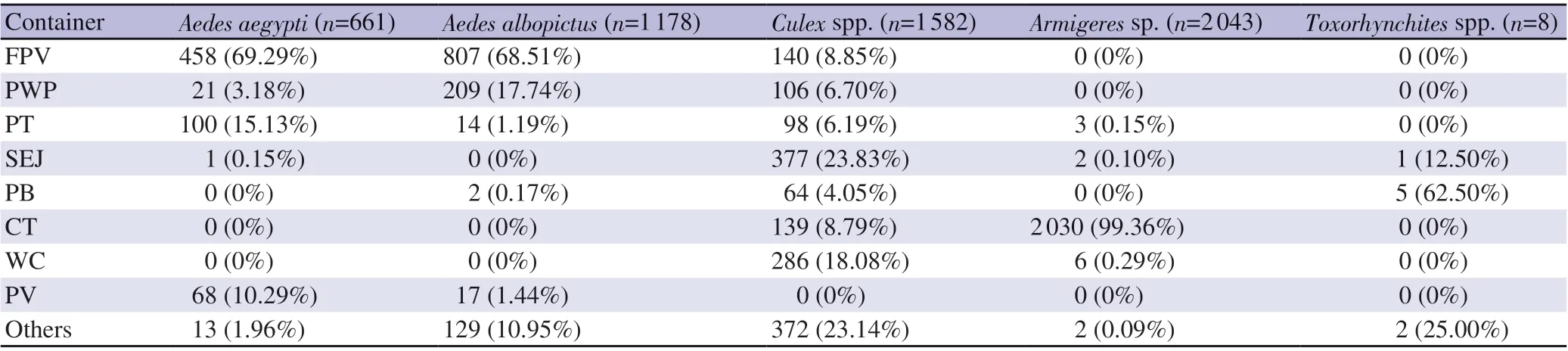
Table 2. Different mosquito larval species found in a variety of the water containers.
2.3. DNA extraction
The mosquito larvae from 57 containers collected from different areas covering different types of containers were chosen for DNA extraction. The larval samples were homogenized in liquid nitrogen and DNA were then extracted according to the manufacturing by using HiPurA™ Multi-Sample DNA Purification Kit (Himedia,India). The DNA concentration was quantitated via NanoDrop™2000/2000c spectrophotometers (Thermo Fisher Scientific,USA) before proceeding to polymerase chain reaction (PCR)amplification.rDNA was at 443 bp in size, and the PCR products of wsp gene were ranged in 590-632 bp. These PCR products were proceeded for DNA sequencing (Biobasic, Canada) for wsp gene confirmation.
2.5. Bioinformatics analysis
The sequence alignment was generated using 4 peaks program(B.V. Gerberastraat, the Netherlands). Each sequence was checked and edited manually for 16s rDNA. The modified DNA sequence was submitted to the BLASTN on NCBI database, whereas the reverse DNA sequence was converted to reverse complement sequence before DNA sequencing analysis.
2.4. PCR amplification and DNA sequencing
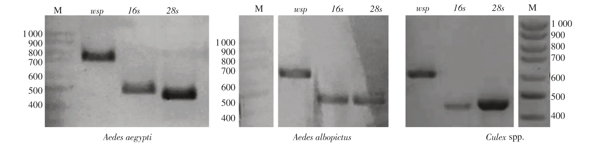
The larvae samples collected from each location were screened for the presence of Wolbachia by PCR amplification as previously described[33,34]. The gene encoding for Wolbachia surface protein was amplified with the wsp 81F and wsp 691R primers. The primer sequences used in this study are shown in Table 1. PCR was conducted in a 25 μL reaction volume using (KOD One™, Toyobo, Japan). The PCR was carried out on C1000 Touch™ Thermal Cycler (Bio-Rad,USA) with the appropriate condition including a pre-denaturation for 5 min at 95 ℃, followed by 35 cycles of denaturation for 30 s at 95 ℃,annealing for 45 s at 55 ℃, and extension for 90 s at 72 ℃, and a final extension for 10 min at 72 ℃. All genomic DNA samples used for wsp gene detection were also amplified for 16s rDNA and 28s rDNA gene sequence as a positive control for the presence of bacterial and eukaryotic (mosquito) genomic DNA, respectively. The PCR products received from 16s rDNA detection appeared at 438 bp, whereas 28s
3. Results
3.1. Mosquito breeding sites
The association of the mosquito breeding sites, and the study areas was investigated. The breeding sites in the academic buildings from the most to least were FPV (63.50%, 87/137), PT (8.03%, 11/137),PWP (7.30%, 10/137), OT (5.84%, 8/137), CT (4.38%, 6/137),PV (2.92%, 4/137), SEJ (2.19%, 3/137), PB (2.19%, 3/137), FCV(2.19%, 3/137), FGV (0.73%, 1/137), and WC (0.73%, 1/137),respectively. The breeding sites in the residential areas were PWP(33.33%, 16/48), SEJ (25.00%, 12/48), FPV (22.92%, 11/48), PT(6.25%, 3/48), CS (4.17%, 2/48), FCV (4.17 %, 2/48), PV (2.08%,1/48), and BO (2.08%, 1/48) respectively. These data revealed that FPV was the main breeding site of mosquitos in both academic buildings and residential areas.

Table 3. The number of households, containers, and larval indices of Aedes species during 2017 and 2018.

Figure 1. The breeding site distribution of Aedes spp. A: the breeding site distribution of Aedes aegypti. B: the breeding site distribution of Aedes albopictus.FPV: flower plastic vases; PWP: plant water pot; PT: plastic tank; SEJ: small earthen jars; PB: paint bucket; CT: cement tank; WC: waste containers; PV:pottery vases; OT: others.
3.2. The prevalence of mosquito larval species
A total of 5 472 mosquito larvae were collected. Five species were identified in which the abundance from the most to least were Armigeres sp. (37.34%, 2 043/5 472), Culex spp. (28.91%, 1 582/5 472), Ae.albopictus (21.53%, 1 178/5 472), Ae. aegypti (12.08%, 661/5 472),and Toxorhynchites spp. (0.15%, 8/5 472), respectively.
3.3. The breeding sites specific for each mosquito species
The different mosquito larval species were found in both academic buildings and residential areas in a variety of the water containers as shown in Table 2. The three major breeding sites of Ae. aegypti were FPV, PT, and PV. Ae. albopictus were also found in both academic buildings and residential areas, and mostly in FPV and PWP whereas Culex spp. were randomly distributed in a variety of containers (SEJ, WC, FPV, CT, PWP, PT, PB, and OT). Armigeres sp.is the most abundant mosquito larvae found mostly in cement tank.Toxorhynchites spp. were rarely seen in all containers.
3.4. Breeding sites for dengue virus vectors
Breeding sites of Aedes spp. as the major vectors of the dengue fever illustrated that, Ae. aegypti were mainly distributed in academic buildings in which FPV was the most abundant breeding site found indoors and the second most abundant was PWP found outdoors (Figure 1A). Ae. albopictus have the major breeding sites in the containers found in FPV located in indoors of the academic buildings (Figure 1B), while Ae. albopictus were distributed mainly in both indoors (FPV) and outdoors in the residential areas (PWP).
3.5. Larval indices for dengue fever risk indication
Our results reflected that in the period of 2017, the HI and BI for Ae. aegypti were 26.67% and 80.00%, respectively, and that for Ae.albopictus were 36.67% and 96.67%, respectively. In the period of 2018, the HI and BI for Ae. aegypti were 40.00% and 260.00%,respectively and that for Ae. albopictus were 60.00% and 384.00%,respectively (Table 3).
3.6. Prevalence of Wolbachia infection in different mosquito larval species
In this study, wsp gene which encodes for the Wolbachia surface protein was detected from DNA extracted from the mosquito larvae samples performed by PCR. The results showed that the wsp gene presented in Ae. aegypti, Ae. albopictus, and Culex spp. ranged in 590-632 bp in size, indicating that Wolbachia resided in some of these mosquito larvae (Figure 2). Confirmation of wsp gene by sequencebased analysis showed high-scoring alignments (more than 200) and 100% identity with wsp gene of Wolbachia endosymbiont resided in Aedes spp. and Culex spp. The abundance of Wolbachia in our study revealed that 61.40% of total mosquito larvae were infected with Wolbachia. The highest proportion of Wolbachia was seen in the larvae of Culex spp. (86.21%) when compared to other mosquito larvae, followed by Ae. albopictus (69.23%) and rarely found in Ae.aegypti (9.09%) (Table 4). Wolbachia infection in Toxorynchites spp.was not detected.

Table 4. Prevalence of Wolbachia infection in different mosquito larval species.
4. Discussion
Climate change could bring mosquito-borne diseases to the areas where these diseases had previously not seen. Every year, there is a fluctuation of climate and weather that could lead to the evolution or shifts of mosquito species. Some of the mosquito species including Ae. aegypti and Ae. albopictus are the main vectors for dengue transmission. The high-risk population during primary infection of dengue fever is found in the patient age ranged in 9-20 years old especially children in tropical and subtropical areas who have high chances to expose to these mosquito species[21,22,35].
In our study, we established the distribution of mosquito larvae in containers found in residential areas and academic buildings of Suranaree University of Technology to investigate the association between the areas and container types which could reflect the water conditions whether they are favored for mosquito larvae’s survival and breeding. The FPV was the main mosquito breeding site in the academic building areas and the PWP followed by the FPV was the main spots in the residential areas. Mosquitoes that preferred to breed via FPV might be due to stagnant water. Previous studies elsewhere have shown faster rates of mosquito evolution when temperature and CO2level were higher. In Southeast Asia,Ae. aegypti is the main vector for dengue virus breeds in stagnant water and commonly found indoors, while Ae. albopictus is commonly found outdoors that could be a result of environmental adaptation[36,37]. However, our study showed that indoors were the main breeding sites for both Ae. aegypti and Ae. albopictus.
We also reported that Armigeres sp. was the most prevalent mosquito species in the campus areas of Suranaree University of Technology. This species could lead to the high infection risk of lymphatic filariasis in these areas. The high breeding rate of this species might be a result of being well-adapted to live in any clogged waterways such as CT and other natural habitats. Culex spp., Ae. albopictus, and Ae. aegypti were also widely distributed in the campus areas which brought the most concern of many mosquitos-borne diseases including dengue fever. In addition, our result indicated that the period from 2017 to 2018 are denguesensitive determined by the HI and BI which were greater than 5%and 20%, respectively. The results reflected that the HI and BI are related with dengue endemic in our study areas, which is agreed with Preechaporn et al. who found the Aedes larvae in the highest proportion in three topographical areas (mangrove, rice paddy and mountains)[38].
Wolbachia are distributed ranging from 40%-70% in all types of insects[39-41] including butterflies, bees, beetles, and some mosquito species worldwide. Wolbachia have been found to mediate dengue virus interference depending on several factors such as elevation of the basal immunity and increase in longevity of mosquitoes[42].Wolbachia alone was found to be able to inhibit viral replication,dissemination, and transmission in transinfected Ae. aegypti in experimental studies. Based on the evidence from Cardona-Salgado et al., Wolbachia found in Ae. albopictus did not affect the replication of dengue virus but was able to reduce the viral infection of mosquito salivary glands and limit viral transmission[43].Previous studies from Kittayapong et al. reported that Wolbachia have been found to occur naturally in Ae. albopictus[24] but not in Ae.aegypti which is the main vector of the dengue virus. Another study on Wolbachia distribution in Ae. albopictus conducted in Malaysia showed Wolbachia infection rate ranging from 60% to 100%[44] and a study of the distribution of Ae. albopictus collected from different locations in Peninsular Malaysia reported that Wolbachia infection was widespread in Ae. albopictus population, both in female and male mosquitos[35]. There is evidence of vertical transmission of Wolbachia from mother to offspring of Ae. albopictus population[24]. Another study has shown for the first time that Wolbachia is present in Ae.albopictus and Ae. aegypti larvae from Kuala Lumpur, Malaysia. In Thailand, although there are some studies on Wolbachia distribution in insects and mosquito adults, but there have been no studies to date on Wolbachia in mosquito larvae[24,25,27]. To the best of our knowledge,this study is the first on the detection of Wolbachia in the larvae in Thailand.
Our study showed that the wsp gene existed in Ae. aegypti, Ae.albopictus, and Culex spp., indicating that Wolbachia resided in some of these mosquito larvae. This study revealed that 61.40% of total mosquito larvae were infected with Wolbachia. The highest proportion of Wolbachia infection was seen in the larvae of Culex spp. and the infection rate was found in Ae. albopictus more than Ae.aegypti. No detection of Wolbachia was found in Toxorynchites spp.These observations indicated that Toxorynchites mosquito larvae may be either physiologically unable to support Wolbachia infection or seldom encounter Wolbachia horizontal transmission events. However,larger numbers of the samples in these groups may be required.
Wolbachia did not affect the replication of dengue virus in Ae.albopictus but was able to reduce the viral infection in the mosquito salivary glands and therefore limit viral transmission, suggesting the role of Wolbachia in naturally restricting the transmission of dengue virus in Ae. albopictus[45]. Therefore, scientists have attempted to transinfect Wolbachia into Ae. aegypti and release these mosquitos containing the endosymbiont Wolbachia to the field that would be beneficial for control of dengue fever and other vectorborne diseases[46]. Moreover, there is no evidence on the harm of Wolbachia to human, animals, or the environment. A previous study showed that Wolbachia bacteria did not cause diseases in people or animals (for example, fish, birds, cats, and dogs)[42].
The limitation of our study was that we did not detect wsp gene in all larvae samples. We detected approximately 36% from all larvae samples. Therefore, there might be some incomplete data represented in this report. Another limitation was that we did not submit for an ethic approval for animal (mosquito). Therefore, this is our flaw about performing this project.
In conclusion, the campus areas of Suranaree University of Technology located in Northeast of Thailand was found to be at high risk of endemic mosquito-borne diseases, especially dengue fever, with the higher risk found in indoors rather than outdoors of academic buildings. This is the first study on the distribution of endosymbiont bacteria, Wolbachia in mosquito larvae in Thailand that we found the highest proportion of Wolbachia in Culex spp. and Ae. albopictus but very few in Ae. aegypti. Therefore, transfection of Wolbachia in mosquito larvae as a purpose of suppression of viral transmission could be used as a potential strategy for a biocontrol of mosquito-borne diseases in the future.
Conflict of interest statement
The authors declare that they have no conflict of interest.
Acknowledgements
We would like to acknowledge Suranaree University of Technology, through the Suranaree University of Technology Research and Development for funding. We also thank the Center for Scientific and Technological Equipment, Suranaree University of Technology, Thammasat University Research Unit in Nutraceuticals and Food Safety and Research group in Medical Biomolecules,Faculty of Medicine, Thammasat University.
Funding
The authors received no extramural funding for the study.
Authors'contributions
TS performed the experiments and data analysis, reviewed final version to be published. SC developed concept, designed experimental studies, and performed the experiments. PM performed data analysis, wrote manuscript, and designed data visualization, and revised the manuscript. PC performed data analysis and designed data visualization. MJ developed concept, designed experimental studies, wrote and edited manuscript, reviewed final version to be published. All authors read and approved the manuscript.
杂志排行

Asian Pacific Journal of Tropical Medicine的其它文章
- Unravelling the situation of malaria misdiagnosis in India: Its adverse impact and management strategies
- A rare presentation of Guillain-Barre syndrome with GQ1b positivity: A case report
- Genetic variation of sand flies (Diptera: Psychodidae) in Gampaha and Kurunegala districts of Sri Lanka: Complementing the morphological identification
- Prevalence and factors associated with belief in COVID-19 vaccine efficacy in Indonesia: A cross-sectional study
- Conventional treatments and non-PEGylated liposome encapsulated doxorubicin for visceral leishmaniasis: A scoping review
- Hurdles in achieving the goal of malaria elimination by India