Cough as a neurological sign: What a clinician should know
2022-06-16MohammedAlBiltagiAdelSalahBediwyNerminKamalSaeed
Mohammed Al-Biltagi, Adel Salah Bediwy, Nermin Kamal Saeed
Mohammed Al-Biltagi, Department of Pediatrics, University Medical Center, King Abdulla Medical City, Arabian Gulf University, Manama 26671, Bahrain
Mohammed Al-Biltagi, Department of Pediatrics, Faculty of Medicine, Tanta University, Tanta 31512, Al Gharbia, Egypt
Mohammed Al-Biltagi, Department of Pediatrics, University Medical Center, Dr. Sulaiman Al Habib Medical Group, Manama 26671, Bahrain
Adel Salah Bediwy, Department of Chest Disease, Faculty of Medicine, Tanta University, Tanta 31512, Alghrabia, Egypt
Adel Salah Bediwy, Department of Chest Disease, University Medical Center, King Abdullah Medical City, Arabian Gulf University, Manama 26671, Bahrain
Adel Salah Bediwy, Department of Chest Diseases, University Medical Center, Dr. Sulaiman Al Habib Medical Group, Manama 26671, Bahrain
Nermin Kamal Saeed, Department of Pathology, Salmaniya Medical Complex, Ministry of Health, Kingdom of Bahrain, Manama 26671, Bahrain
Nermin Kamal Saeed, Department of Pathology, Irish Royal College of Surgeon, Busaiteen 15503, Almuharraq, Bahrain
Abstract Cough is a common respiratory complaint driving patients to seek medical advice. Besides being a fundamental respiratory sign, it is also a crucial neurological sign. There are three main types of coughs: Reflex cough (type I), voluntary cough (type II), and evoked cough (type III). Cough is a reflex predominantly mediated by control centers in the respiratory areas of the brainstem, modulated by the cerebral cortex. Cough reflex sensitivity could be increased in many neurological disorders such as brainstem space-occupying lesions, medullary lesions secondary to Chiari type I malformations, tics disorders such as Tourette's syndrome, somatic cough, cerebellar neurodegenerative diseases, and chronic vagal neuropathy due to allergic and non-allergic conditions. Meanwhile, cough sensitivity decreases in multiple sclerosis, brain hypoxia, cerebral hemispheric stroke with a brainstem shock, Parkinson's disease, dementia due to Lewy body disease, amyotrophic lateral sclerosis, and peripheral neuropathy as diabetic neuropathy, hereditary sensory and autonomic neuropathy type IV, vitamin B12, and folate deficiency. Arnold's nerve ear-cough reflex, syncopal cough, cough headache, opioids-associated cough, and cough-anal reflex are signs that could help diagnose underlying neurological conditions. Cough reflex testing is a quick, easy, and cheap test performed during the cranial nerve examination. In this article, we reviewed the role of cough in various neurological disorders that increase or decrease cough sensitivity.
Key Words: Cough reflex; Neurological disorders; Cerebral disorders; Cerebellar disorder; Vagal neuropathy;Parkinsonism
INTRODUCTION
Cough is a forced expiratory effort against a closed glottis that opens suddenly with the expulsion of secretion and foreign particles out of the airways, producing a distinctive sound. Cough is one of the most common complaints driving patients to seek medical advice. It is one of the essential respiratory protective mechanisms, alerting to the presence of a potential or actual respiratory tract lesion, and helps to clear secretions and foreign particles from the airways[1]. There are three main types of coughs according to the central control mechanisms: Reflex cough (type I), voluntary cough (type II), and evoked cough (type III), which follows the urge to cough[1,2].
Both reflex and voluntary cough initiate similar mechanisms of cough motor behavior. Cough is a reflex predominantly mediated by control centers in the respiratory areas of the brainstem, modulated by the cerebral cortex (Figure 1). Cough production passes through three harmonized phases: Inspiratory, compression, and expiratory. It starts with contraction of the inspiratory muscles (drawing air into the lungs), closure of the glottis (which generates a subglottic pressure), and abduction of the vocal folds with a forced expiration (enforcing the glottis to open) with expelling of the secretions out. However, the cough reflex is under the voluntary control of the higher neurologic centers, such as the cerebral cortex, which has a vital role in initiating and inhibiting cough[3]. The reflex has afferent sensory nerve fibers (mainly branches of the vagus nerve), which carry the afferent impulses diffusely to the medulla to reach the upper brain stem and pons. Other brain parts are integrated with the proper function of the cough center in the medulla as the pontine respiratory group, the lateral tegmental field, and deep cerebellar nuclei, which play a role in the pattern of cough generation, and regulation. The efferent fibers carry the signals from the cough centerviathe vagus, phrenic, and spinal motor nerves to the diaphragm, abdominal wall, and muscles[4]. As the cough reflex is a reflex, it could affect or be affected by different neurological disorders (Table 1). Both reflex and volitional coughs could be tested in various neurological and otolaryngological conditions. Other methods can test the sensitivity and efficiency of the cough reflex. The sensitivity can be assessed by the concentration or the duration at which the cough can be evoked when exposed to variable concentrations and/or durations of nebulized aerosols of a tussive substance (such as citric acid, L-tartaric acid, or capsaicin). However, considerable variabilities in the used methods are present while performing the test[5-7]. A group of Japanese scientists developed a device to measure cough strength while testing the cough reflex to assess cough efficiency and strength. They added an electronic spirometer to an ultrasonic nebulizer through a special pipe with a double lumen. The spirometer measures the peak cough flow of the provoked involuntary cough[8].
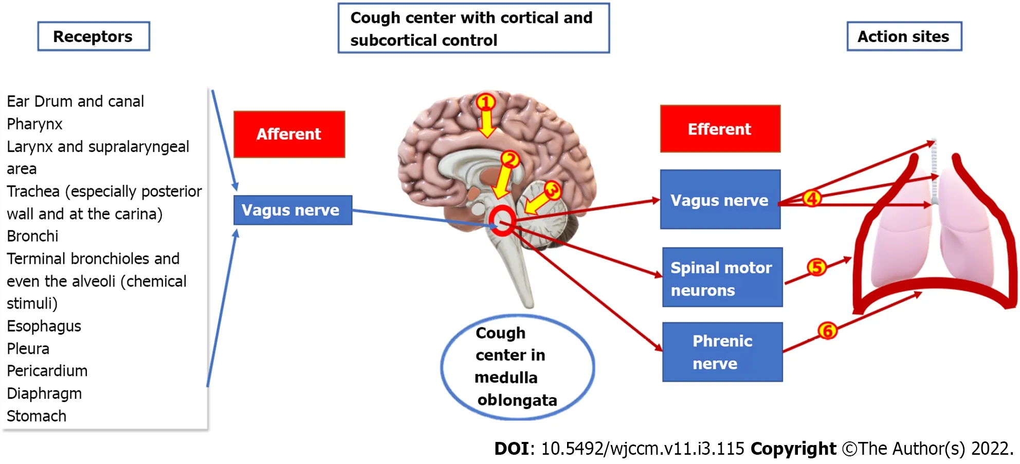
Figure 1 Cough reflex. The cough center lies in the medulla oblongata in the brainstem. Cough receptors project through the vagus nerve to relay neurons in the solitary nucleus, which project to other parts of the respiratory network, especially the pre-Bötzinger complex. Higher brain centers (cerebral cortex[1]) provide voluntary control over cough, e.g., cough inhibition. However, voluntary coughing does not seem to activate medullary systems. Subcortical centers[2] receive signals from other receptors and other emotional stimuli acting through the hypothalamus. Cerebellum[3] also has control over the cough center. The cough center starts the cough by signaling to the effector organs through the vagus nerve to the larynx, trachea, and bronchi[4], spinal motor neurons[5] to the expiratory muscles, and the phrenic nerve[6] to the diaphragm.
NEUROLOGICAL CONDITIONS ASSOCIATED WITH INCREASED COUGH REFLEX SENSITIVITY
Various neurological diseases could associate with increased cough reflex sensitivity, including cerebral and cerebellar disorders, neuromyelitis optica spectrum disorder (NMOSD), and vagal neuropathy (Table 1).

Table 1 Neurological conditions associated with increased cough reflex sensitivity and its mechanism
Cerebral disorders
The urge-to-cough (UTC) is a cognitive sensation needed to initiate and inhibit the reflexive cough stimuli lower than what is usually required to evoke a motor cough. Cough is mediated by the cerebral cortical or subcortical regions and activates multiple brain regions such as the insula, anterior midcingulate cortex, primary sensory cortex, orbitofrontal cortex, supplementary motor area, and cerebellum[9]. Cough, without an apparent medical etiology, is refractory to medical management, underlying a possible psychiatric or psychological basis was previously called psychogenic, habit, or tic cough. Nowadays, the term "psychogenic" is replaced by "somatic" cough, and the term "habit" was replaced by "tic" cough, according to the Diagnostic and Statistical Manual of Mental Disorders, fifth (DSM-5) edition[10]. The exact prevalence of somatic cough syndrome is not well known due to scarcity and discrepancies in studies. However, it affects about 3% to 10% of children suffering from a chronic cough with unknown causes and about 3.02% of Chinese in-patients with chronic cough[11].
The differentiation between somatic and non-somatic chronic cough is occasionally challenging because patients with chronic cough are more prone to psychomorbidities such as anxiety and depression, which can trigger a chronic cough. Diagnosis of somatic cough syndrome should only be made if the patient meets the DSM-5 criteria, independent of the presence or absence of the nocturnal cough or a cough with a barking/honking quality. Some categories of patients with somatic cough disorders (as children) may benefit from non-pharmacological trials of hypnosis or suggestion therapy or combinations of reassurance, counseling, or referral to a psychologist and/or psychiatrist[12]. Tic cough is a form of vocal or phonic tics characterized by sudden, brief, intermittent, involuntary, or semivoluntary cough. It may be associated with other motor or vocal tics such as throat clearing, sniffing, grunting, squeaking, screaming, barking, blowing, and sucking sounds[13]. To diagnose the cough as a tic, we depend on core tic criteria such as suppressibility, distractibility, suggestibility, variability, presence of a premonitory sensation, and whether the cough is single or a part of many tics[14]. Tourette's syndrome is a well-described neuropsychiatric disorder characterized by involuntary motor and phonics tics such as coughing, grunting, and wheezing. These phonic tics can be misdiagnosed as respiratory tract disorders such as asthma and upper and lower respiratory system infections. A careful history and thorough neurologic assessment are needed to reach a proper diagnosis. Behavior therapy, psychotherapy, deep brain stimulation, botulinum (Botox) injections, antiepileptics, and antidepressants are possible therapeutic options[15]. When the chronic cough is associated with cerebral manifestations such as truncal ataxia, nystagmus, or incoordination, a central cause in the cough center or higher controlling area should be suspected. Primary central reasons for chronic cough are scarce. A cough may be the initial symptom in patients with Chiari I malformations due to lesions in the dorsal medullary region of the brainstem. A space-occupying brainstem lesion involving the cough center or compressing on the efferent fibers can be a rare cause of chronic cough[16].
NMOSD
NMOSD is a rare autoimmune disease of the central nervous system with inflammation of the long segments of the spinal cord inflammation (myelitis) and optic nerve (severe optic neuritis) with attacks of intractable vomiting and hiccoughs due to autoimmune-mediated lesion affecting the postrema area and medullary floor of the fourth ventricle[17]. An uncontrollable cough may be an added key manifestation aiding the diagnosis of NMOSD, as described in many case reports. The cough is caused by autonomic dysregulation secondary to loss of parasympathetic innervation, which originates predominantly in the nucleus ambiguous of the medulla oblongata[18].
Cerebellar disorders
The neurons in the ventrolateral medulla that create cough and respiratory patterns interact with neural networks in the cerebellum-rostral interposed nucleus, rostral fastigial nucleus, and infra-cerebellar nucleus. The deep cerebellar nuclei are engaged in neural activities necessary for breathing and coughing. For this reason, a dramatic reduction in the cough frequency is observed after cerebellectomy or lesion of the interposed nucleus[19]. In neurodegenerative disorders associated with cerebellar degeneration, there is a reduction in the frequency of coughing episodes that coincides with cerebellar atrophy. However, in a rare type of autosomal dominant cerebellar ataxia (Spinocerebellar ataxia type 5), episodes of spasmodic cough begin 10 to 30 years earlier than the onset of ataxia. It could also be associated with spasmodic dysphonia and tremor. A study from Portugal showed that the prevalence of spasmodic cough is about 2.7% in all the families with documented autosomal dominant cerebellar ataxia. Both spasmodic cough and dysphonia can be caused by laryngeal hyperreactivity and vagal dysfunction. These cough bursts could be considered reliable markers for familial neurodegenerative disease if a previously diagnosed case exists in the family[20].
Vagal neuropathy
The prevalence of chronic cough in vagal neuropathy differs according to the underlying pathology. It is prevalent with laryngeal disorders such as laryngeal sensory neuropathy, postviral vagal neuropathy, and irritable larynx. On the other hand, it is rare with hereditary sensory neuropathy and Vitamin B12deficiency[21]. Cough reflex hypersensitivity manifests by coughing spells, frequently triggered by low threshold stimuli which the patient faces during his usual daily activities such as exposure to a changing temperature, aerosols, perfumes, odors, or during talking or laughing. Cough reflex hypersensitivity is observed in all respiratory diseases (either acute or chronic) when the cough is a predominant feature. At the same time, neuroinflammation is one of the important underlying reasons for cough reflex hypersensitivity[22]. Cranial nerves, including the vagus nerve, can be affected by neuropathic inflammatory processes. The vagus nerve extensively innervates the respiratory and digestive tracts. Vagus nerve dysfunction can trigger cough[23].
Chronic neuropathy of the laryngopharyngeal nerve, a branch of the vagus nerve, presents with symptoms of laryngeal irritation such as chronic cough, stridor, throat irritation, dysphonia, and foreign body sensation in the throat. There is increased cough reflex sensitization with abnormal neuropathic responses to the receptor stimuli in patients suffering from laryngeal neuropathy. Laryngopharyngeal neuropathy can result in changes in the afferent branches of the laryngeal and digestive reflex arch. Consequently, various stimuli like acids can trigger the symptoms. This laryngopharyngeal neuropathy may be associated with paradoxical vocal fold movement as a part of an irritable larynx syndrome where afferent reflex hypersensitivity is a common mechanism[24]. A vagal nerve neuropathy can also impair other motor branches of the vagus nerve, causing paresis or even paralysis of the vocal folds, paradoxical vocal fold movement, or other sensory branches inducing chronic cough and other symptoms such as throat tickling sensation, sore throat, laryngeal paraesthesia, and laryngospasm. These symptoms may be exacerbated and provoked by talking, laughter, irritating inhalants, and laryngeal palpation[25].
Vagus nerve dysfunction can follow viral infections, irritant exposure, or complicated chronic conditions such as asthma. In asthma, elevated substance P and neurokinin A levels in the induced sputum samples reflect airway neuronal activation. Furthermore, neuropeptide calcitonin-gene-related peptide (NCRP) levels in bronchoalveolar lavage from children with chronic cough are positively correlated with capsaicin cough reflex sensitivity. There is an increased expression of NCRP in the nerves supplying the airways in patients with chronic cough[26]. In conditions with intractable coughs, such as idiopathic pulmonary fibrosis, there are high levels of the nerve growth factor in the patients' airways which has significant neuroinflammatory consequences and is one of the factors responsible for cough chronicity[27]. Vitamin B12 deficiency can cause sensory neuropathy resulting in pharyngeal and laryngeal dysfunction, triggering a chronic cough. Vitamin B12 supplementation can improve the histamine threshold and significantly increase the cough threshold in patients with chronic cough due to vitamin B12 deficiency but has no significant effect on subjects without deficiency[28]. Vitamin B12 deficiency-related cough should be in mind in patients treated with proton pump inhibitors or cytotoxic medications.
Behavioral therapy and medical management are needed to treat the hypersensitive cough reflex. Practicing respiratory retraining and learning how to do cough suppression strategies and techniques could help the patients cut the vicious circle of cough by loop suppression of the reflex. A superior laryngeal nerve (SLN) block is another method to help relieve chronic cough due to hypersensitive cough reflex. SLN block can be done as an outpatient service, where a combination of triamcinolone acetonide, lidocaine, and epinephrine is injected into the SLN internal branch at the level of the thyroid membrane. If injection of both sides is needed, we should do one side at a time[29]. Gabapentin, a wellknown antiepileptic drug, showed efficacy in controlling epilepsy and various painful conditions such as pruritus, diabetic neuropathy, fibromyalgia syndrome, hiccups, hot flashes, neuropathic pain, and restless leg syndrome. It was also successful in treating some cases of chronic refractory cough. It works by modulating the release of excitatory neurotransmitters, which act by interacting with gammaaminobutyric acid (GABA) receptors or N-methyl-D-aspartate receptors. Gabapentin is a valuable and safe drug in treating sensory neuropathic cough. Successful control of the cough by Gabapentin can help to confirm the diagnosis of sensory neuropathic cough. Tricyclic antidepressants, amitriptyline, and desipramine can also be used to treat this type of cough, but they are not as safe as Gabapentin, especially in old age[30]. Considering chronic cough as a neuropathic disorder, just like chronic neuropathic pain, will significantly change the potential strategies for diagnosing and managing chronic cough[31].
NEUROLOGICAL CONDITIONS ASSOCIATED WITH DIMINISHED COUGH REFLEX SENSITIVITY
Being a reflex predominantly involves the brainstem and is modulated by the cerebral cortex; cough can be diminished in several neurological disorders affecting the peripheral and central nervous systems. Diminishing cough reflex (dystussia) is associated with a high risk of developing pneumonia and increased morbidity and mortality rates in these diseases (Table 2).
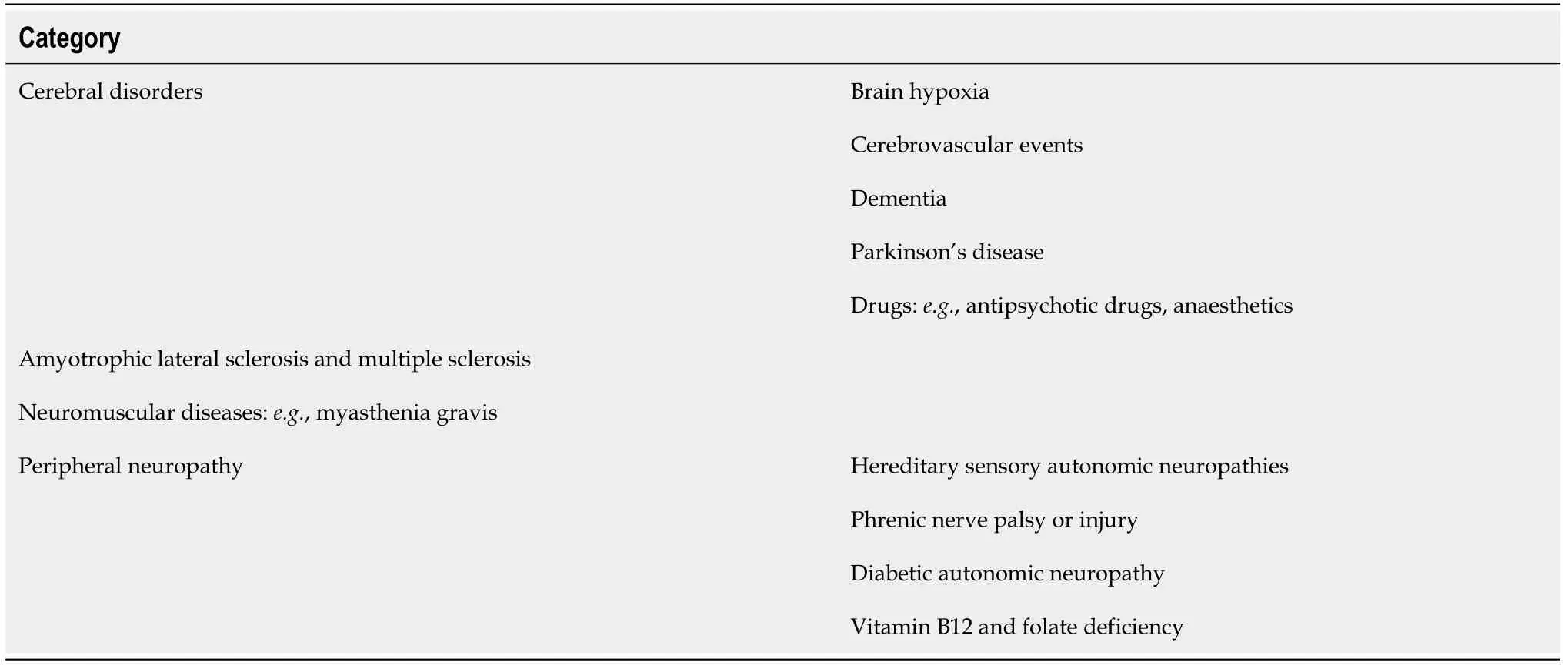
Table 2 Neurological conditions associated with diminished cough reflex sensitivity
Brain hypoxia and cerebrovascular events
The central nervous system (CNS) is significantly affected by hypoxia, which can depress cough through different mechanisms and decrease the sensitivity of the peripheral cough receptors and the rostral and caudal parts of the solitary nucleus. This nucleus is the recipient of all visceral afferents and an essential part of the regulatory centers of internal homeostasis through its multiple projections with cardiorespiratory and gastrointestinal regulatory centers[32]. The depressive effect of the hypoxia on the solitary nucleus is mediated by the GABA-mediated pathway. GABA is the chief inhibitory neurotransmitter and can down-regulate the cough reflex sensitivity. Therefore, Baclofen, a GABA agonist, can decrease the cough sensitivity to capsaicin in healthy individuals[33]. In addition, hypoxia can increase CNS levels of endogenous opioids, thus reducing the cough sensitivity by inhibiting the central component of the cough. Hypoxia can occur in many cardiovascular diseases. The hypoxia-related impairment of the cough increases the morbidity and mortality rates in these diseases[34]. Cough reflex can be assessed in a comatose patient as a part of the Brainstem Responses Assessment Sedation Score in the intensive care unit by observing the patient's response to a tracheal suctioning. It is considered positive if any contraction of abdominal muscles is observed[35].
Cortex has control over the cough. The ability to voluntarily produce and suppress a cough is an example of the cortical control of the cough. Reduced strength of the voluntary cough may increase the risk of aspiration and other pulmonary consequences due to inadequate clearing of the aspirated material from the airway, as seen in patients with brainstem or cerebral stroke associated with an abnormal laryngeal cough reflex[36]. Many patients with cerebral hemispheric stroke showed a temporary or long-lasting malfunction of the laryngeal cough reflex (Known as "brainstem shock"). This shock is characterized by a generalized transient or permanent neurological malfunction of one or more vital neurological functions, including the respiratory drive, reticular activating system, or the laryngeal cough reflex.
Consequently, many patients with significant or minor hemispheric strokes may develop impaired consciousness and need intubation due to reduced respiratory drive. Addingtonet al[37] showed the importance of the stroke location in determining the effect of stroke on the laryngeal cough reflex and consequently on the pneumonia risk. They showed that the brainstem and cerebral hemispheric infarcts are more liable to affect the laryngeal cough reflex than basal ganglionic or cerebellar infarcts[37]. Danielset al[38] showed that 67% of their patients with stroke did not show cough response, and 38% had suffered from aspiration[38]. Therefore, adding cough sensitivity testing to the clinical evaluation of the swallowing function will significantly reduce the aspiration pneumonia risk in patients with cerebral or brainstem stroke[7]. It also helps in monitoring the recovery from stroke and evaluating the postsurgical recovery of the laryngeal cough reflex after extubation and following general anesthesia[39].
Patients with Lewy body disease-related dementia have decreased cough reflex sensitivity and central respiratory chemosensitivity, with decreased insula activation associated with UTC[9]. Patients with Parkinson's disease also have reduced intensity of voluntary and reflex cough efforts with a slightly higher cough threshold. Fontanaet al[39] found that a motor rather than a sensory component of the cough reflex is primarily involved, especially in the early stages, primarily due to impairment in the central activation of motor units and reduced neural drive to expiratory muscles. The impaired central activation reflects the presence of bradykinesia which is one of the critical functional disorders in these patients[36]. Parkinsonism is associated with decreased Dopamine and other neurotransmitters production in substantia nigra, impairing substance P production in vagal sensory nerve C-fibers in the cervical ganglia. The low level of substance P weakens the swallowing reflex and suppresses the cough reflex causing frequent aspiration[40]. About 20% of deaths in patients with Parkinsonism were related to pneumonia, probably because of the impaired cough reflex and upper airway muscle dysfunction[41]. In the same way, multiple sclerosis, with its characteristic disseminated demyelination patches in both the brain and spinal cord, can affect the voluntary cough efficiency and respiratory muscle power due to bulbar dysfunction and corticospinal tract damage in the spinal cord. The degree of impairment of cough reflex has an inverse correlation with the patients' degree of disability[42].
Motor neuron diseases
Motor neuron disease is a chronic degenerative neurological disorder affecting the corticospinal tracts, motor nuclei in the brainstem, and the anterior horn cells of the spinal cord. It reduces the capacity of efficient cough. There is a hyperactive cough reflex in its early stages due to inflammatory mediators such as bradykinin and prostaglandins. As the disease progresses, there is continuous damage-causing cough desensitization. Various combinations of upper and lower motor neuron dysfunction may increase the need to cough but, unfortunately, impair the efficiency of both the voluntary and reflex types of coughs[43]. Amyotrophic lateral sclerosis is characterized by upper (UMN) and lower motor neuron (LMN) degeneration which negatively impacts the ability of respiratory and laryngeal musculature to work in harmony during the cough phases. The rigidity due to UMN degeneration and weakness due to LMN degeneration led to abnormal cough flow and impaired airway clearance abilities, causing different pulmonary sequelae, such as poor secretion management, recurrent pneumonia, and even respiratory failure[44]. Voluntary cough testing detects the presence of dysphagia and impaired airway defense physiologic capacity and secretion management. Constant assessment of voluntary cough function provides rapid detection of respiratory deterioration, permitting appropriate implementation of cough assist, non-invasive ventilation, and respiratory training before significant function degradation[45].
Neuromuscular diseases
Neuromuscular diseases are associated with increasing breathing disorders, including swallowing dysfunction, cough impairment, and frequent choking. In myasthenia gravis, cranial nerves impairment and bulbar weakness could be the initial symptoms causing frequent aspiration and, consequently, increasing the coughing frequency. However, if the patient develops a respiratory failure, the associated hypoxia causes peripheral and central impairment of the cough reflex sensitivity[46]. Phrenic nerve palsy or injury is associated with decreased cough reflex[47].
Peripheral neuropathy
Since cough is a defensive reflex, it could be affected by diseases targeting the peripheral nerves. Consequently, vagotomy or anesthesia-induced vagal block abolishes cough[48]. Hereditary Sensory Autonomic Neuropathies (HSAN) are rare hereditary peripheral neuropathies characterized by the loss of large myelinated and unmyelinated fibers resulting in decreased pain sensation and its associated consequences. Congenital insensitivity to pain with anhidrosis (CIPA) is HSAN type-IV; it occurs due to a mutation in the gene encoding for the neurotrophic tyrosine kinase receptor type I, called theNTRK1gene[49]. Both pain and cough can be evoked experimentally by stimulating nociceptive C-fibers and faster-conducting A-δ-fibers. Consequently, CIPA can impair both pain and cough. Few cases reports described this association[50,51].
Diabetes-related autonomic neuropathy is one of the most typical complications of diabetes mellitus (DM). Meanwhile, the vagus nerve is one of the first nerves damaged in DM. Different studies showed a significant increase in the cough threshold with cough reflex impairment. Ciljakovaet al[52] found a robust negative correlation between cough reflex sensitivity and heart rate variability as an indicator of diabetic autonomic neuropathy[52]. Down-regulation of the cough reflex may start very early in the pathogenesis of diabetes. Varechovaet al[53] found decreased cough reflex sensitivity in children with Type-I DM with subclinical autonomic neuropathy. Testing those children for reduced cough reflex could reflect the presence of autonomic dysfunction and its impact on respiratory and general health[53]. Cough reflex sensitivity could also decrease with aging, during sleep, cranial nerve conduction abnormalities due to vitamin B12 and folate deficiency, and inhibition of dopamine receptors by antipsychotic drugs[40].
HOW CAN COUGH HELP TO DIAGNOSE NEUROLOGIC DISORDERS?
When a chronic cough is present, the underlying lesion should be investigated.
Arnold's nerve ear-cough reflex
In Arnold's nerve ear-cough reflex, the cough is triggered by mechanical stimulation of the external auditory meatus through the auricular branch of the vagus nerve (Arnold's nerve), which supplies the external auditory canal, middle ear, and auditory tube. The test is done using a cotton swab on a stick to stimulate the ear by placing the swab 3 to 5 mm into the external auditory canal and rotating for 2 to 3 s. We consider the test positive if the patient coughs within 10 s. The test should be performed on both sides, as many persons may only have one affected side. The test is positive in 2% of healthy children and adults, 3% of children, and 25% of adults with chronic cough. A positive reflex is more common in women than men and is unilateral in over 90% of patients[54].
Interestingly, hair within the ear canal can stimulate Arnold's nerve and trigger the urge to cough (Oto-tricho-tussia). Such patients can be easily treated by removing the hair[55]. This effect can be applied to any foreign body or earwax impaction in the auditory canal. Consequently, examining the external auditory canal should be a routine in patients with chronic cough, especially in old age[56]. The high prevalence of positive Arnold’s nerve reflex in patients with chronic cough suggests that chronic cough is a neuropathic condition due to a disorder or alteration in the vagus (vagal hypersensitivity) that could be secondary to sensory nerve damage caused by the inflammatory, infective, or allergic factors. It is usually accompanied by other neuropathic features such as throat irritation (laryngeal paraesthesia). Cough is triggered upon exposure to non-tussive triggers such as cold air and eating (allotussia or UTC). The low prevalence of positive reflex in children with chronic cough (3%) compared to the adults (25%) indicates that the hypersensitivity of this reflex may be acquired, possibly by a viral infection[57]. A positive Arnold’s nerve reflex can be reversed after successful therapy of chronic cough. However, a positive Arnold’s nerve reflex is not a valid predictor of the cause of chronic cough but can trigger the need to investigate it[58].
Holmes-Adie syndrome
Holmes-Adie syndrome is another rare cause of tendon areflexia, unilateral or bilateral tonic pupils with slow reaction to near direct light, and chronic cough; due to autonomic dysfunction affecting some cranial nerves, including the vagus nerve. Autonomic dysfunction is a frequent finding for this condition; attributed to lesions in both afferent and efferent sympathetic and parasympathetic neurons. Airways reflux secondary to vagal dysfunction is a possible etiology of cough in these patients. The patients present with anisocoria, abnormal deep tendon reflexes, patchy hyperhidrosis or anhidrosis, and chronic cough[59]. Many patients with sensory neuropathic cough were relieved by neuralgianeuromodulator drugs, such as amitriptyline, desipramine, Gabapentin, pregabalin, oxcarbazepine, and others, when other potential causes of chronic cough have been ruled out. These medications may help reduce or abolish cough by diminishing the nerve-ending "misfires" caused by sensory neuropathic cough[60].
Cough syncope
Cough syncope is a temporary impairment or loss of consciousness with facial congestion and cyanosis; it typically occurs within seconds of a coughing paroxysm, followed by a rapid recovery. Cough syncope originally can mimic epilepsy. It was previously considered a form of epilepsy "known as laryngeal epilepsy" because of the associated jerking movements. However, many studies showed regular brain electrical activity during the episodes. It typically occurs in middle-aged and older, overweight, or muscularly built male smokers with a history of chronic obstructive lung disease. These persons are more prone to create a very high intrathoracic pressure associated with cough-induced syncope and fainting[61]. As it is mainly an adult disease, cough syncope was rarely reported in children, particularly under ten years[62]. The exact mechanism of cough syncope is debatable. Cough markedly elevates the intrathoracic pressures, diminishes the cardiac output, and decreases the systemic blood pressure and cerebral perfusion. At the same time, cerebrospinal fluid (CSF) pressure increases causing reduced brain perfusion; or a cerebral concussion-like effect due to rapid CSF pressure elevation. Another theory suggests that the cough initiates a neurally-mediated reflex vasodepressorbradycardia. Elimination of cough eliminates the resultant syncopal episodes[63].
The patient may have a fixed upward deviation of the eyes during the syncopal episode, which should not be confused with epilepsy. EEG shows temporary slowing during the attack but no seizure discharges. It is always accompanied by a coughing paroxysm. During the attack, the face becomes plethoric rather than cyanotic, and the entire episode lasts less than a minute. An aura never precedes it and is very rarely followed by post-ictal confusion/headache. Cough syncope frequently occurs at night while prone, whereas epilepsy can develop in any position[64]. Cough syncope is associated with a high incidence of pulmonary, cardiac, and neurologic disorders. Numerous CNS disorders were reported to be associated with cough syncope, including cerebral tumors (meningioma, glioblastoma), herniation of cerebellar tonsils (Type 1 Arnold-Chiari malformation), hydrocephalus, carotid and vertebral arterial occlusive disease, basilar invagination, autosomal dominant hereditary sensory neuropathy, and medullary infarction[65].
Cough headache
Cough-triggered headaches are uncommon, with a lifetime prevalence of 1%. Headache can be triggered by a rapid increase in the intra-abdominal, intra-thoracic, and intracranial pressure, caused by coughing, sneezing or straining in patients with low pain threshold[66]. It is either primary or symptomatic. Primary cough headache (previously known as benign cough headache or Valsalva maneuver headache) is currently defined as a headache with sudden onset, occurring only in association with coughing, straining, and/or Valsalva maneuver. It lasts from one second to 30 min and is unrelated to other disorders[67]. It is more frequent in males over 40 years, and usually bilateral, but sometimes unilateral. Pain is of moderate-to-severe intensity and is usually located in the frontotemporal regions, but sometimes presents with different patterns such as toothache. The pain can be triggered by Valsalva maneuvers but never by physical exercise. Nausea, vomiting, photo- and phonophobia are uncommon[68].
Underlying disorders can be detected in 40% of cases with symptomatic cough headaches. These lesions may involve but are not limited to Chiari type I malformation, obstructive hydrocephalus, posterior fossa structural lesions (as arachnoid cysts, dermoid tumors, meningiomas, or Os odontoideum), spontaneous low CSF pressure or leak, subdural hematoma, multiple brain metastases, acute sphenoid sinusitis, pneumocephalus, pneumococcal meningitis, or non-ruptured cerebral aneurysm[69]. Symptoms are more common than those observed with the primary type, depending on the underlying abnormality. The headache is increasing in intensity with variable durations and locations. The pain may be pressing, explosive, bursting, stabbing, dull, electrical, lancinating, or having a mixed nature. Headache duration ranges from seconds to several weeks[65]. Headache can be triggered by a cough and other factors such as laughing, exertion, weightlifting, defecation, or rapid body or head postural changes. Posterior fossa symptoms are common, such as dizziness, unsteadiness, facial and upper limb numbness, vertigo, and syncope. The mechanism of headache is due to raised intracranial pressure, evidenced by the disappearance of the headache after surgical correction of the lesion[70].
Opioids-associated cough
Opioids are well known to have a central antitussive action. However, some opioids such as Alfentanil, Fentanyl, and Sufentanil can elicit a brief tussive effect in about 50% of the patients (especially smokers) within a few seconds from the rapid bolus intravenous injection. This tussive effect is due to the chemical stimulation of opioid receptors in the smooth muscles in the trachea, bronchi, and bronchioles. This pulmonary chemoreflex is unlikely to be mediated by the vagus nerve, as it is not affected by atropine pretreatment. Instead, pretreatment with inhaled β-2 adrenergic agonists considerably decreases the rate of cough related to the intravenous opioid injection. This opioid-associated cough is usually self-limited. It is also related to circulation time and could serve as a clinical landmark for veinto-brain time or cardiac output[71].
Cough-anal reflex
The anal wink in response to cough or sniff is a significant clinical sign during a neurological examination. It could be elicited by asking the patient to voluntarily cough or sniff while observing the anus. This reflex is not affected by transection of the spinal cord while being lost in cauda equina lesions. It is easier to be done and more convenient to the patient than the classic anal reflex. It is a promising tool and is better to be included in the neurological examination[72].
OTHER RESPIRATORY SYMPTOMS THAT COULD HAVE NEUROLOGICAL PATHOLOGY
Many other respiratory symptoms and signs could have underlying neuropathologies. Intractable sneezing and hiccup could be seen in patients with NMOSD[73]. However, a diminished sneezing reflex or difficulty initiating sneezing or the urge to sneeze is an uncommon neurological symptom. A runny nose and hypo or hyper-reflexive rhinopathy could indicate autonomic nervous system dysfunction[74]. Nasal discharge may be observed in Parkinson's disease, dementia, and Alzheimer's disease or arise from their treatment[75]. CSF rhinorrhea is observed in head trauma and can be easily distinguished by a simple glucose dipstick test[76]. Throat clearing, dysphonia, and vocal fatigue can be observed in many patients with postviral vagal neuropathy[77]. However, a detailed discussion of these symptoms is out of the scope of this review.
TREATMENT OF NEUROLOGICAL DISEASE-RELATED COUGH
Treatment of cough secondary to neurogenic disorder is mainly directed to treat the cause. A suggested guideline for managing chronic neuropathic cough is demonstrated in Figure 2. Different modalities could be used to treat these coughs after trying to treat the original neurogenic disorder. Speech and language therapy, behavior modification, biofeedback, self-hypnosis, bed-sheet technique, and differential reinforcement can help treat somatic cough[21]. We can also try combined codeine and guaifenesin or empiric therapy with Gabapentin, Pregabalin, Nortriptyline, or Amitriptyline[78]. Botulinum toxin injection into the thyroarytenoid muscles or vocal cords may help to relieve chronic cough secondary to a neuropathic disorder[79]. Referral to a Specialist cough clinic could be an excellent choice to reach a definitive treatment for chronic cough not responding to the previous management. Enrolment in ongoing clinical trials could also be a valid option. Transcutaneous electrical nerve stimulation is a relatively new electroanalgesia method that helps relieve neuropathic pain disorders, including refractory chronic neuropathic cough, which is physiologically like other neuropathic pain conditions. Michalowskiet al[80] studied the tolerability and feasibility of using Transcutaneous electrical nerve stimulation to treat neuropathic cough[80]. Other new modalities and novel therapeutic agents are under trial, especially those working on the brainstem and cerebral cortex.
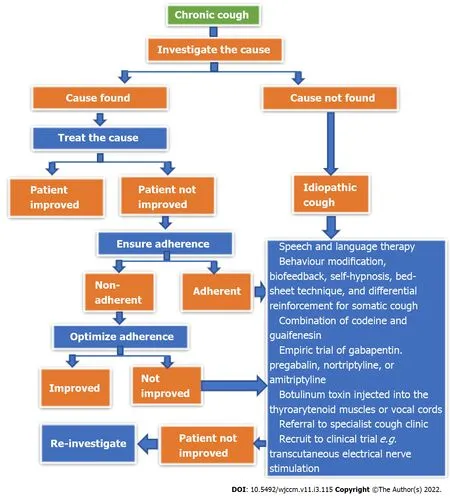
Figure 2 Proposed guidelines for the treatment of chronic cough.
CONCLUSION
A cough is a crucial neurological sign, the same as a critical respiratory sign. Cough reflex sensitivity could be increased or decreased in many neurological disorders. Cough reflex testing is quick, easy, and cheap tests can be performed during the cranial nerve examination.
ACKNOWLEDGEMENTS
We thank the anonymous referees for their valuable suggestions.
FOOTNOTES
Author contributions:Al-Biltagi M, Bediwy AS, and Saeed NK did the research, collected the data, and wrote and revised the manuscript.
Conflict-of-interest statement:No conflict of interest.
Open-Access:This article is an open-access article that was selected by an in-house editor and fully peer-reviewed by external reviewers. It is distributed in accordance with the Creative Commons Attribution NonCommercial (CC BYNC 4.0) license, which permits others to distribute, remix, adapt, build upon this work non-commercially, and license their derivative works on different terms, provided the original work is properly cited and the use is noncommercial. See: https://creativecommons.org/Licenses/by-nc/4.0/
Country/Territory of origin:Bahrain
ORCID number:Mohammed Al-Biltagi 0000-0002-7761-9536; Adel Salah Bediwy 0000-0002-0281-0010; Nermin Kamal Saeed 0000-0001-7875-8207.
S-Editor:Fan JR
L-Editor:A
P-Editor:Fan JR
杂志排行

World Journal of Critical Care Medicine的其它文章
- Presentation and outcome of myocardial infarction with nonobstructive coronary arteries in coronavirus disease 2019
- Plasma D-dimer level in early and late-onset neonatal sepsis
- Stress cardiomyopathy in critical care: A case series of 109 patients
- Need for oxygen therapy and ventilatory support in premature infants in a hospital in Southern Brazil
- Critical care practices in the world: Results of the global intensive care unit need assessment survey 2020
- Diuretic combinations in critically ill patients with respiratory failure:A systematic review and meta-analysis