Transcription regulators differentiate mesenchymal stem cells into chondroprogenitors,and their in vivo implantation regenerated the intervertebral disc degeneration
2022-06-02ShumailaKhalidSobiaEkramAsmatSalimRasulChaudhryIrfanKhan
Shumaila Khalid,Sobia Ekram,Asmat Salim,G.Rasul Chaudhry,Irfan Khan
Shumaila Khalid,Sobia Ekram,Asmat Salim,lrfan Khan,Dr.Panjwani Center for Molecular Medicine and Drug Research,International Center for Chemical and Biological Sciences,University of Karachi,Karachi 75270,Sindh,Pakistan
G.Rasul Chaudhry,Department of Biological Sciences,Oakland University,Rochester,MI 48309,United States
Abstract BACKGROUND Intervertebral disc degeneration(IVDD)is the leading cause of lower back pain.Disc degeneration is characterized by reduced cellularity and decreased production of extracellular matrix(ECM).Mesenchymal stem cells(MSCs)have been envisioned as a promising treatment for degenerative illnesses.Cell-based therapy using ECM-producing chondrogenic derivatives of MSCs has the potential to restore the functionality of the intervertebral disc(IVD).AIM To investigate the potential of chondrogenic transcription factors to promote differentiation of human umbilical cord MSCs into chondrocytes,and to assess their therapeutic potential in IVD regeneration.METHODS MSCs were isolated and characterized morphologically and immunologically by the expression of specific markers.MSCs were then transfected with Sox-9 and Six-1 transcription factors to direct differentiation and were assessed for chondrogenic lineage based on the expression of specific markers.These differentiated MSCs were implanted in the rat model of IVDD.The regenerative potential of transplanted cells was investigated using histochemical and molecular analyses of IVDs.RESULTS Isolated cells showed fibroblast-like morphology and expressed CD105,CD90,CD73,CD29,and Vimentin but not CD45 antigens.Overexpression of Sox-9 and Six-1 greatly enhanced the gene expression of transforming growth factor beta-1 gene,BMP,Sox-9,Six-1,and Aggrecan,and protein expression of Sox-9 and Six-1.The implanted cells integrated,survived,and homed in the degenerated intervertebral disc.Histological grading showed that the transfected MSCs regenerated the IVD and restored normal architecture.CONCLUSION Genetically modified MSCs accelerate cartilage regeneration,providing a unique opportunity and impetus for stem cell-based therapeutic approach for degenerative disc diseases.
Key Words: Intervertebral disc degeneration;Human umbilical cord;Transcription factors;Mesenchymal stem cells;Gene expression;Regeneration
lNTRODUCTlON
Severe lower back pain(LBP)is the major disability responsible for physical discomfort,emotional distress,and significant decline in social life.Intervertebral disc(IVD)degeneration is a consequence of LBP affecting 80% of the world’s population and economy[1].IVD provides cushioning between vertebrae and absorbs pressure placed on the spine.It is an avascular and aneural site consisting of central nucleus pulposus(NP)containing a limited number of notochondral cells and a high volume of proteoglycans and glycosaminoglycans(GAGs)[2],with surrounding concentric rings of annular fibrosus(AF)[3]and cartilaginous endplate(CEP)[4].The disc nourishes with the nutrients and metabolites provided by CEP,which is the prime region for controlled diffusion[5].The main function of IVDs is to transmit load between the spinal column and body weight,and helps in body flexion and torsion[6].Degeneration of IVD starts with the disintegration of resident cells which is ultimately followed by a reduction in the proteoglycan and water content.The degenerative process intensifies with age,injury,and genetic factors[7].These factors decrease the synthesis of extracellular matrix(ECM)in the NP region[8].Additionally,loss of notochondral cells disturbs the balance of anabolic and catabolic processes,resulting in disc degeneration[9].These drastic changes also release cytokines and accelerate the secretion of matrix metalloproteinases.Current approaches to rejuvenate the tissue infrastructure and improve biochemical homeostasis of IVD using pharmacological and conventional or surgical treatments do not provide long-lasting relief from LBP caused by degenerative disc disease(DDD)[10,11].
Stem cell therapy has been considered a promising therapeutic option for degenerative diseases,including DDD.Stem cells can be isolated from various sources like bone marrow,umbilical cord,adipose tissues,etc.[12].Mesenchymal stem cells(MSCs)are the most preferred population because of their proliferation,differentiation,and immunomodulatory potential[13].The concept of inducing differentiation of MSCs through the overexpression of chondro-specific genes before injecting them into IVD disease(IVDD)model is novel which is likely to significantly improve the microenvironment for the regeneration of the disc[14].One of the significant advantages of the stem cell-based gene therapy approach is that it can exhibit a long-lasting effect.However,the genetically modified cells need to be analyzed for safety and effectiveness.Nevertheless,such genetic modifications have been explored for targeting traditional inheritable genetic disorders,such as cystic fibrosis,hemophilia,and hypercholesteremia,and to treat acute and chronic diseases[15].
Murine MSCs have been transduced by the adenovirus-mediatedtransforming growth factor beta-2(TGFβ2)gene that led to the synthesis of proteoglycans and downregulation of markers for hypertrophy[16],whileBMP2is responsible for cartilage production when transducedviaretroviral vector promoting chondrogenesis[17].Gene overexpression proved to be a powerful tool to differentiate MSCs into chondroprogenitors(CPCs)which,upon transplantation,healed the degenerated region of IVD[18].Similarly,collagen synthesis was upregulated in the osteoarthritis imperfecta mice model by transduction of bone marrow MSCs with retroviral-mediated transcription ofprocollagen alpha 2[19].
The novel role ofSix-1and other transcription factors,includingpitx1,andttf1,for differentiation of MSCs,resulted in cell progression towards the early stages of cartilage differentiation[20].Additionally,different genes have been targeted by researchers to treat IVDs.For example,IL-1andBMP2cotransducedviaadenovirus vector showed positive biological activity by enhancing ECM at the site of inflamed articular cartilage[21].Similarly,whenSoxtri-genes(Sox-9,Sox-6,Sox-7)were electroporated to MSCs for transplantation in the IVDD model,it enhanced chondrogenesis and suppressed hypertrophy of MSCs compared to the normal non transfected MSCs[22].
In the present study,we hypothesized that damaged IVD could be regenerated,and its normal physiological function can be restored if MSCs are preconditioned by overexpressing chondrogenic transcription factorsSix-1andSox-9or by their synergistic combination,as well as by MSCs pre-differentiated into CPCs,transplanted into the damaged disc in the form of induced chondro-progenitor cells(iCPCs).
MATERlALS AND METHODS
Isolation and propagation of human umbilical cord derived MSCs
The study protocol(IEC-009-UCB-2015)was approved by the institutional ethical committee on human subjects.Human umbilical cord samples(n= 20)were collected from Zainab Panjwani Memorial Hospital following cesarean section.Formal ethical consent was obtained from the donor parents.The cord was washed with phosphate buffered saline(PBS)to remove blood clots.This was followed by mincing the cord tissue into approximately 1-3 mm pieces.The minced tissue was then transferred to a T-75 culture flask containing 12 mL complete medium(Dulbecco’s Modified Eagle’s Medium[DMEM])supplemented with 10% fetal bovine serum,1 mmol/L L-glutamine,1 mmol/L sodium pyruvate,and 1% penicillin-streptomycin,and kept in a humidified incubator at 37 °C,maintained at 5% CO2.The growth medium was refreshed every 3rdday.After 14 d,cell outgrowth was observed from explants.Upon reaching 70%-80% confluency,sub-culturing was performed.
Immunocytochemistry for characterization of MSCs
Cells at passage 2(P2)were grown on the coverslip,fixed with 4% paraformaldehyde for 15 min at room temperature(RT)and permeabilized with 0.1% Triton X-100 in PBS.Blocking was performed with 2%bovine serum albumin(BSA)and 0.1% Tween-20 in PBS for 30 min at RT.The primary antibodies against CD73,CD105,Vimentin,CD29,and CD90 at recommended dilutions were added and incubated at 4 °C overnight.The cells were washed three times with PBS and then subjected to secondary antibodies,Alexa fluor-546 or 488 at a dilution of 1:200 for 1 h at 37 °C.Nuclei were stained with DAPI for 10 min.Finally,cells were mounted with an aqueous mounting medium and visualized under a fluorescent microscope(NiE,Nikon,Japan).
Immunophenotyping of MSCs
Cells were analyzed for the presence of MSC specific markers by flow cytometry using standard protocol.Briefly,the cells were washed with PBS and blocked using a blocking solution(2% BSA).This was followed by incubation with primary antibodies against CD45,Vimentin,CD105,and CD73 at RT for 2 h,and then with the secondary antibody,Alexa fluor 546.Cells were analyzed by flow cytometer(FACS Celesta,Becton Dickinson,Franklin Lakes,NJ,United States).
Tri-lineage differentiation of MSCs
To validate that the isolated cells from human umbilical cord tissues are MSCs,the differentiation potential of these cells into adipogenic,osteogenic,and chondrogenic lineages was assessed.Cells at passage P2were seeded in a 6-well plate and grown in DMEM until they reached 60%-70% confluency,then DMEM was replaced with adipogenic induction medium(1 μM dexamethasone,10 μM insulin,and 200 μM indomethacin),osteogenic differentiation medium(0.1 μM dexamethasone,10 μM βglycerophosphate,and 50 M ascorbate phosphate),and chondrogenic medium(1 μM dexamethasone,10 ng insulin,20 ng TGFβ1 and 100 μM ascorbic acid).Cells in the induction media were cultured for 3 wk.Cells were stained with Oil Red O,Alizarin Red S,and Alcian blue stains for the detection of adipogenic,osteogenic,and chondrogenic differentiation,respectively,and observed under a brightfield microscope.Images were captured with a CCD camera(TE2000,Nikon).
Amplification and isolation of plasmid vectors
Plasmid constructs forSox-9andSix-1in the form ofE.colistab cultures were obtained from Addgene(www.addgene.org;plasmid No.62972 and 49263,respectively).E.coliwas grown in Luria broth and plasmid DNA was isolated by using a maxiprep plasmid DNA isolation kit(Thermo Scientific,Waltham,MA,United States)according to the manufacturer’s instructions.Plasmid DNA was quantified using a nano-drop spectrophotometer and resolved on 1% agarose gel to check purity.
Transfection of human umbilical cord derived MSCs by electroporation
To transfect MSCs with plasmids(pcDNA3.1 HA-rnSox-9and MSCV-Six-1tagged GFP),MSCs were suspended in sterile R buffer containing 30 μg plasmids and were electroporated at 1200 volts;10 ms and 1 pulse with Neon Transfection System(Thermo Scientific).MSCs were transfected separately withSox-9andSix-1,and co-transfected with 15 μg each ofSox-9andSix-1plasmids.Each subset of transfected MSCs was cultured in a basic growth medium for 48 h,followed by incubation in a chondrogenic induction and standard growth media till day 21.MSCs grown in the chondrogenic induction medium were used as positive control,while non-electroporated MSCs in the basal medium was used as negative control.
Evaluation of transfection efficiency
After 48 h of electroporation,MSCV-Six-1GFP labeled plasmid was analyzed for GFP expression under fluorescent microscope.HA-rnSox-9and MSCV-Six-1transfected MSCs were analyzed for protein expression by immunocytochemical staining.To evaluate the transfection efficiency,the fluorescent intensity of Six-1 and Sox-9 expressing cells was quantified with Image J software and plotted with MS excel.
Protein expression analysis
Normal and transfected MSCs cultured in the basal and chondro-induction media for 21 d were evaluated for chondrogenic protein expression by immunocytochemical analysis as described in the above section for the presence of chondrogenic markers Six-1,Sox-9,TGFβ1,TGFβ2,and aggrecan,as well as a stem cell potency marker Stro-1 at the recommended dilutions.Phalloidin labeled with Alexa fluor 488 or 546 was used to visualize the cellular cytoskeleton.Slides were observed under a fluorescent microscope(NiE,Nikon).Fluorescent cells were quantified with Image J software and plotted with Microsoft Excel.
Gene expression dynamics
RNA was isolated using 1 mL TRIzol for lysis,followed by 200 µL of chloroform for aqueous phase separation,and centrifugation at 12000 rpm for 10 min.The aqueous phase was collected and 1 mL of absolute ethanol was added for overnight incubation at -20°C.The suspension was centrifuged,and the pellet was washed with 70% ethanol.Finally,the pellet was dissolved in 20 µL nuclease-free water and stored at -20°C.Quantification and purity of RNA were determined at 260 nm and 280 nm,respectively.First-strand cDNA was prepared by using 1 μg RNA by RevertAidTM First Strand cDNA synthesis kit(K1622,Thermo Scientific).qPCR amplification was performed in three biological replicates in 96-well plates using qPCR master mix(A600A,Promega,Madison,WI,United States)for genes provided in Table 1.β-actin and GAPDH were used as internal controls.

Table 1 Primer sequences used in the study with their annealing temperatures
Experimental animals
A total of 21 Wistar rats(aged 3-5 mo)were used forin vivoexperiments under the guidelines for the care and use of laboratory animals.Local ethical approval was obtained under protocol number(#20170051)from the Institutional Animal Care and Use Committee of Dr.Panjwani Center for Molecular Medicine and Drug Research,University of Karachi,Pakistan.Animals weighting between 200-250 g were used for experiments to ensure equal size of IVDs to minimize variation in results.
Establishment of needle punctured IVDD model
Rats were anesthetized by injecting a mixture of ketamine hydrochloride(60 mg/kg)and xylazine hydrochloride(7 mg/kg),intraperitoneally.To ensure complete anesthesia,reflexes were checked by tail pinch test.The tail was sterilized with 70% ethanol and three sequential intervertebral disc spaces were manually located and marked.Sterile 21 G × 1-inch needle was penetrated in between the coccygeal vertebrae(Co);Co5/Co6(unpunctured disc or negative control),Co6/Co7(punctured disc or positive control),and Co7/Co8(cells transplanted disc),through the level of an upper region(annulus fibrosus)to the middle section of the disc to aspire nucleus pulposus to induce degeneration.The needle was perpendicularly kept for about 20 s for rapid degeneration.Animals were placed back into their respective cages and observed on daily basis for changes in diet uptake and behavior till 14 d.
Cell labeling for transplantation
Forin vivocell tracking,transfected and non-transfected cells were detached,washed,and centrifugedto obtain a cell pellet.Cells were labeled with red fluorescent lipophilic cationic indocarbocyanine(DiI)membrane labeling dye(V-22885,Vybrant®DiI cell-labeling solution,Invitrogen,Carlsbad,CA,United States)according to the manufacturer’s instructions.The activity of unbound dye was instantly inhibited by adding 5 mL complete medium,followed by washing with PBS and dissolving in 50 μL PBS.To check the labeling efficiency,these cells were observed under a fluorescent microscope and flow cytometer.
Cellular transplantation in the IVDD model
There were seven experimental groups;normal,degenerated,MSC transplanted,Sox-9MSC transplanted,Six-1MSC transplanted,Sox-9andSix-1MSC transplanted(synergistic),and iCPC transplanted groups(n= 3).One million cells suspended in 50 μL PBS were transplanted into the site of injury.After 2 wk of transplantation,animals were decapitated and tail discs were carefully harvested and demineralized in 11% formic acid for 2 h.Tissues were transferred into small molds containing optimal cutting temperature(OCT)medium(Surgipath,FSC22,Leica Microsystems,Wetzlar,Germany),and immediately stored at -20°C to turn it into a frozen block.
Histological analysis
Sectioning of frozen blocks was performed using a cryostat machine(Shandon,Thermo Electron Corporation,UK)with a sharp cutting blade.Ten µm thick sections were cut and loaded on gelatin coated slides,and further classified by hematoxylin-eosin and Alcian blue staining.Images were captured with a bright field microscope(NiE,Nikon).Histological grading was performed and plotted.
Tracking of DiI labeled cells in the transplanted IVDs
Cryosections were observed under a fluorescent microscope to track the presence of transplanted DiI labeled cells.To check the viability,long term survival and distribution of the transplanted cells in the IVDD model,the sections were stained with Alexa fluor 488 labeled phalloidin to stain the cytoskeleton protein F-actin.Images were captured with fluorescent microscope.Fluorescent intensity was measured with Image J software and plotted with Microsoft Excel.
Statistical analysis
All statistical evaluations were performed using IBM SPSS version 21.Each experiment was run in triplicate and presented as mean ± SD.Multiple comparative analysis was performed using One Way-ANOVA and Bonferroni post hoc test,consideringP< 0.05,P< 0.01,andP< 0.001 as statistically significant.
RESULTS
Isolation,proliferation,and characterization of MSCs from a primary culture of human umbilical cord tissue
Cell growth observed after the 15thday of explant culture is termed P0cells,as shown in Figure 1A.Fibroblast-like cells of the homogenous population were detached upon reaching 70% confluency and sub-cultured;this is termed as P1cells propagated further until passage P2.These cells showed expression of stem cell markers CD73,CD105,Vimentin,CD29,and CD90 as depicted in Figure 1B.The immunophenotypic analysis was performed using flow cytometry to analyze the expression of CD45,Vimentin,CD105,and CD73,as shown in Figure 1C.The isolated cells were analyzed for tri-lineage differentiation by culturing in the induction media for 3 wk;MSCs differentiated into osteogenic,adipogenic,and chondrogenic lineages revealed respectively by Alizarin Red S which indicated mineral deposits,Oil Red O,which positively stained oil droplets,and Alcian blue which stained ECM of chondrocytes,as shown in Figure 1D.
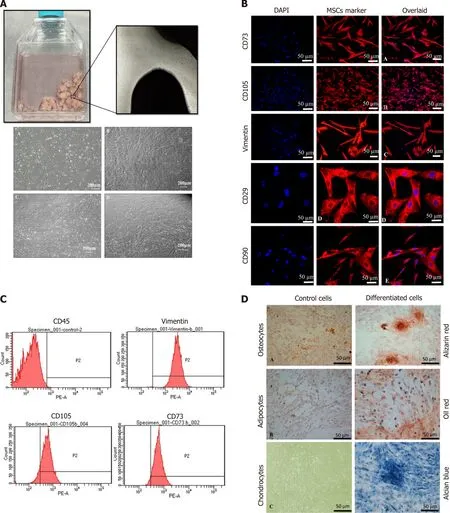
Figure 1 Characterization of human umbilical cord mesenchymal stem cells.A:Culture of human umbilical cord mesenchymal stem cells(hUCMSCs)showed spindle-shaped fibroblast-like morphology at passage P1 to P4;B:MSCs showed positive expression of CD73,CD105,Vimentin,CD29,and CD90.Nuclei were stained with DAPI;C:Histogram of MSCs with specific markers.MSCs showed negative expression of CD45,while positive expression for Vimentin,CD105,and CD73;D:Tri-lineage differentiation of hUC-MSCs.Alizarin Red stained calcium deposits produced by osteocytes,Oil red O-stained lipid vacuoles produced by adipocytes,and Alcian blue stained proteoglycans and glycosaminoglycans secreted by chondrocytes.
Transfection efficiency of MSCs
Sox-9andSix-1transfected MSCs were characterized for transfection efficiency.MSCs were successfully transfected as shown by the expression of Sox-9 and Six-1 proteins after 48 h of transfection compared to control,as shown in Figure 2.
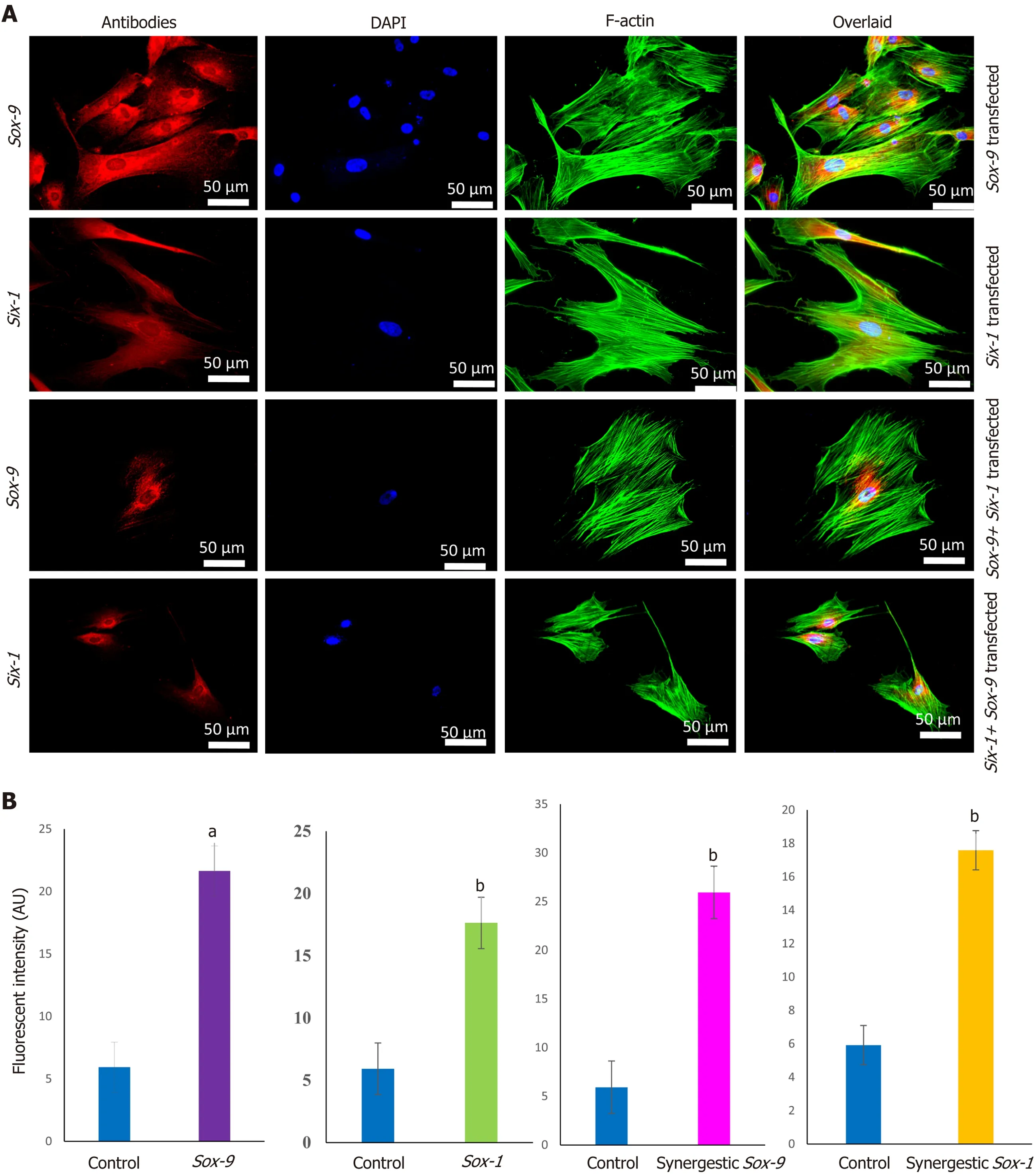
Figure 2 Transfection efficiency of human umbilical cord mesenchymal stem cells.A:Positive expression of Sox-9,and Six-1 proteins was observed after 48 h of electroporation,as analyzed by immunocytochemical staining;B:Quantification of fluorescent intensities showed that Sox-9 transfected cells expressed Sox-9 protein,Six-1 transfected mesenchymal stem cells(MSCs)expressed Six-1 protein,and Sox-9+Six-1 transfected MSCs expressed Sox-9,and Six-1 proteins.aP < 0.05 vs control;bP < 0.01 vs control.
Characterization of differentiated transfected MSCs
MSCs analyzed by immunocytochemical staining indicated that they did not express chondro-markers Sox-9,TGFβ2,and aggrecan as shown in Figure 3A,whereas MSCs cultured in chondrogenic medium expressed TGFβ2 and aggrecan as shown in Figure 3B.The later cells are termed iCPCs.The cellular cytoskeleton of F-actin was stained with Alexa fluor 488/546 labeled phalloidin.After day 21 of transfection,striking differences in the morphological features of the differentiated cells were noted.The transfected cells completely lost their fibroblast-like appearance.Cells displayed a broad and polygonal shape similar to the iCPCs as shown in Figure 4.Transfected MSCs after 21 d of culture in the normal and chondro-induction media were immunostained for the expression of Sox-9 and Six-1 proteins,as shown in Figure 5.The fluorescent intensity was quantified,and the results showed that MSCs transfected withSox-9,Six-1,and their combination expressed chondrogenic markers following 21 d of culture in the basal medium.Similarly,the transfected MSCs in the chondro-induction medium also expressed chondrogenic markers Sox-9 and Six-1 after 21 d of culture as shown in Figure 5.
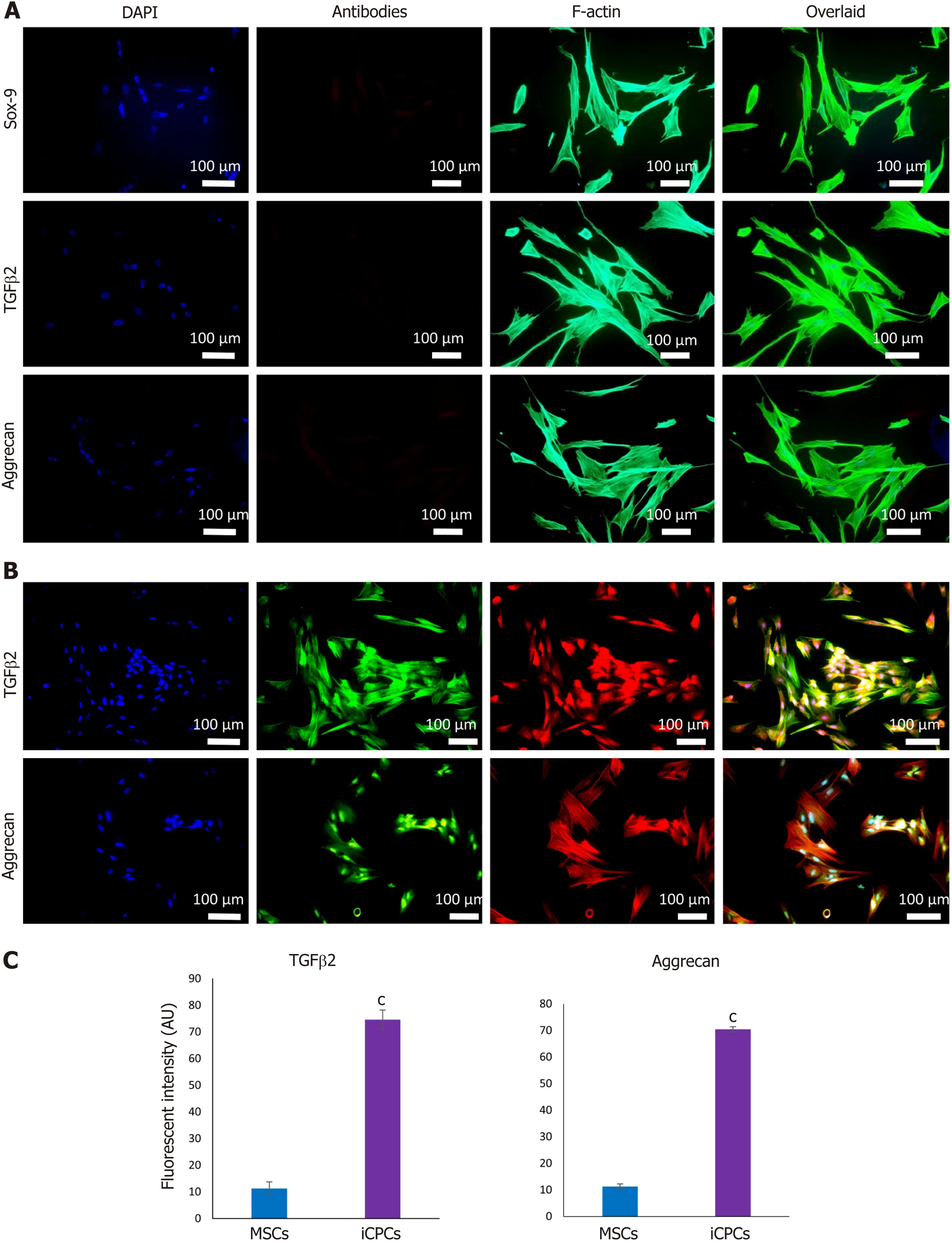
Figure 3 Expression of chondrogenic markers in human umbilical cord mesenchymal stem cells and induced chondro-progenitor cells.A:Human umbilical cord(hUC)-mesenchymal stem cells(MSCs)did not express Sox-9,transforming growth factor beta-2(TGFβ2),and aggrecan,which indicates that MSCs are negative for the expression of early and late chondrogenic markers;B:Induced chondro-progenitor cells expressed aggrecan,and TGFβ2,showing that MSCs were differentiated into chondrocytes;C:The quantification of fluorescent intensities for aggrecan and TGFβ2 showed their significantly higher expression in differentiated cells as compared to control.cP < 0.001 vs MSCs.
Stemness of MSCs
Immunostaining of MSCs showed positive expression of stemness marker Stro-1 in the control cells,whereas transfected MSCs lost the expression of Stro-1 after 21 d of culture in normal and chondroinduction media.The immunofluorescence quantification showed a significant reduction in Stro-1 protein expression in all the transfected and differentiated cells,as shown in Figure 6.
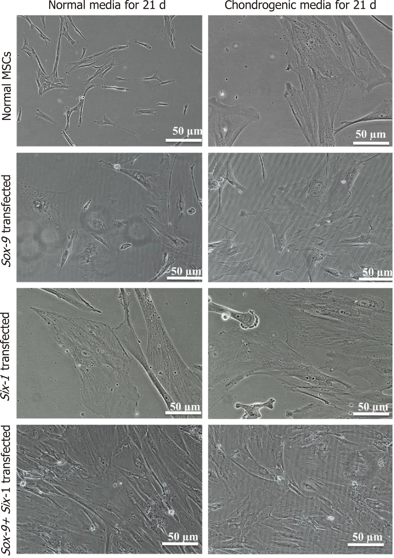
Figure 4 Morphology of transfected human umbilical cord mesenchymal stem cells.Transfected human umbilical cord mesenchymal stem cells(MSCs)after 21 d of culture in the normal and chondro-induction media under phase contrast microscope showed broad and polygonal morphology which is similar to the morphology of induced chondroprogenitor cells,indicating that differentiation is induced in the transfected cells.
Gene expression dynamics of Sox-9 and Six-1 transfected MSCs
To check the transcriptional changes of transfected MSCs in comparison to the control MSCs,fold change regulation(2-ΔΔCt)of chondro-specific genes was analyzed.Expression ofTGFβ1,BMP,Sox-9,andSix-1at 48 h showed that these genes were significantly up-regulated inSox-9,Six-1and their combination(Sox-9 + Six-1)groups as shown in Figure 7.Expression ofSox-9was significantly up-regulated at day 21 post-transfection in the basal medium inSox-9and the combination group,while no significant difference was observed in theSix-1transfected group.The expression ofSix-1has shown no effect in any of the transfected groups after 21 d of transfection.BMP,aggrecan,andTGFβ1were significantly up-regulated in theSox-9,Six-1,and the combination group at 21 d of transfection in the basal and chondro-induction media,as shown in the Figure 7.
Histological examination
Immunohistochemical staining after 2 wk of transplantation showed that the implanted cells homed,distributed,and integrated into the IVD.The quantification of fluorescently labeled cells in the IVD sections showed that the transfected MSC group has significantly higher fluorescent intensity than normal MSCs.The sections were stained for F-actin to visualize the cellular cytoskeleton,which showed that the DiI labeled implanted cells were stained with phalloidin,as evident by co-localization of red and green fluorescence in Figure 8.H and E staining of the degenerated intervertebral disc displayed complete shrinkage of the NP region with fissuring morphology at NP-AF interface compared to normal IVD.Cells surrounded by the matrix in healthy NP were not present in the degenerated IVD.Transfected MSC and iCPC transplanted IVD sections showed better cellularity than degenerated IVDs as shown in Figure 9.However,the partial deformity was observed in the MSC transplanted IVD sections.Alcian blue staining of IVDD showed negligible presence of glucosaminoglycans in the degenerated and MSC transplanted groups.In contrast,transfected MSC and iCPC transplanted IVDDs showed a significant amount of glucosaminoglycans as shown in Figure 9.Histological scoring showed that transfected MSCs better regenerated the IVD as compared to normal MSCs.
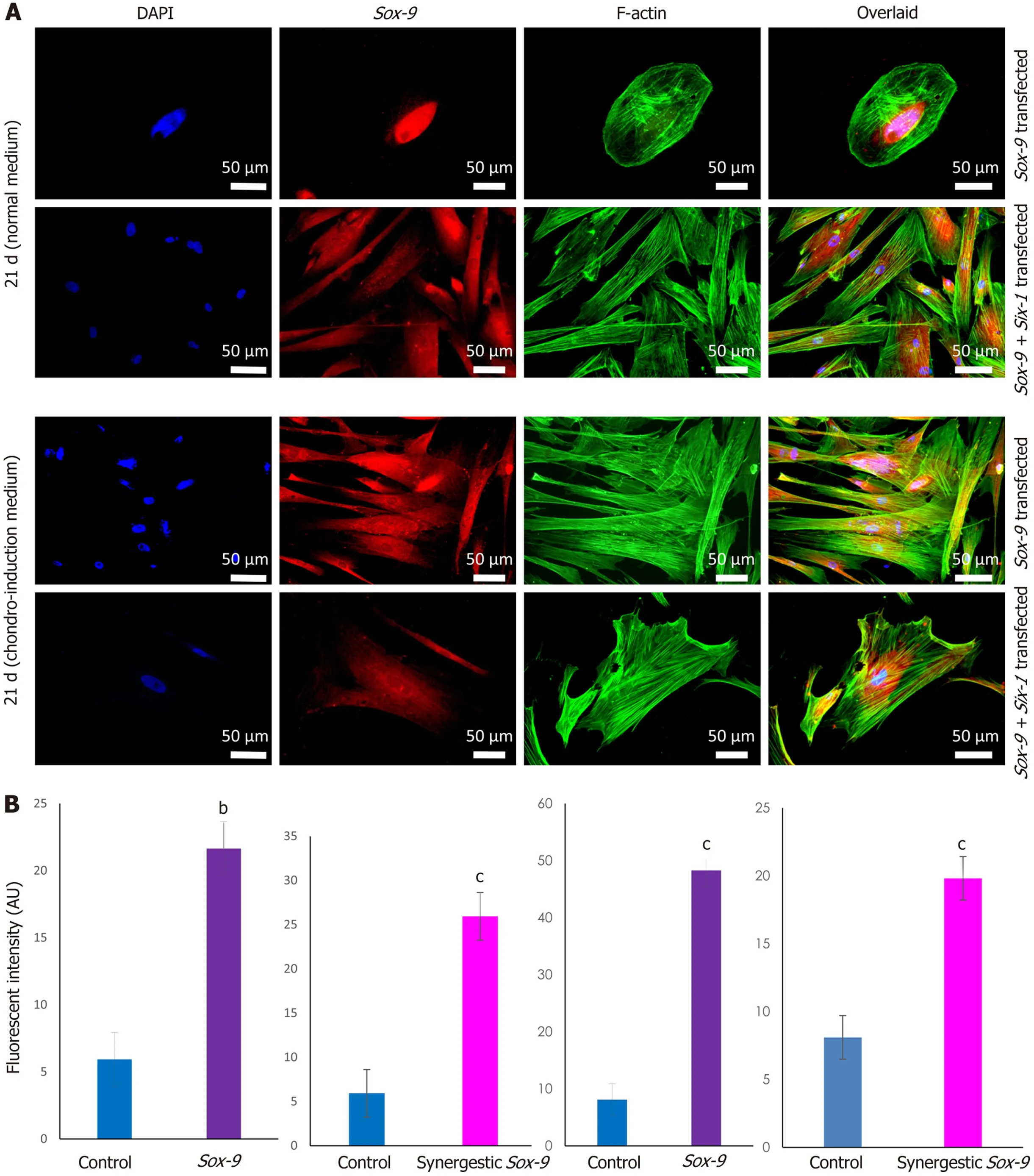
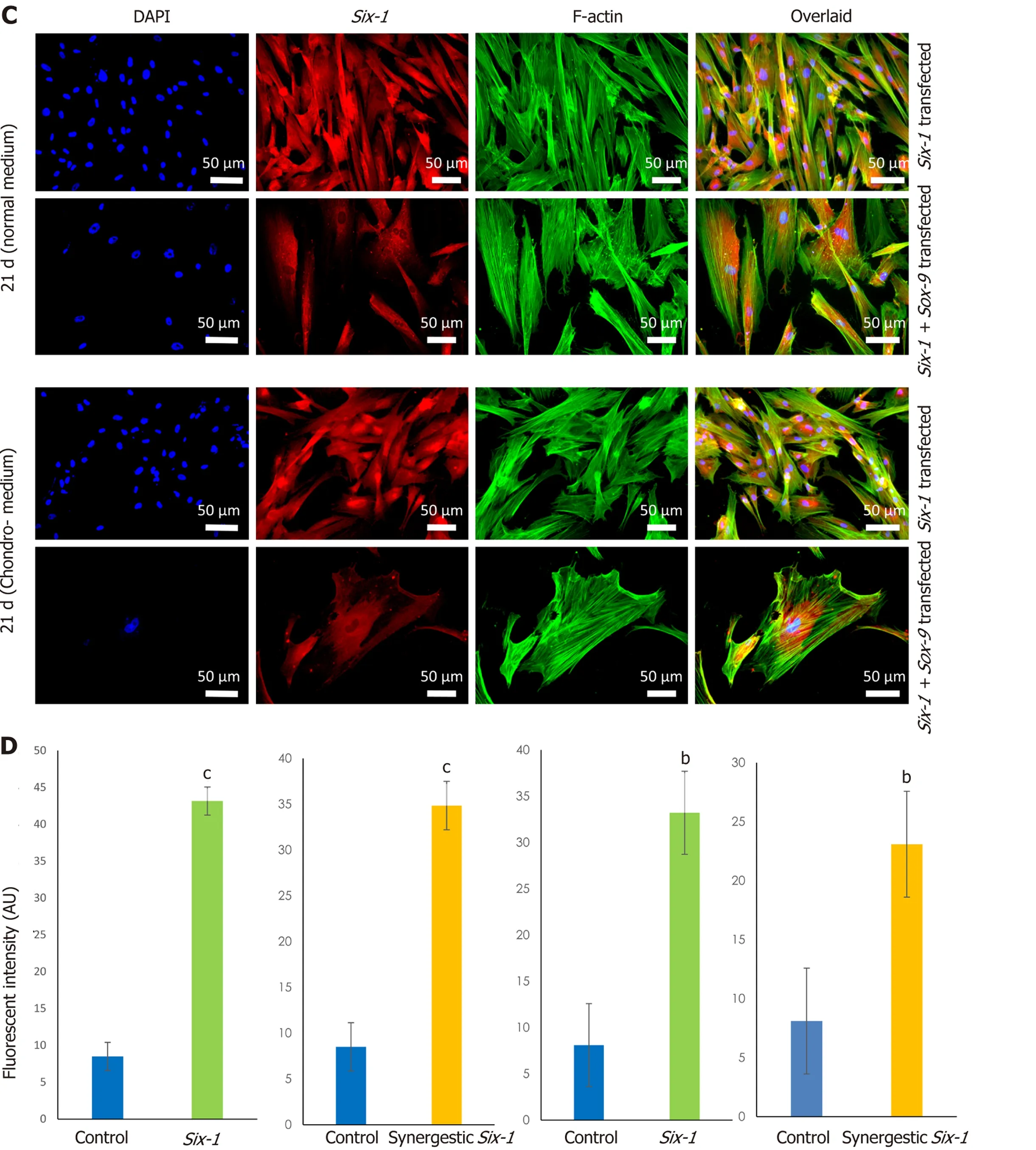
Figure 5 Expression of Sox-9 and Six-1 proteins in transfected human umbilical cord mesenchymal stem cells.Human umbilical cord mesenchymal stem cells transfected with Sox-9,Six-1 or both in combination(Sox-9 + Six-1)were cultured in the normal and chondro-induction media for 21 d.Cells were stained for the expression of Sox-9 and Six-1 proteins by immunocytochemical staining.Alexa fluor 488 labeled phalloidin was used to visualize F-actin of the cellular cytoskeleton.A,B:The expression of Sox-9 in Sox-9 and synergistic transfected group in normal and chondro-induction media,and their fluorescent intensities,respectively;C,D:The expression of Six-1 in Six-1 and synergistic transfected group in normal and chondro-induction media,and their fluorescent intensities,respectively.bP < 0.01 vs control;cP < 0.001 vs control.
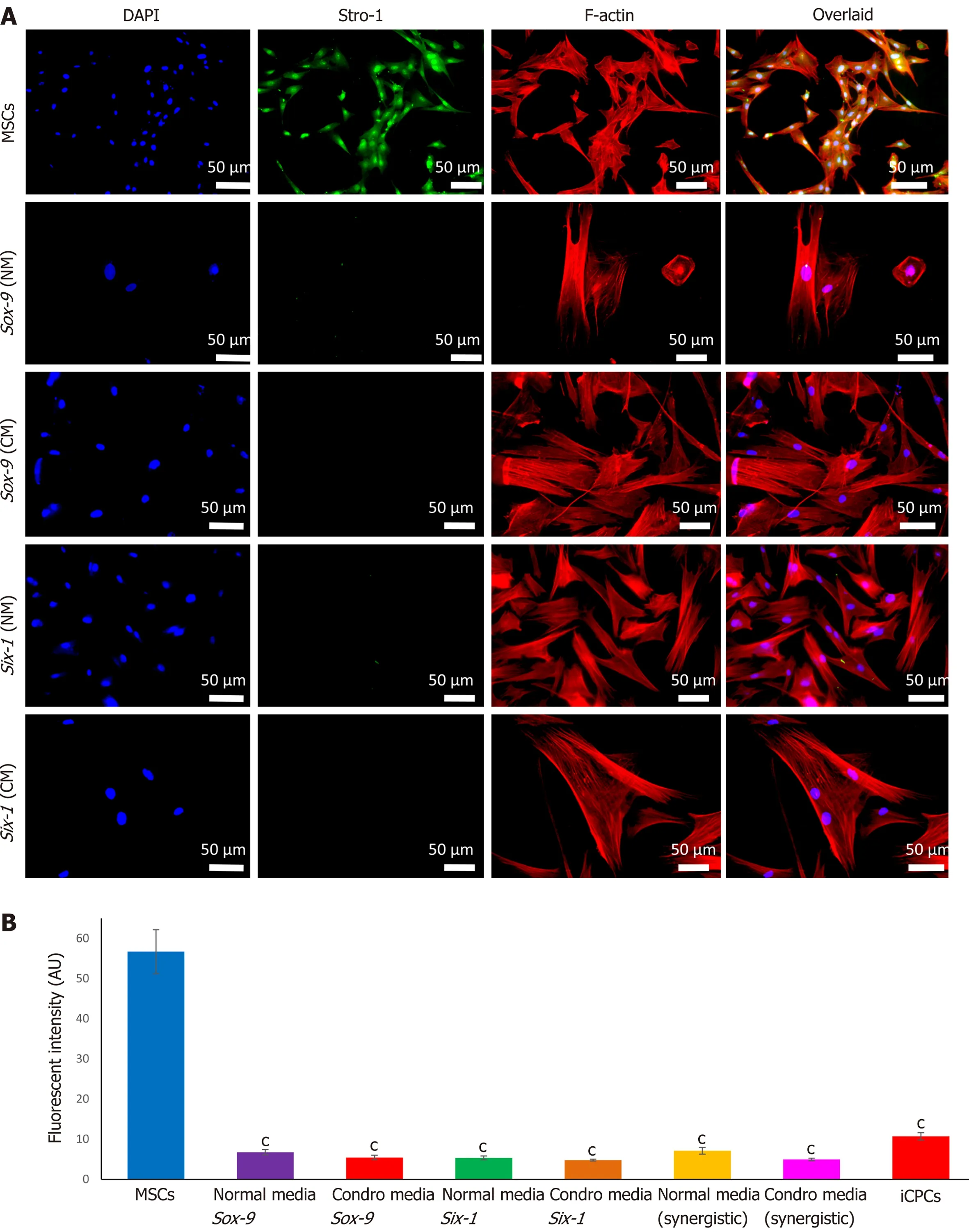
Figure 6 Stemness of transfected human umbilical cord mesenchymal stem cells.A:After 21 d of in vitro culture in normal and chondro-induction media,transfected cells were immunocytochemically stained for the expression of mesenchymal stem cell(MSC)stemness marker Stro-1.Alexa fluor 546 labeled phalloidin was used to visualize F-actin of the cellular cytoskeleton.The nuclei were stained with DAPI;B:The fluorescent intensity of Stro-1 was quantified and showed the expression of Stro-1 protein.cP < 0.001 vs MSCs.iCPCs:Induced chondro-progenitor cells.
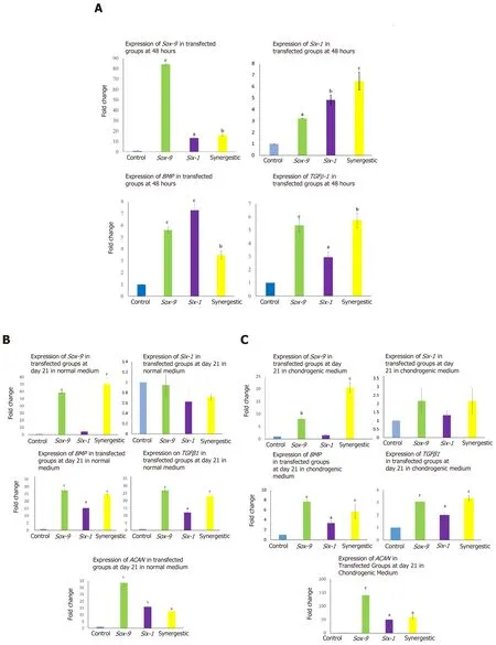
Figure 7 Gene expression analysis of transfected human umbilical cord mesenchymal stem cells via quantitative polymerase chain reaction.A:Bar graphs with significant transcriptional expression of transforming growth factor beta-1 gene(TGFβ1),BMP,Sox-9,and Six-1 at 48 h posttransfection;B,C:Significant expression of TGFβ1,BMP,Sox-9,and aggrecan observed at 21 d post-transfection shows long term sustainability in the expression pattern.However,the expression was time-dependent,Six-1 was significantly downregulated in both normal and chondro-induction media at day 21.aP < 0.05 vs control;bP < 0.01 vs control;cP < 0.001 vs control.
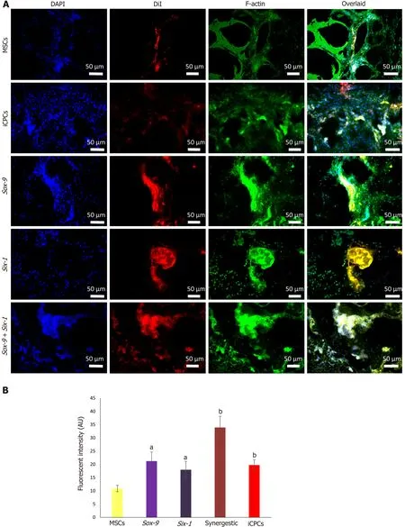
Figure 8 Tracking of transplanted cells in rat intervertebral disc degeneration model.A:Tracking of the DiI-labeled normal and transfected mesenchymal stem cells(MSCs),and induced chondro-progenitor cells transplanted disc indicated the co-localization of red fluorescence originating from the DiIlabeled cells and green fluorescence from Alexa fluor 488 labeled phalloidin(F-actin),confirming their distribution,and homing in the intervertebral discs;B:Fluorescence intensities of the group transplanted with transfected cells showed significantly high fluorescence compared to normal MSCs.aP < 0.05 vs MSCs;bP <0.01 vs MSCs.iCPCs:Induced chondro-progenitor cells.
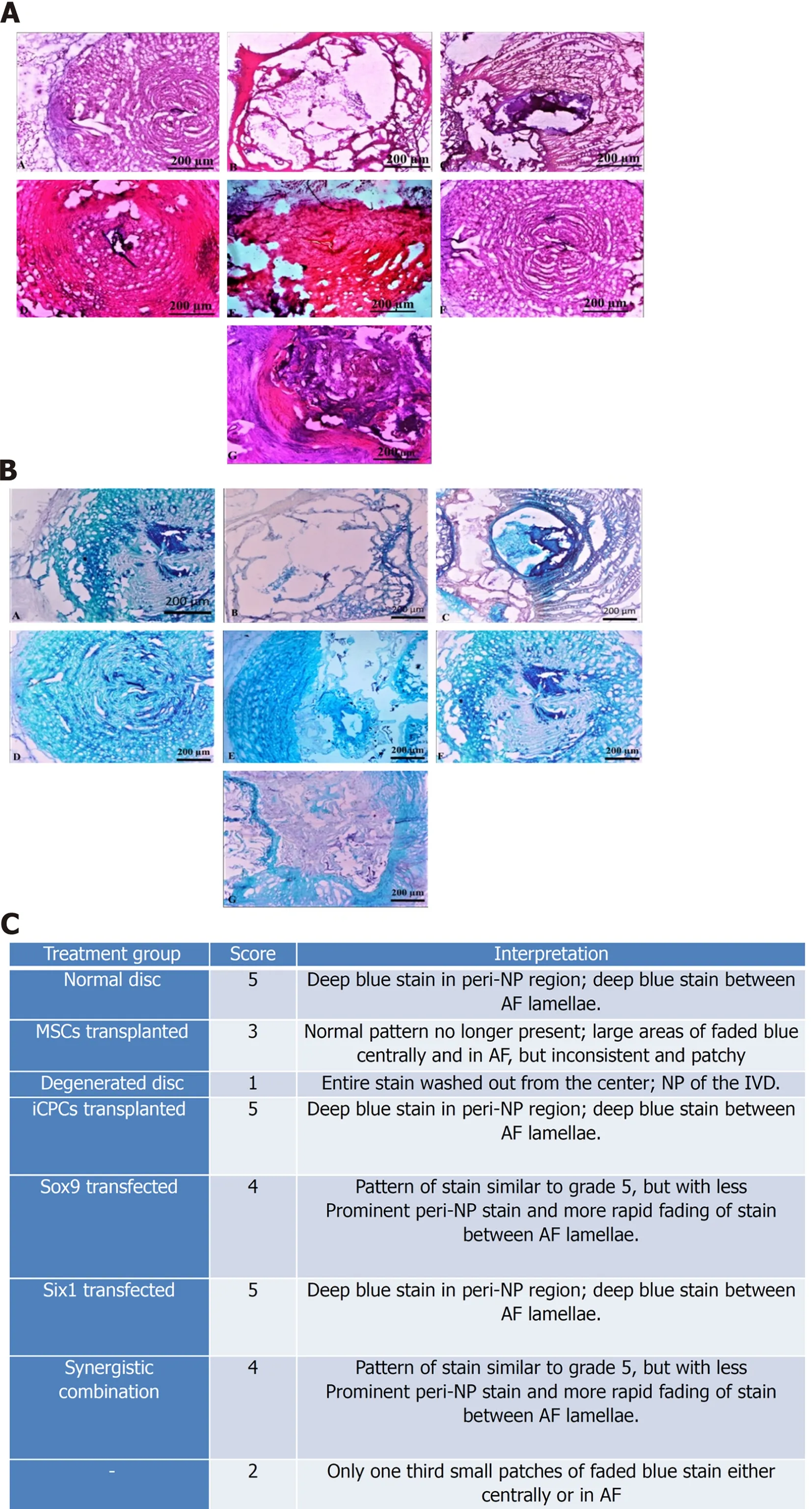
Figure 9 Histological examination of intervertebral disc.A:(A-G)Bright field imaging of intervertebral discs(IVDs)showing nucleus pulposus(NP)content of the healthy(Co6/7),and degenerated disc(Co5/6)treated with normal mesenchymal stem cells(MSCs),transfected MSCs,and induced chondro-progenitor cells(iCPCs)(Co5/6)IVDs,in each group.Bright-field microscopic images of IVDs were captured from the cryosections stained by hematoxylin and eosin;normal healthy disc,degenerated disc,and degenerated disc transplanted with normal MSCs,Six-1 transfected MSCs,Sox-9 transfected MSCs,iCPCs,and Sox-9 + Six-1 transfected MSCs,respectively;B:(A-G)Bright-field microscopic images of IVDs were captured from the cryosections stained by Alcian blue displayed glycoproteins secreted by the transplanted cells compared to the degenerated disc;normal healthy disc,degenerated disc,and degenerated disc transplanted with normal MSCs,Six-1 transfected MSCs,Sox-9 transfected MSCs,iCPCs,and Sox-9 + Six-1 transfected MSCs,respectively;C:Histological grading with a score for regeneration.AF:Annular fibrosus.
DlSCUSSlON
Discogenic pain arising from the degenerated intervertebral disc is considered one of the leading causes of chronic low back pain.Analgesics and physiotherapy are the only treatment options,which only reduce the symptoms;pathological progression of intervertebral disc degeneration cannot be precluded by these methods.A normal healthy disc is an avascular tissue with a consistent cell density of 5.5 × 10³cells/mm3that greatly reduces with age and injury.In disc degeneration morphology,the disc water content and matrix composition is significantly reduced[23].Once the damage is initiated,it ultimately leads to cell loss in the nucleus pulposus region of the disc,which leads to deterioration as cartilage tissue has limited mending capability.Because cartilage in the intervertebral disc contributes to overall body movements and flexion,dysfunction in the disc prominently affects the body motions,including flexion and bending.It may cause severe back pain leading to a decline in the quality of life[24].Recently,advances in understanding disc biology led to the interest in fixing the degeneration of disc by gene therapy combined with stem cells which may support the regeneration by overcoming the drawbacks of the self-renewal process[25].
This study was focused on determining the role of specific chondrogenic transcription factorsSox-9andSix-1in differentiating the MSCs into chondrocytesin vitro.Transfected MSCs after transplantation were analyzed for their enhanced role in homing,production of the extracellular matrix,and regeneration of the degenerated intervertebral disc.
Human umbilical cord tissue was used as a source of MSCs.Cord explants were cultured,and MSCs were isolated and grown in culture.They were passaged to obtain a pure population and their proliferative ability was analyzed.Further,cells showed typical spindle shape fibroblast-like morphology and positive expression of specific surface markers CD73,CD105,and Vimentin,as reported in other studies[26].Harvested cells also exhibited significant expression of Stro-1 marker.Since Stro-1 is a well reported MSC marker,once MSCs are differentiated,they lose its expression[27].Stro-1 is not expressed in iCPCs[28],which is in agreement with our findings.The presence of MSC antigens CD73,CD105,and Vimentin were also confirmed by flow cytometric analysis,while CD45 which is a hematopoietic marker was used as a negative control;similar observations were also reported previously[29,30].Additionally,human umbilical cord cells showed great potential of differentiation into chondrocytes,adipocytes,and osteocytes[31].The differentiated cells showed irregular,broad-shaped morphology and presence of proteoglycans for chondrocytes,calcium deposits for osteocytes,and lipid molecules for adipocytes.Successfulin vitromultilineage differentiation proved that the isolated cells were MSCs.All these morphological,cytological,and biochemical properties were in agreement with previous studies and strongly favored the existence of a pure MSC population[32].
On day 21,the morphology of transfected cells showed a large,broad,and flat shape,similar to the morphology of iCPCs,unlike non-transfected MSCs that showed typical adult stem cell features.Similar morphological differences ofSox-9transfected cells at different days were also reported in a prior investigation[33].We also observed that the differentiation of cells has significantly reduced the expression of Stro-1,compared to normal MSCs,in agreement with previous findings[28].
Gene expression dynamics of normal and transfected MSCs and MSCs induced to chondrocytes showed that the chondrogenic markers,Sox-9,Six-1,BMP,andTGFβ2were significantly upregulated,indicating that the cellular differentiation towards chondrogenic lineage has been initiated.The mRNA expression is downregulated after sufficient protein expression is achieved or once the early induction into a particular lineage is initiated.Prior studies reported that theSix-1mRNA expression is downregulated after few hours.On day 21,it did not show any change;however,the Six-1 protein was found to be at a significant level[34].Transcription factors have been reported to induce differentiation in human umbilical cord derived MSCs(hUC-MSCs)into chondrocytes[35,37].Sox-9andSix-1are welldocumented transcription factors to initiate chondrogenesis[38,39].It has been found that overexpression ofSox-9andSix-1induced the transcription ofBMP2,TGFβ1,Sox-9,andSix-1after 48 h of transfection which efficiently directed the fate of MSCs towards chondrocytes[40].In the differentiated cells,Six-1expression downregulates,and aggrecan which is a late chondrogenesis marker,is reported to be upregulated[34,41].Sox-9andSix-1triggered hUC-MSCs to undergo morphological changes and induce differentiation in MSCs[42].
To elucidate the effect of chondrogenic transcription factors,transfected MSCs,non-transfected MSCs,and iCPCs were analyzed for the regeneration of the intervertebral disc after 2 wk of transplantation.Fluorescently labeled cells were tracked in the harvested disc with fluorescence microscopy,which showed that MSCs transfected withSox-9 and Six-1,as well as the co-transfected MSCs,did better homing,integration,distribution,and differentiation into notochordal cell types and regenerated the degenerated disc.To analyze the potential of differentiated MSCs,a histological assessment was performed for cellularity and ECM and GAG content.H and E staining of the degenerated intervertebral disc displayed NP ossification,reduced cell density,and diminished extracellular matrix in the form of shrunken shape.It has been documented that in the degenerated disc,interconnection with AF is lost and fissuring manifestation in the NP area is prominent[43,44].The disc transplanted with MSCs only exhibited slight improvement in preserving structural integrity.However,disc injected with transfected MSCs showed remarkable difference in contrast to the IVDD.The overall appearance resembled the normal intervertebral disc IVD morphology.Alcian blue staining showed significant amount of GAG content in the disc transplanted with transfected MSCs,which is in agreement with previous findings[10,45].Additionally,xenogenic transplanted MSCs were selectively investigated by measuring specific chondrogenic markers which contribute to restore IVD integrity[46].
Tracking of labeled cells with DiI showed better survival,homing and integration of transfected hUCMSCs into the punctured rat model of IVDD.Regeneration of NP region showed better delivery of cells to the site of injury.Significantly,the effect of transfected cells on nucleus pulposus content was more noticeable than non-transfected hUC-MSCs.Immunohistochemistry revealed co-localized expression of Sox-9 and Six-1 with actin(used as internal control).Previous reports have shown similar findings that xenogenic MSCs home and integrate in the IVD[47,48].The studies showed that the disc transplanted with genetically modified MSCs did home,survive,and were functionally active in the NP area[49].
The findings in the current study elucidated that gene modification or overexpression in stem cells has immense potential for the regeneration of degenerated disc,compared to normal MSCs.Genetically modified MSCs survived long and better regenerated the degenerated NP of the disc.The enforced expression ofSix-1,Sox-9,and their synergistic(Six-1+Sox-9)co-transfection enhanced the chondrogenic differentiation of human cord MSCs into chondrogenic lineage in the normal growth medium.
CONCLUSlON
Overexpression of the chondrogenic transcription factors in hUC-MSCs accelerated their differentiation potential into chondroprogenitor cells.The synergistic effect ofSox-9andSix-1transcription factors led the MSCs to differentiate into chondrogenic cells in the basal medium and produced the same effect as the chondro-induction medium.Thein vivoimplantation of these transfected cells leads to their better homing,integration,and differentiation into NP cells of the IVD.Histological observation and grading score showed that cellular transplanted group significantly regenerated the degenerated disc.This approach could be further developed for treating DDD.
ARTlCLE HlGHLlGHTS
Research background
Genetic manipulation is now considered the safest and promising approach in the field of regenerative medicine.Mesenchymal stem cells(MSCs)are the potential candidates for the clinical use of genetically engineered stem cells.They are non-immunogenic,proliferative,and possess multiple lineage differen tiation potential.Gene modification of MSCs using chondrogenic transcription factors may lead to the use of this strategy to treat debilitating diseases related to cartilage.
Research motivation
Genetic modification of MSCs offers a competent source for the sustained translation of therapeutic proteins.Transfection of human umbilical cord derived MSCs(hUC-MSCs)using chondro-specific transcription factors may lead to enhance chondrogenesis and efficient regeneration of cartilage-related injuries.
Research objectives
The main objective of the study was to analyze the effect of transcription factorsSox-9andSix-1in chondrogenesis by transfecting them into hUC-MSCs and determining successful intervertebral disc(IVD)regeneration.
Research methods
MSCs were isolated from cord tissue and characterized using their specific markers.MSCs were transfected using transcription factorsSox-9andSix-1.Cell differentiation was analyzed at the transcriptional and translational levels,while the regeneration potential of transfected MSCs was observed by transplanting them into the degenerated rat IVD model.Post-transplantation histology and cell tracking analysis were performed to evaluate cartilage regeneration.
Research results
In vitroanalysis showed that transcription and translation of chondrogenic markers were significantly higher in transfected MSCs at 24 h,and 21 d in comparison to control MSCs.Transfected MSCs at the site of degenerated IVD differentiated into chondrocytes which secreted chondro-proteins.The cells homed and regenerated the injured cartilage as that of normal cartilage,as evident from immunohistological and histological analyses.
Research conclusions
Genetic modification of hUC-MSCs with two chondrogenic transcriptional factorsSox-9andSix-1enhances their chondrogenic differentiation.Their synergistic effect on MSCs accelerated the regeneration of degenerated cartilage with complete restoration of tissue architecture.
Research perspectives
The present manuscript offers a promising therapeutic approach to revolutionize the treatment of cartilaginous defects and spinal cord injuries.The outcomes discussed in this study accentuates the advances towards the clinical translation of such approaches.
ACKNOWLEDGEMENTS
We acknowledge International Center for Chemical and Biological Sciences,University of Karachi,Karachi-75270,Pakistan for providing imaging facility.
FOOTNOTES
Author contributions:Khalid S performed the experiment and wrote the first draft of manuscript;Ekram S helped in experimentation and data acquisition;Salim A and Chaudhry GR evaluated the data and helped in manuscript preparation;Khan I designed the experiment,evaluated and analyzed the data,secured the funding,and finalized the manuscript.
Supported byHigher Education Commission Pakistan,No.7083.
lnstitutional review board statement:IEC approval for the protocol ICCBS/IEC-009-UCB-2015/protocol/1.0.Informed consent was obtained from donor parents for the use of umbilical cord tissue in research.
lnstitutional animal care and use committee statement:Approval for Animal Study Protocol,No.2018-0016.
Conflict-of-interest statement:No conflict of interest.
Data sharing statement:No extra data to share.
ARRlVE guidelines statement:ARRIVE guidelines were followed.
Open-Access:This article is an open-access article that was selected by an in-house editor and fully peer-reviewed by external reviewers.It is distributed in accordance with the Creative Commons Attribution NonCommercial(CC BYNC 4.0)license,which permits others to distribute,remix,adapt,build upon this work non-commercially,and license their derivative works on different terms,provided the original work is properly cited and the use is noncommercial.See:https://creativecommons.org/Licenses/by-nc/4.0/
Country/Territory of origin:Pakistan
ORClD number:Shumaila Khalid 0000-0002-4523-6936;Sobia Ekram 0000-0003-0073-8729;Asmat Salim 0000-0001-5181-0458;G Rasul Chaudhry 0000-0003-1692-8420;Irfan Khan 0000-0003-1878-7836.
S-Editor:Wang JL
L-Editor:Filipodia
P-Editor:Zhang YL
杂志排行
World Journal of Stem Cells的其它文章
- Abnormal lipid synthesis as a therapeutic target for cancer stem cells
- Extracellular vesicles from hypoxia-preconditioned mesenchymal stem cells alleviates myocardial injury by targeting thioredoxininteracting protein-mediated hypoxia-inducible factor-1α pathway
- Anti-fibrotic effect of adipose-derived stem cells on fibrotic scars
- Physical energy-based ultrasound shifts M1 macrophage differentiation towards M2 state
