Therapeutic efficacy observation of warm needling moxibustion plus Tuina for knee osteoarthritis
2022-04-18GONGXufang龚旭芳CHENJianqiang陈剑强
GONG Xufang (龚旭芳), CHEN Jianqiang (陈剑强)
Zhejiang Rongjun Hospital, Jiaxing 314000, China
Abstract
Keywords: Acupuncture Therapy; Warm Needling Therapy; Tuina; Massage; Osteoarthritis, Knee; Pain Measurement; Visual Analog Scale; Electromyography
Knee osteoarthritis (KOA) is a chronic joint disease characterized by degenerative changes of knee cartilage and secondary bone hyperplasia. Pain is mostly the first symptom of KOA patients, and it often occurs after a long time of semi-squatting position or walking. With the development of the disease, the lasting time and degree of pain gradually increase, especially during climbing stairs, squatting, and standing up. Meanwhile,morning stiffness, joint swelling, and joint motor dysfunction may occur. In the later stage of the disease,with the cartilage degeneration, meniscus damage,osteophyte hyperplasia, and weakening of muscle and ligament strength can lead to knee deformity. According to the report, the prevalence of KOA is 3.8% worldwide,and the prevalence is higher in females than in males,and KOA has been listed as the 11th leading cause of disability[1]. The prevalence of KOA in China is about 8.1%, and there are obvious gender and regional differences, that is, there are more women than men,more in the inland than in coastal areas, and more in rural areas than in cities. At the same time, age, obesity,trauma and gender are closely related to the incidence of KOA[2-4].
KOA is a progressive degenerative disease, and there is no specific treatment at present. Therefore, the treatment mainly aims at relieving pain, improving joint function, and delaying disease progression. Currently,treatments for KOA in Western medicine mainly include health education, medications, repair treatment and reconstruction treatment. Among them, health education and conservative drug treatment are preferred in the early stage, but there is no specific protocol of drug treatment, and there are certain toxic side effects and drug dependence[5].
In recent years, traditional Chinese medicine (TCM),especially external treatment of TCM, has achieved significant clinical effects in treating KOA and has the advantages of simple operation and few adverse reactions. Warm needling moxibustion is an external therapy that combines moxibustion and acupuncture,with effects of warming meridians for dispelling cold,and harmonizing Yin and Yang. Tuina (Chinese therapeutic massage) therapy can unblock meridians and collaterals, release tissue adhesions, and then improve local blood circulation, relieve pain, and improve joint activities. In this study, we observed the clinical efficacy of warm needling moxibustion plus Tuina for KOA.
1 Clinical Materials
1.1 Diagnostic criteria
This study referred to the diagnostic criteria for KOA in theGuidelines for Diagnosis and Treatment of Osteoarthritis(2018): (1) recurrent knee pain in the past 1 month; (2) X-ray examination showed narrowing of the joint space, subchondral osteosclerosis, and/or cystic changes, and osteophyte formation at the edge of the joint; (3) aged ≥50 years; (4) morning stiffness≤30 min; (5) bone crepitus during movement.Combined with clinical manifestations and X-ray examination, KOA could be diagnosed if patient met items (1) + (2), or (1) + (4) + (5), or (1) + (3) + (4) + (5).
1.2 Inclusion criteria
Those who met the above diagnostic criteria, aged 50 to 65 years old; Kellgren-Lawrence (KL) grades 0-Ⅲ on X-ray imaging examination; 2-8 points scored by visual analog scale (VAS); signed informed consent.
1.3 Exclusion criteria
Patients with severe primary diseases of the internal medical system or mental diseases; those who received other therapies at the same time; those with ulceration at the treatment sites that may affect the treatment.
1.4 Elimination and dropout criteria
Those who had adverse events or special physiological changes that was not suitable for continuing the trial; those who dropped out halfway;patients with incomplete data that would affect the efficacy observation and statistics.
1.5 Statistical methods
All data were statistically analyzed by the SPSS version 20.0 statistical software. Measurement data were expressed as mean ± standard deviation (x±s),andt-test was applied. Counting data were analyzed by Chi-square test.P<0.05 was considered to indicate a statistically significant difference.
1.6 General data
A total of 60 patients with KOA were enrolled from the Acupuncture and Tuina Clinic of Zhejiang Rongjun Hospital, between June 2017 and June 2019. All patients were randomly divided into a control group and an observation group by the random number table,with 30 cases in each group. By statistical analysis, there were no significant differences in the general data between the two groups (P>0.05), indicating that the two groups were comparable (Table 1).
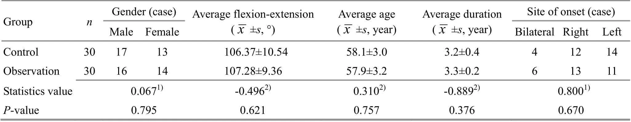
Table 1. Comparison of general data between the two groups
2 Treatment Methods
2.1 Control group
Patients in the control group were treated with warm needling moxibustion.
Acupoints: Futu (ST32), Xuehai (SP10), Dubi (ST35),Zusanli (ST36), Yanglingquan (GB34), Yinlingquan (SP9),and Ashi points.
Methods: The patient took a sitting position, with the knee flexion of 90° and the affected knee skin fully exposed. After routine disinfection with 75% ethanol,the physician punctured the acupoints with sterile disposable acupuncture needles of 0.25 mm in diameter and 40 mm in length. After the arrival of Qi,even reinforcing-reducing manipulation was performed.And then a moxa stick of about 1 cm in length and about 1.2 g in mass was placed onto each needle handle and ignited. After the moxa sticks burned out,the ash was removed, and the needles were retained for 15 min before withdrawal. The treatment was performed once every other day, and 10 times in total.
2.2 Observation group
The observation group was treated with Tuina therapy after receiving the same warm needling moxibustion as in the control group.
Therapeutic manipulation for injured soft tissue: The patient took a prone position. The physician stood on the side of the affected knee, and applied Gun-Rolling manipulation to the back of the thigh, popliteal fossa,and calf for about 5 min, focusing on the popliteal fossa.And then Dian-Digital pressing and An-Pressing manipulations were used to Yinmen (BL37), Weizhong(BL40), Weiyang (BL39), and Chengshan (BL57), for 30 s for each acupoint. Then the patient changed to a supine position. The physician stood on the side of the affected knee. Firstly applied Gun-Rolling, An-Pressing, Rou-Kneading, Na-Grasping, and Nie-Pinching manipulations to the quadriceps femoris and around patella for about 5 min. And then applied one-thumb Tui-Pushing manipulation around the patella to make local penetrating heat. Then Dian-Digital pressing and An-Pressing manipulations were used to Liangqiu (ST34),Xuehai (SP10), Dubi (ST35), Yanglingquan (GB34),Yinlingquan (SP9), Zusanli (ST36), and Taixi (KI3) in turn.And then Tui-pushed and An-Pressed Dubi (ST35) and Neixiyan (EX-LE4) with the thumb and index finger to make the patient feel soreness and numbness. Then Yun-Circular pushed and Rou-Kneaded the patella about 15 times with one palm root.
Reduction therapy: The patient took a supine position with the knee and hip flexed by 90°. The physician stood on the affected side of the patient, held the patient's knee with one hand and the ankle with the other, shook the knee joint while pulling, fully flexed the knee joint and then straightened it. Finally, the patient was seated with the feet flat on the ground in a neutral position. The physician placed the thumbs of both hands at Dubi (ST35) with the other four fingers surrounding the popliteal fossa, asked the patient to stand up and sit down. During the process of standing up and sitting down, the physician applied upward and backward pressure on both thumbs to adjust the patellofemoral joint, and the remaining four fingers simultaneously applied internal and external rotation to the tibia, to adjust the tibiofemoral joint. The operation was repeated 3 times. And the treatment was performed once every other day, 10 times in total.
3 Results Observation
3.1 Observation items
The VAS, Western Ontario and McMaster Universities osteoarthritis index (WOMAC), and surface electromyography (sEMG) signals of quadriceps femoris were used as the observation indicators and evaluated 1 d before the treatment and after 10 times of treatment.
3.1.1 VAS score
A 10 cm straight line was marked with “0” at one end,and “10” at the other end. 0 point indicated no pain;1-3 points indicated mild pain that was tolerable and did not affect normal life; 4-6 points indicated moderate pain that occasionally affected sleep, and interfered with normal life; 7-9 points indicated severe pain that interfered with normal life, affected sleep, and aggravated after activities; 10 points indicated unbearable pain that seriously affected normal life.After fully explaining to the patients, asked them to score according to their own feelings.
3.1.2 WOMAC score
A total of 24 items were scored in three dimensions:pain, stiffness, and daily function. According to the symptoms, they were divided into none, mild,moderate, severe, and very severe, and the scores were 0, 1, 2, 3, and 4 points, respectively. The full score was 96 points.
3.1.3 Quadriceps femoris sEMG signals
The Viking Quest 4 electromyographic evoked potential system (Thermo Nicolet Corporation, USA)was used to detect the sEMG signals of the quadriceps femoris before and After treatment, mainly measuring the integrated electromyography (iEMG) and median frequency (MF) of the vastus lateralis (VL) and vastus medialis oblique (VMO).
3.2 Criteria of clinical efficacy
Clinically cured: The clinical symptoms and signs disappeared or basically disappeared, and the WOMAC score decreased by ≥95%.
Markedly effective: The clinical symptoms and signs significantly improved, and the WOMAC score decreased by ≥70%, but <95%. X-ray showed marked improvement.
Effective: The clinical symptoms and signs improved,and the WOMAC score decreased by ≥30%, but <70%.
Invalid: The clinical symptoms and signs were not significantly improved, or even became worse.
3.3 Results
3.3.1 Comparison of the total effective rate
After treatment, the total effective rate of the observation group was 90.0%, which was significantly higher than 76.7% of the control group, and the difference between the groups was statistically significant (P<0.05), (Table 2).
3.3.2 Comparison of the WOMAC score
After treatment, the WOMAC score of both groups decreased significantly, and the intra-group differences were statistically significant (P<0.05). The WOMAC score of the observation group was lower than that of the control group, and the difference was statistically significant (P<0.05), (Table 3).
3.3.3 Comparison of the VAS score
After treatment, the VAS score of both groups decreased significantly, and the intra-group differences were statistically significant (P<0.05). The VAS score of the observation group was lower than that of the control group, and the difference was statistically significant (P<0.05), (Table 4).
3.3.4 Comparison of the quadriceps femoris sEMG signals
After treatment, the iEMG value and MF of quadriceps femoris in both groups increased, and the intra-group differences were statistically significant(P<0.05). The iEMG value and MF of quadriceps femoris in the observation group were significantly higher than those in the control group (P<0.05), indicating that the observation group is superior to the control group comparing the improvement of quadriceps femoris function (Table 5-Table 8).

Table 2. Comparison of the total effective rate between the two groups (case)
Table 3. Comparison of the WOMAC score between the two groups (±s point)
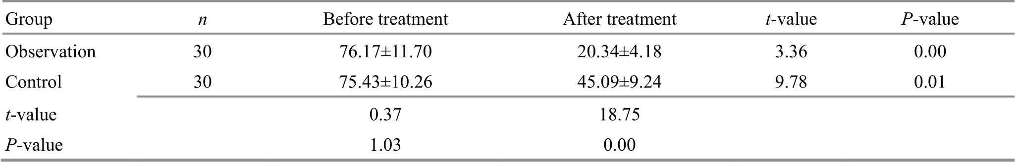
Table 3. Comparison of the WOMAC score between the two groups (±s point)
Note: WOMAC=Western Ontario and McMaster Universities osteoarthritis index
Group n Before treatment After treatment t-value P-value Observation 30 76.17±11.70 20.34±4.18 3.36 0.00 Control 30 75.43±10.26 45.09±9.24 9.78 0.01 t-value 0.37 18.75 P-value 1.03 0.00
Table 4. Comparison of the VAS score between the two groups (±s point)
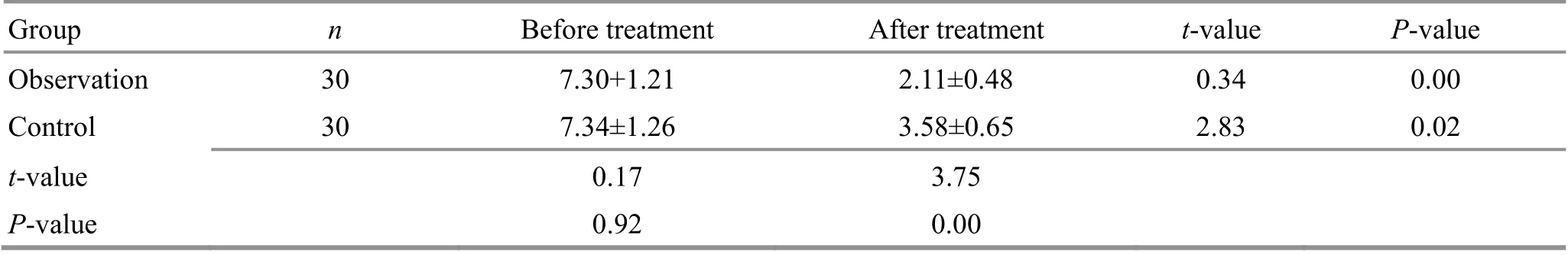
Table 4. Comparison of the VAS score between the two groups (±s point)
Note: VAS=Visual analog scale
Group n Before treatment After treatment t-value P-value Observation 30 7.30+1.21 2.11±0.48 0.34 0.00 Control 30 7.34±1.26 3.58±0.65 2.83 0.02 t-value 0.17 3.75 P-value 0.92 0.00
Table 5. Comparison of the iEMG values of VL between the two groups (±s μV)
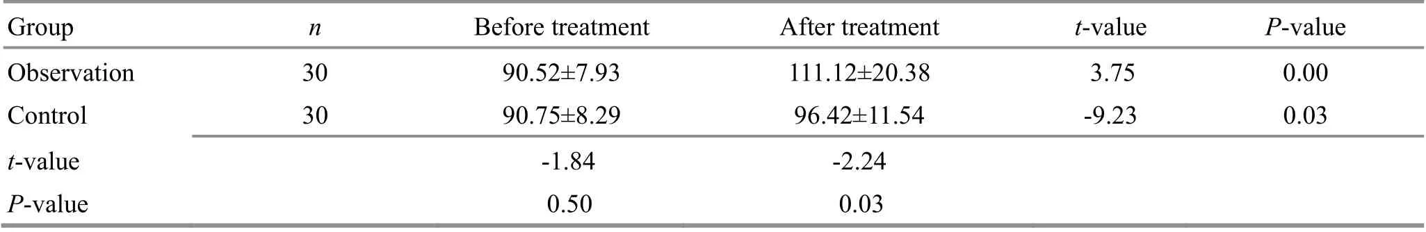
Table 5. Comparison of the iEMG values of VL between the two groups (±s μV)
Note: iEMG=Integrated electromyography; VL=Vastus lateralis
Group n Before treatment After treatment t-value P-value Observation 30 90.52±7.93 111.12±20.38 3.75 0.00 Control 30 90.75±8.29 96.42±11.54 -9.23 0.03 t-value -1.84 -2.24 P-value 0.50 0.03
Table 6. Comparison of the MF of VL between the two groups (±s Hz)
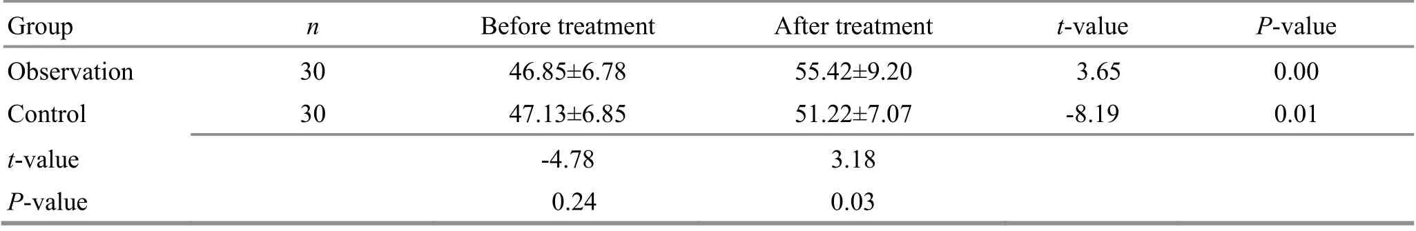
Table 6. Comparison of the MF of VL between the two groups (±s Hz)
Note: MF=Median frequency; VL=Vastus lateralis
Group n Before treatment After treatment t-value P-value Observation 30 46.85±6.78 55.42±9.20 3.65 0.00 Control 30 47.13±6.85 51.22±7.07 -8.19 0.01 t-value -4.78 3.18 P-value 0.24 0.03
Table 7. Comparison of the iEMG values of VMO between the two groups (±s μV)
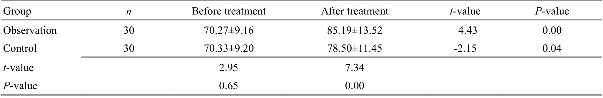
Table 7. Comparison of the iEMG values of VMO between the two groups (±s μV)
Note: iEMG=Integrated electromyography; VMO=Vastus medialis oblique
Group n Before treatment After treatment t-value P-value Observation 30 70.27±9.16 85.19±13.52 4.43 0.00 Control 30 70.33±9.20 78.50±11.45 -2.15 0.04 t-value 2.95 7.34 P-value 0.65 0.00
Table 8. Comparison of the MF of VMO between the two groups (±s Hz)
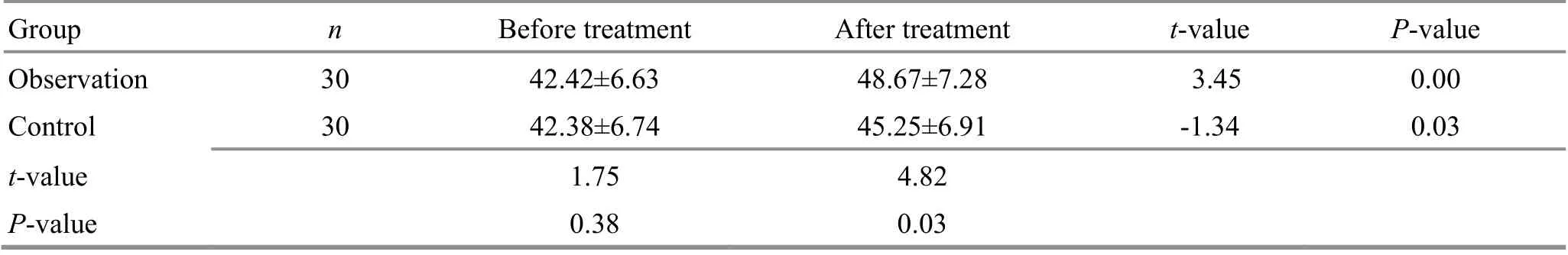
Table 8. Comparison of the MF of VMO between the two groups (±s Hz)
Note: MF=Median frequency; VMO=Vastus medialis oblique
Group n Before treatment After treatment t-value P-value Observation 30 42.42±6.63 48.67±7.28 3.45 0.00 Control 30 42.38±6.74 45.25±6.91 -1.34 0.03 t-value 1.75 4.82 P-value 0.38 0.03
4 Discussion
KOA is the result of complex multi-factors, and the basic cause of its pathogenesis is the imbalance of joint biomechanics. With the increase of age, degenerative changes of joint structure (such as articular cartilage degeneration, meniscus injury, and joint integrity deficiency) and soft tissue around the joint (such as aseptic inflammation of ligament attachment points,synovitis, and changes in muscle strength around the knee) lead to abnormal knee stress, further accelerating articular cartilage degeneration and osteophyte formation, and secondary subchondral bone destruction. At the same time, the release of inflammatory factors activates the signaling pathway,exerts the cascaded amplifying biological effect, and induces the degradation of cartilage matrix and chondrocyte apoptosis, finally resulting in cartilage destruction.
KOA belongs to Bi-Impediment syndrome in TCM,also called He Xi Feng (arthrosis like crane knee) or knee Bi-Impediment. It is mostly caused by invasion of the external pathogens, obstruction of sinews and meridians, and improper work and rest, as well as the insufficiency of essential Qi, and the insecurity of defense-exterior, aging and weakness, deficiency of liver and kidney, and lack of nourishment in sinews and meridians. It is a syndrome of essential deficiency and superficial excess, that is, the deficiency of liver and kidney, and lack of nourishment in sinews and bones are the core; pathogens blocking meridians, and Qi-blood stagnation is the superficiality. Chinese medicine believes that the “sinew-bone imbalance” is the key to the occurrence of KOA. Bone is the main structure of the human body. Strong bones can support the body. Sinews can restrain the bones and smooth the joints. On the one hand, the sinews are attached to the bones to maintain the posture of the human body, and on the other hand, the circulation of Qi and blood in the sinews provides energy for joint movement. The knee is where the sinews are gathered, and it is an important joint of the body. The “sinew-bone imbalance” of the body is most likely to occur in the knee joint. Warm needling moxibustion is combination of moxibustion and acupuncture, and has the dual effects of acupuncture and moxibustion. Xuehai (SP10) has the effects of relaxing tendons, activating collaterals, and promoting blood circulation to relieve pain. It is the key point for unblocking meridians and promoting blood circulation. HAN SN[6]found that acupuncture at Futu(ST32) could stimulate the contraction of the quadriceps femoris muscle, thereby strengthening the strength of quadriceps femoris muscle, improving the microcirculation around the joint, and enhancing the stability of the knee joint, consequently relieving pain and improving functional activities. WANG J,et al[7]found that warm needling moxibustion at Dubi (ST35)could down-regulate the expression of the apoptosisregulated proteins in knee cartilage tissue, thereby promoting the repair of articular cartilage. Zusanli (ST36)is the He-Sea point of the Stomach Meridian. It is a key point for pain relief and health care. Warm needling moxibustion at Zusanli (ST36) can warm meridians and unblock collaterals, dispel wind and eliminate dampness,activate Qi-flow for relieving pain, invigorate the spleen and harmonize the stomach. Modern studies have shown that warm needling moxibustion at Zusanli (ST36)can reduce the prostaglandin E2and nitric oxide levels in knee joint fluid, improve microcirculation, accelerate the absorption of inflammatory factors, eliminate pain,and improve joint functional activities[8-10].Yanglingquan (GB34) is the Influential Point of Tendons of the Eight Influential Points. It can invigorate Yang Qi,activate blood circulation for unblocking collaterals, and promote Qi-flow for relieving pain. Modern medicine believes that warm needling moxibustion at Yanglingquan (GB34) can effectively remove pain-causing substances in KOA patients, increase the blood supply to local tissues of the knee joint and relieve joint pain[11]. Yinlingquan (SP9) is the He-Sea point of the Spleen Meridian, with the effects of clearing dampness-heat, unblocking the meridians and collaterals, and reducing swelling for relieving pain.Studies have shown that acupuncture at Yinlingquan(SP9) and Yanglingquan (GB34) can increase the positive expression of local neuropeptide hormones in the acupoint area and increase the serum content of substance P in rats, thereby regulating the immune and endocrine function of the body[12-13]. Ashi point is mostly a reaction point in a disease, and it is also the best stimulation point for treatment. It has specific effects on various diseases. Tuina is one of the oldest external therapies for human beings. It acts on meridians, acupoints, and specific parts of the human body through manipulations, so as to regulate Zang-Fu organs, unblock meridians, and promote Qi-flow for activating blood circulation, and tendon-regulation and reconditioning. Ancient literature records show that Tuina has the effects of dispelling cold and promoting Qi-flow, activating blood circulation for relieving pain,and unblocking meridians and collaterals. Modern studies have shown that Tuina can inhibit the expression of inflammatory factors interleukin-1β and 5-hydroxytryptamine, improve pain threshold of local tissue, release soft tissue adhesions around the knee joint, improve local blood supply, promote the absorption of intra-articular effusion and tissue repair,inhibit cartilage degeneration, and can enhance the muscle strength, improve joint stability, restore joint biomechanical balance, so as to achieve the balanced state of “bone-restoration and tendon-softening”[14-16].The quadriceps femoris is one of the important structures for maintaining the stability of the knee joint.In this study, we found that the MF and iEMG values of sEMG in the observation group were higher than those in the control group, indicating that warm needling moxibustion plus Tuina can effectively increase the quadriceps femoris muscle strength and improve knee joint functions.
In summary, warm needling moxibustion plus Tuina can effectively relieve pain in the joints, improve joint function, and increase the EMG conduction velocity of the quadriceps femoris. In addition, the operation is simple, safe, and without toxic or side effects, thus worthy of clinical application.
Conflict of Interest
The authors declare that there is no potential conflict of interest in this article.
Acknowledgments
This work was supported by Science and Technology Planning Project of Traditional Chinese Medicine, Zhejiang Province (浙江省中医药科技计划项目, No. 2017ZA154).
Statement of Informed Consent
Informed consent was obtained from all individual participants.
Received: 30 January 2021/Accepted: 13 May 2021
猜你喜欢
杂志排行
Journal of Acupuncture and Tuina Science的其它文章
- Effects of electroacupuncture on angiogenesis and cortical VEGF and BDNF expression in rats with focal cerebral ischemia
- Reduction of serum level of interleukin-2 and pruritus severity after acupuncture at Quchi (LI11) in hemodialysis patients: a placebo-controlled randomized clinical trial
- Acupuncture plus naloxone hydrochloride in the treatment of coma after surgery for cerebral hemorrhage: a randomized controlled trial
- Clinical observation of pediatric Tuina plus oral Chinese medication for pediatric anorexia due to spleen failing in transportation
- Protocol-optimizing study of combining Tuina and horse-riding squat exercise for knee osteoarthritis
- Therapeutic efficacy of acupuncture plus Tuina for spastic cerebral palsy and discussion of its mechanism