Structure and Properties of a Cd(II) Metal-organic Framework Based on a Newly Designed Heterotopic Tripodal N-Donor Ligand①
2022-03-12HUANGJieFenCHENYiHoLIANGZhenHuZHENGShengRunCAOJun
HUANG Jie-Fen CHEN Yi-Ho LIANG Zhen-Hu ZHENG Sheng-Run② CAO Jun
a (School of Chemistry, South China Normal University, Guangzhou 510006, China)
b (School of Materials Science and Hydrogen Energy & Guangdong Key Laboratory for
Hydrogen Energy Technologies, Foshan University, Foshan, Guangdong 528000, China)
ABSTRACT In this paper, a Cd(II) metal-organic framework (MOF), Cd-DIBT (HDIBT = 5-(3΄,5΄-di(1Himidazol-1-yl)-[1,1΄-biphenyl]-4-yl)-1H-tetrazole), has been constructed based on a newly designed heterotopic tripodal ligand containing both imidazolyl and pyrazolyl groups. The Cd-DIBT exhibits a new three-dimensional(3,3,9)-connected trinodal network topology with point symbol of (42·6)(43)2(48·615·812·10) (namely scnu) based on binuclear secondary building blocks (SBUs). Staggered 1D channels were observed in such framework and was estimated to have 5487 Å3 potential solvent area (56%). The stability study reveals that the framework is unstable and easily transforms into amorphous MOF after the removal of guest molecules. In addition, the Cd-DIBT shows a ligand-centered luminescence.
Keywords: heterotopic tripodal ligand, metal-organic framework, topology, crystal structure;
1 INTRODUCTION
The construction of metal-organic frameworks (MOFs)has still gained a lot of attention because of their structure diversity, as well as their potential applications in many fields including energy, environment, biomedicine, etc[1-6].According to the types of coordination groups on the ligands,most ligands can be classified into the nitrogen-containing ligands, carboxylic acid ligands or nitrogen-containing carboxylic acid ligands. Among them, nitrogen-containing tripodal ligands with three coordinating groups are one of the effective ligands that have been used for the assembly of MOFs and discrete metal-organic cage due to their multidentate coordination modes and strong coordination ability towards most of the transition metal ions[7,8]. In addition, some of the N-donors are neutral, which makes them easier to form cationic frameworks when compared with those based on carboxylic acid ligands[9-11]. At present,most of the reported nitrogen-containing tripodal ligands possess three identical coordination donors (namely homotopic tripodal ligands), while ligands that possess three different N-donor groups (namely heterotopic tripodal ligands) have been relatively less explored[12-16]. Recently,we have focused on the design and synthesis of MOFs based on heterotopic tripodal ligands containing both imidazolyl and tetrazolyl groups[12-15]. The imidazolyl and tetrazolyl groups exhibit similar geometries but have different coordination abilities, coordination modes and charge neutrality. The tetrazolyl group exhibits weaker coordination ability but with varied coordination modes, which is usually applied to form multi-nuclear SBUs in the assembly of MOFs. In contrast, the 1-substitued imidazolyl group displays simple monodentate coordination but stronger coordination ability, which may be applied to connect the SBUs to form the framework. Heterotopic ligands containing both imidazolyl and tetrazolyl groups may exhibit different coordination chemistry from its corresponding homotopic ligands and are still deserved to be explored. Thus, MOFs based on two imidazolyl-tetrazolyl heterotopic tripodal ligands, 5,5΄-(5-(1H-imidazol-1-yl)-1,3-phenylene)-bis(2Htetrazole) (H2IPBT) and 5-(3,5-di(1H-imidazol-1-yl)phenyl)-2H-tetrazole (HDIPT) have been studied by our group recently. Herein, as part of a continuous study, a newly designed heterotopic tripodal N-donor ligand that can be seen as the extension of HDIPT, namely, 5-(3΄,5΄-di(1Himidazol-1-yl)-[1,1΄-biphenyl]-4-yl)-1H-tetrazole (HDIBT)(Scheme 1), was employed to construct a new Cd(II) CP.The crystal structure and fluorescence property were investigated for this new species.
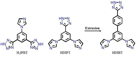
Scheme 1. Structures of some imidazolyl-tetrazolyl heterotopic tripodal ligands
2 EXPERIMENTAL
2. 1 Materials and methods
Ligand HDIBT was obtained from Shanghai Kylpharm Co., Ltd through customized service. All other reagents were commercially available and used without further purification; Infrared spectra (IR) were recorded on a Nicolet FT-IR-170SX spectrophotometer. Powder X-ray diffraction(PXRD) data were taken on an Ultima IV X-ray powder diffractometer at 40 kV and 40 mA for a CuKαradiation (λ=1.5406 Å). Thermogravimetric analysis (TG) was performed on NETZSCH STA 449 C under air atmosphere at a heating rate of 10 °C/min. The photoluminescence emission spectra were carried out on a Hitachi F-4600 spectrophotometer.
2. 2 Synthesis of Cd-DIBT
A mixture of Cd(NO3)2·4H2O (30.8 mg, 0.1 mmol),HDIBT ligand (35.4 mg, 0.1 mmol), and DMF (6 mL) was sealed in a 10 mL teflon-lined stainless-steel autoclave,which was mixed by sonication and heated at 90 °C for 96 hours under autogenous pressure. Then, the mixture was slowly cooled down to room temperature during 24 hours.Colorless crystals of Cd-HDIBT were obtained. Yield 63%(based on the HDIBT). IR (KBr, ν/cm-1): 3120(m), 2915(w),1659(m), 1603(s), 1501(s), 1445(m), 1384(vs), 1242(m),1116(m), 1070(s), 1014(m), 935(m), 845(m), 758(m),642(m).
2. 3 X-ray structure determination
A single crystal of Cd-DIBT was collected at 298 K on a Rigaku SuperNova Dual Atlas diffractometer using mirror monochromatized CuKαradiation from a high-flux microfocus source. The multi-scan absorption correction was performed by SCALE3 ABSPACK scaling algorithm program[17]. The structure was solved by direct methods and refined by full-matrix least-squares techniques employing the SHELXT[18]and SHELXL[19]program packages,respectively. All non-H atoms were refined anisotropically.All the other hydrogen atoms were produced theoretically.The contributions of distorted solvent and anions in the channels were removed by SQUEEZE[19]. Selected bond lengths and bond angles are provided in Table 1. Crystal data for C57H39Cd2N24(Mr= 1284.92 g/mol): monoclinic system, space groupC2/c,a= 16.7214(5),b= 23.9705(7),c= 24.4712(8) Å,β= 91.419(3)°,V= 9805.5(5) Å3,Z= 4,T=298(2) K,μ(CuKα) = 1.54184 mm-1,Dc= 0.870 g/cm3, 71065 reflections measured (6.45°≤2θ≤147.12°), 18406 unique(Rint= 0.0499,Rsigma= 0.078) which were used in all calculations. The finalR= 0.0626 (I> 2σ(I)) andwR= 0.1714 (all data).

Table 1. Selected Bond Lengths (Å) and Bond Angles (°)
3 RESULTS AND DISCUSSION
3. 1 X-ray crystal structure
X-ray single-crystal diffraction analysis showed that the Cd-DIBT crystallizes in monoclinic space groupC2/c. The asymmetric unit of Cd-DIBT contains one Cd(II) ion, one and a half of DIBT-ligands, and some undefined solvent molecules and anions in the channels. In order to meet the requirement of electrical neutrality, a counter anion is required in the channel. However, due to the serve disorder,it is difficult to determine anions and solvent molecules from the single crystal diffraction data. Therefore, the presence of anions is determined from the IR spectrum. As shown in Fig. 1,the IR spectrum of Cd-DIBT is similar to that of the HDIBT ligand. The slight shift of the peak position is due to the formation of coordination bonds. However, there are two peaks that are clearly different from the ligand. The first is the strong peak at 1384 nm, which is the typical peak of nitrate anion; the second is that at 1659 nm, which is assigned to the stretching vibration of C=O bond in the DMF molecule.
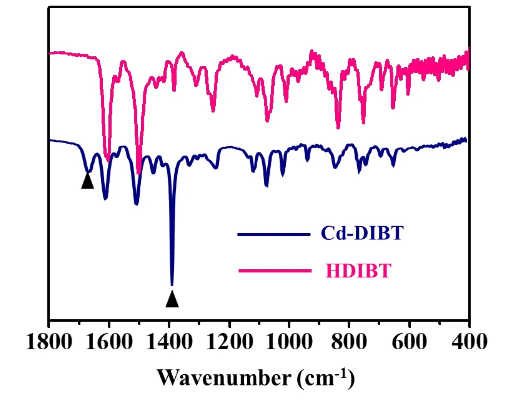
Fig. 1. Spectra of HDIBT ligand and Cd-DIBT
As shown in Fig. 2, the metal Cd(II) ion adopts a distorted octahedral coordination geometry which is coordinated with three tetrazole N atoms (Ntz) and three imidazole nitrogen atoms (Nim). Although the coordination groups are different,the bond lengths of Cd-Ntzand Cd-Ntzare comparable, as shown in Table 1. The two DIBT-ligands adopt similarµ4-coordination modes, in which the tetrazolyl group bridges two Cd(II) ions, and the two imidazole nitrogen atoms coordinate with the other two Cd(II) ions, respectively. The difference between them is mainly in the conformation,which can be seen by the three dihedral angles between the three coordinating groups and their adjacent benzene ring(please list which angles) are 46.34o, 27.76o, 19.33oand 25.44o, 25.44o, 4.80o, respectively. On the other hand, two Cd(II) ions are jointed by three tetrazolyl groups to form a binuclear SBU, which is often observed in MOFs based on tetrazolyl-based ligands[12-14]. The distance between the adjacent Cd(II) ions in the SBU is 4.0676(1) Å. The coordination bonds between the Cd(II) ions and imidazolyl groups connect the binuclear SBUs into a 3D framework, as shown in Fig. 2c. There are 1D small channels in two mutually perpendicular directions, so that the framework become porous and has 5487 Å3potential solvent area(56%)[20].
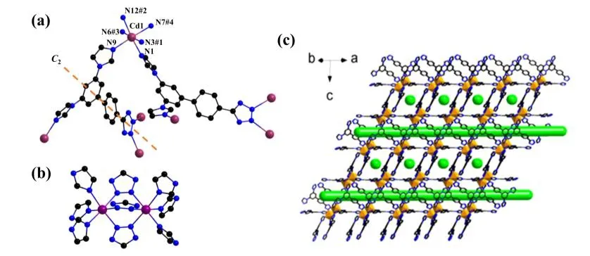
Fig. 2. (a) Coordination environment of Cd(II) and coordination modes of DIBT- ligand. Symmetry codes: #1: -x + 3/2, -y + 1/2, -z;#2: -x + 3/2, y + 1/2, -z + 1/2; #3: x, -y, z + 1/2; #4: -x + 1, -y, -z. (b) Binuclear SBU. (c) 3D framework of Cd-DIBT.1D channels in two directions perpendicular to each other are shown in green
In order to get a more clear idea about the structure of Cd-DIBT, the connectivity between the binuclear and the ligands is investigated by topological analysis. As shown in Fig. 3, the binuclear SBU connecting to nine DIBT-ligands can be regarded as a nine-connected node with point symbol(48·615·812·10). On the other hand, both the DIBT-ligands can be regarded as a three-connected node because they both connect to three binuclear SBUs. However, the point symbols are different to be (42·6) and (43), respectively. In addition, the ratio of 9-connected and the two 3-connected nodes is 1:2:3,thus the framework topology can be denoted as a new (3,3,9)-connected trinodal network topology with point symbol of(42·6)(43)2(48·615·812·10) as indicated by TOPOS[21](Fig. 2),which is namedscnu.

Fig. 3. Nine- and three-connected nodes and the topology network of Cd-DIBT
3. 2 Stability
The PXRD of the as-synthesized bulky sample was measured and the result is shown in Fig. 4. The simulated PXRD basically matches with the simulated data, indicating the major product is Cd-DIBT. The observed broaden peak suggests that the crystallinity is not very high, which may result from the framework collapses when the guest solvent is released. In order to investigate its stability, the PXRD was measured after one week and two weeks. We found that the diffraction peak is further weakened, leaving only the peak at about seven degrees after a week. Then, the sample was transformed into completely amorphous after two weeks.Although stability is necessary to realize many functions of crystalline MOF, recent studies on amorphous MOF also show that amorphous may also have better applications in some special areas[22-25]. The TG curve of Cd-DIBT (Fig. 5)shows that about 11% weight loss occurs below 150oC,which may be attributed to the release of solvent molecules in the channels. Then, the framework is stable up to about 310oC. The major weight loss happened above that temperature, indicating a decomposition of the framework.
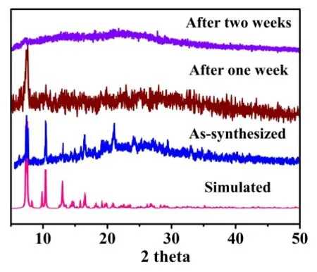
Fig. 4. PXRD of the simulated, as-synthesized and therefrom stored in air samples

Fig. 5. TG curve of Cd-DIBT
3. 3 Photoluminescent property
Previous studies have shown that Cd(II) MOFs exhibit photoluminescent properties[26-29]. Therefore, the photoluminescent property of Cd-DIBT in the solid state has been measured. As shown in Fig. 6, the maximum emission peak is at 450 nm upon excitation at 338 nm. The emission band of Cd-DIBT is comparable with the emission of HDIBT at 450 nm (λex= 328 nm). Thus, the emission of Cd-DIBT may be assigned to the ligand-centered emission because thed10ions are difficult to oxidize or reduce. The red shifts of the excitation might be due to the molecular orbital and conformation of Cd-DIBT tuned by the coordination of Cd(II)ions.

Fig. 6. Excited and emission curves of HDIBT and Cd-HDIBT at room temperature
4 CONCLUSION
In summary, a new Cd(II) MOF based on a newly designed imidazolyl-tetrazolyl heterotopic tripodal ligand was prepared and characterized. Crystal structure study reveals that the new MOF is a high-connected framework based on binuclear SBU. The framework contains 1D channels and easily changes into amorphous MOF when the guest molecules are exchanged or lost. FL study reveals that the MOF exhibits ligand-centered emission. Further studies on the construction of new MOFs based on this new ligand and properties by using the crystal to amorphous transition of Cd-DIBT are in progress in our laboratory.
杂志排行
结构化学的其它文章
- Synthesis, Crystal Structure and Fungicidal Activity of 3,4-Dichloro-5-(6-chloro-9-(4-fluorobenzyl)-9H-purin-8-yl)isothiazole①
- Synthesis, Crystal Structure and Antifungal Activity of New Furan-1,3,4-oxadiazole Carboxamide Derivatives①
- Syntheses, Crystal Structures, Anticancer Activities and DNA-Binding Properties of the Dibutyltin Complexes Based on Benzoin Aroyl Hydrazone①
- Synthesis, Crystal Structure, Fungicidal Activities and Molecular Docking of Acyl Urea Derivatives Containing 2-Chloronicotine Motif①
- Molecular Design and Property Prediction of High Density 4-Nitro-5-(5-nitro-1,2,4-triazol-3-yl)-2H-1,2,3-triazolate Derivatives as the Potential High Energy Explosives①
- A New Borate-phosphate Compound CsNa2Lu2(BO3)(PO4)2:Crystal Structure and Tb3+ Doped Luminescence①
