Workers’ Cerebrocortical Activity in Hot and Humid Condition:An Electroencephalogram Study
2022-03-03YixuanWeiYifeiXuShuWangLongzheJinTianqiDing
Yixuan Wei,Yifei Xu,Shu Wang,Longzhe Jin,Tianqi Ding
Abstract:Hyperthermal environments can harm workers’ health and safety.However,it is difficult to include effective protection into standards because heat-related impacts vary significantly according to individual workers and multiple factors.Studies suggested obvious relationship between environment condition and bio-electricity signal,including electroencephalogram (EEG) signal.We used a detector with 64 electrodes to perform dedicated EEG measurements of nine individual subjects to analyze human cerebral activity under hyperthermal (35℃,80% RH) and standard conditions (25℃,30% RH).Amplitude changes of the frequency wavebands were analyzed using statistical analysis.Seven participants showed increasing beta activity due to high temperature and high humidity in the primary somatosensory cortex (electrode C3) and the temporopolar region (electrode FT 8).The amplitude value of alpha wave is increased from 0.194 to 0.213 while the amplitude value of beta wave is increased from 0.144 to 0.160.Value is decreased due to hyperthermal environment for most people.The results of this study could be used to inform the development of wearable equipment to monitor the health of on-site workers,which is fundamental to improve worker safety and wellbeing.
Keywords:confined space;electroencephalogram (EEG);hyperthermal environment;heat exposure;workers’ health
1 Introduction
Rapid economic and industrial development has compelled many countries to recognize the importance of workers’ health and safety.Research and development of protective equipment and enhancement of safety awareness have become priorities for many nations.Despite this emphasis,confined space fatality remains a significant cause of workplace deaths in many countries [1,2].The main reason for fatality in these spaces is the unfavorable natural ventilation that contributes to the accumulation of toxic gases,hypoxia,hyperthermal,and humid environments.Recently,investigators have examined the mechanisms by which“extreme ”environments affect human health.However,these theories have been proposed mainly for medical use [3,4],while the industrial workplace currently lacks the implementation of technology for status monitoring and early warning.The prevention of potential harm to workers’ health and life requires manned experiments to demonstrate the relationship between extreme environment and worker symptoms.
Extreme thermal environments can harm workers’ health and safety.High temperatures are widely found in the workplace,such as in iron,steel,and nonferrous foundries; construction;food canneries;chemical plants;and mining sites [5].A cross-sectional study reported that the injury prevalence of workers exposed to extreme thermal environments in several countries ranged from 9.2% to 49% [6].Extreme thermal environments also commonly occur in confined workplaces such as heating pipe networks in municipal construction.Workers must enter these confined spaces and perform multiple activities under heat stress,for example,maintenance,inspection,cleaning,etc.Although many countries have proposed relevant standards to protect workers in confined spaces [7,8],most only have general requirements and vague statements concerning industry safety management.
Considerable literature has grown around the theme of physiological response to heat exposure [9].Heat-related effects not only negatively impact working productivity [10] but also cause impairments such as heat fatigue,heat syncope,heat cramps,exhaustion,and heatstroke[11,12].A decrease in cognitive function and perceptual motor skills could occur along with unsafe behaviors,leading to injuries and illness in extremely hot environments [13]. The risks to workers’ health may vary according to multiple factors [14] (e.g.,environment,age,disability,diseases,workload,etc.) and increase due to the global warming trend.According to Hoehne et al.[15],the risk induced by outdoor heat exposure shows large contracts among different cities,demographic groups,and work types.In addition,heat tolerance may vary between workers because of congenital and acquired factors.Despite several standards mentioning the“administrative management and recommended working environment”of occupational health and safety,significant research findings have not been specifically addressed in legislation and policies[16,17],such as the evaluation of maximum acceptable work duration in extreme thermal conditions [18] and health hazards of working in a hot and humid environment. Therefore,sensitivity analysis and individual monitoring should be conducted to avoid further heat-related impairment[19].
Cerebral activity is more sensitive to heat stress than peripheral response and has been analyzed in the central nervous system [20].Previous studies identified changes in physical parameters under extreme thermal conditions including maximum oxygen uptake (CO2max),middle cerebral artery blood flow velocity (MCAV),and global cerebral blood flow (gCBF).Most findings showed that intense exertion in heat leads to attenuation of MCAV and gCBF,while the oxygen extraction in the brain remains high [21,22].However,the reasons for the reduction in cerebral perfusion are complex and unclear [23].Overall,these changes suggest cellular homeostasis and the continuous need for brain energy,which is captured by electroencephalograms (EEGs)[24].However,the irreversible damage to tissue caused by heat stress is not fully understood,especially in humans [25].
Many studies have analyzed the effects of temperature and humidity on cerebral activity[26].Chang et al.reported no significant difference in EEG activity when the subjects’ hands were stimulated in 40°C–43°C water,while the theta band power decreased during cold stimulation [27].The 90 min thermal experiments (35°C and 80%) conducted by Tiago-Costa et al.demonstrated amplitude decreases in alpha and beta activities in both the left and right occipital areas [28].Analysis of changes in cerebral activity and fatigue state during intense exertion in a heated environment indicated a decrease in beta activity and an increase in the ratio of alpha to beta index [29].Similar results were reported by Kacem et al.in which EEG power showed a shift and core temperature increases in female subjects when they reached exhaustion [30].However,their EEG measurement was relatively simple,with only one or two positions on the scalp used for EEG recording. Zhu et al. examined cognitive activity under varying temperature and humidity based on the EEG signal.Although some studies indicated that subjects were less alert at higher humidity [31],the subjects did not perform differently on cognitive tests under hyperthermal conditions.In addition to hot conditions,Periard et al.also considered the hypoxic impact in dual-exercise experiments,which showed increased alpha and beta activities at the beginning of the standard and hypoxic conditions,which then decreased.In addition,the alpha activity was lower under the hot condition than under standard conditions [32].Furthermore,recent advancements in sensing technologies has promoted the wearable device for enhancing people’s well-being and safety.For example,Jebelli proposed a model to recognize construction workers’ stress at the site based on simple wearable EEG device [33].J M Morales et al.assess the validity of a single-channel EEG device to monitoring the mental fatigue of drives [34].A new photoplethysmography (PPG) sensor suite was adopted by Brian and Bradford to measure multiple physiological parameters in reduced oxygen environments [35].In healthcare section,realtime monitoring of heart rate,respiratory frequency and other physical parameters was realized by computational efficient wearable sensor networks [36] and fog computing assisted wearable sensor platform [37].
A review of literatures revealed a few key points:
①Although extensive studies have been carried out to protect workers’ health and safety in confined spaces,damage due to heat exposure is rarely considered as an important physical hazard to workers.
②The physiological response to heat exposure shows large contracts among different people,regions,and work types.Hence,developing wearable equipment for early warning of health damage during heat exposure may be an effective solution for individual monitoring. However,most existing products were developed for medical use and few studies have reported research and development in this context.
③Many studies have analyzed the correlation between cerebral activity and mental state as well as the impact of temperatures and humidity on EEG data.However,whether heat exposure causes increased brain activity is unclear due to different experimental procedures.The change in cerebral activity is likely caused by multiple factors including environmental change,mental fatigue,or peripheral fatigue.
Hence,effective monitoring of worker status and forecasting workers’ behavior remain formidable tasks,as they involve uncertain and complex interactions between the physical environment and stochastic occupant behavior.The objective of this study was to analyze the cerebral activity of humans under hyperthermal conditions.Unlike the aforementioned studies,which used simple measurements,dedicated EEG measurements with 64 electrodes have been used to explicitly identify changes in cerebral activity due to atmospheric temperature and humidity with limited artifacts.In addition to the analysis of cerebral activity,photoplethysmography(PPG) signals have also been measured to determine cardiovascular responses under passive heat stress.Finally,we summarized the results of existing studies and found both heat-induced increase and heat-induced decrease in the EEG data.Overall,this paper focused on identifying EEG changes during heat exposure that could be fundamental for the development of wearable equipment for health monitoring and early warning.
The remainder of this paper is structured as follows.In Section 2,we introduce the experimental participants,procedures,and EEG measurements.The EEG activities in regions and selected electrodes under heat exposure are described in Section 3.In Section 4,we discuss the cardiovascular response under hyperthermal conditions and compare our results with those of other studies.Finally,in Section 5,we draw conclusions.
2 Materials and Methods
2.1 Participants
Nine undergraduates from the University of Science &Technology Beijing voluntarily participated in this study.They were asked to avoid strenuous exercise 72 h before their participation and to refrain from consuming caffeine or alcohol for 24 h.The physical and mental condition of all participants was good because a great quality of sleep was required before the experiment.Their characteristics were (mean +SD) as follows:age 19.5±1.5 years,body mass 57.2±12.8 kg,height 168±10 cm.The participants were fully informed of the experimental procedures before providing written informed consent.They were also required to complete a Medical History Questionnaire before being admitted to the study.The protocol was approved by the ethics committee.
2.2 Experiments
The participants arrived at the laboratory at the same time of the day.Upon arrival,they changed into lab gowns and rested in a seated position in temperate conditions (25±2°C,30% relative humidity [RH]) while being instrumented with the 64-channel EEG cap.The thermal resistance of clothing was assumed to be 1 clo because the participants changed into the lab-gown.In this experiment,we collected EEG signals of participants under two environmental conditions:standard (25℃,30% RH) and extreme (35℃,80% RH).The participants were required to sit in the climate chamber for 15 min in each condition.
The experimental environment was provided by the climate chamber,the internal and external parts of which are shown in Fig.1.A temperature controller,humidity controller,gas-filling device,and various sensors were installed in the chamber.The emergency stop button was set to ensure participant safety in the chamber.When the emergency button was triggered,an alarm would sound and fresh air would be supplied to the chamber immediately.

Fig.1 Experimental conditions
2.3 EEG Measurement
EEG data were collected and recorded using an EEG-cap (Electro-Cap International,Inc.,USA)with 64 Ag-AgCl electrodes arranged according to the international 10–20 standard.In the preparation,each electrode was referenced to the reference electrode CPz,and GND served as the ground electrode.The electrodes were filled with electrogel for signal transduction.The recording impedance of all electrodes was checked and did not exceed 20 kΩ.
The EEG signals of the participants in the standard condition were first measured and collected.The electrode cap,amplifier,and recording computer were brought into the climate chamber by the participants.During the recording,the participants remained seated and avoided unnecessary movement.After 10 min of measurement,the participants left the chamber and relaxed for a while.We then adjusted the internal environment to hyperthermal conditions and allowed the participants to enter the chamber for another recording.
2.4 EEG Analysis
EEGLAB (version 14.1) was used to preprocess the data sets.The EEG data sets contained raw data for 62 electrodes.First,the data were downsampled to 200 Hz,filtered (low cutoff:1 Hz,high cutoff:30 Hz) with a Gaussian filter design,and re-referenced to the average reference of M1 and M2.Second,for each segment channel,inspection and artifact removal were conducted based on the raw data.Unusable channels were topographically interpolated (interpolation type:spline) to maintain the original number of electrodes.Then,a 2-min EEG data segment during the recording was selected for analysis.Third,independent component analysis (ICA) was used to remove artifacts such as eye twitch artifacts.The epoch was analyzed by fast Fourier transform,and three frequency wavebands were obtained:theta (4–8 Hz),alpha (8–12 Hz),and beta (12–25 Hz).The amplitudes of the frequency wavebands were then extracted. To identify the differences in EEG data sets between standard and extreme conditions,paired-sample t-tests were performed for the amplitude of frequency wavebands.
3 Results
3.1 Regional Changes in EEG Activity
This study observed no significant differences in EEG data between male and female participants.Therefore,each participant was treated as an individual case.Significant effects of atmospheric temperature and humidity on EEG activity were observed in the regional changes of the frequency wavebands. The amplitude changes(p<0.05) in theta,alpha,and beta waves are shown in Tab. 1. Seven of nine participants showed significant changes in the low-frequency Hz waveband in the middle frontal (BA 46),frontopolar (BA 10),and granular frontal (BA 9) regions.Although the electrodes showing significant differences varied between participants,frontal AF 7 showed the most pronounced theta wave changes among the seven participants.Specifically,in the hot and humid condition,increased activity was observed in Participants 1,3,5,7,and 9,while Participants 2 and 6 showed decreased activity.Eight participants showed significant alpha wave differences in the left frontal area (BA 45 and 8),except for Participant 5.The main effect of high temperature and humidity on the alpha wave band for Participant 5 was in the parietal area (BA 7,39,and 40).In general,stronger alpha activity was associated with extreme conditions for seven participants.However,Participants 2 and 5 showed significantly decreased alpha wave amplitude.
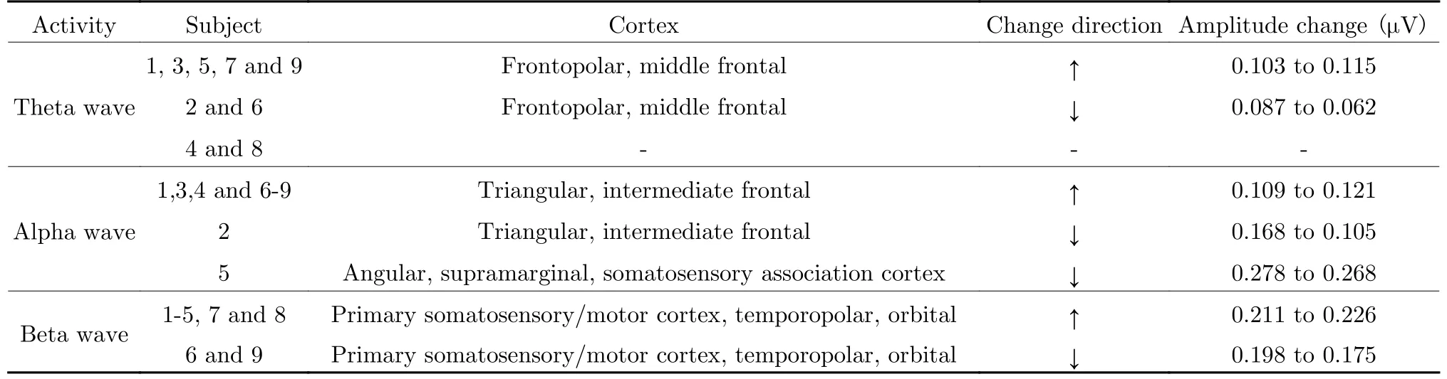
Tab.1 Brain structure showing regional activities (p <0.05) in the hot and humid condition
With regard to high-frequency Hz wavebands,the regional activities of all participants were centralized in electrodes C3 and FT8,which is more accordant than theta and alpha wavebands.As shown in Tab.1,significant changes in the beta wave were observed in the right hemisphere of the orbital and temporopolar areas as well as in the left hemisphere of the primary somatosensory and motor cortex.In addition to the primary somatosensory area,the temporopolar region (electrode FT8) was also connected to the perception of the temperature and humidity.In this study,seven of nine participants showed significant increases in beta wave amplitude,except for Participants 6 and 9.In addition,the amplitude change of frequency waveband from normal condition to hot and humid condition is p resented in Tab.1.
3.2 EEG Activities of Selected Electrodes
In the manned experiment in our study,most participants showed increased and larger variance in EEG activity in the hot and humid condition compared to that for the standard condition.The results in Tab.2 showed that alpha waveband >theta waveband >beta waveband,where >indicates“larger mean value and standard deviation”.Specifically,Participants 4 and 8 tended to show EEG activity with larger fluctuations in all frequency wavebands and conditions.In contrast,Participant 6 showed predominately beta activity only,while the low-frequency wavebands were relatively small (Avg.<0.1,SD<0.20).In addition,Participant 7 tended to have weaker theta activity (Avg.<0.1,SD<0.02) than did other participants. With regard to the alpha waveband,the mean value and standard deviation of Participant 1 were extremely small(Avg.<0.1,SD<0.04).
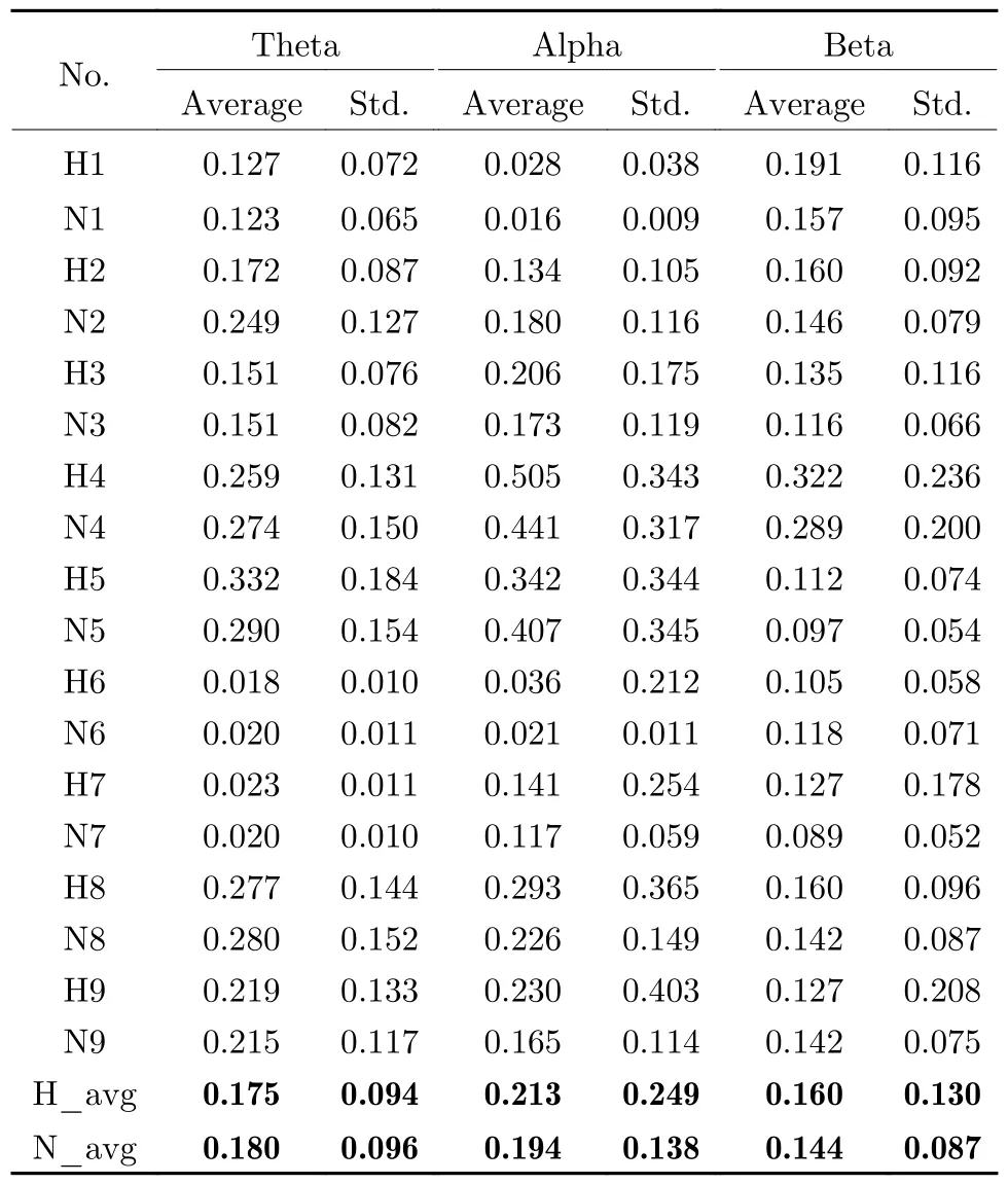
Tab.2 Statistical analysis of frequency waveband amplitudes under constant and extreme conditions
Our results provide objective evidence of increased beta wave activity due to high temperature and high humidity,as reflected by electrodes C3 and FT8.Originally,the frontoparietal Ushaped region was referred to as the homunculus,which was first proposed by Penfield in 1937 and revised several times [38,39].As shown in Fig.2,the topographical organization described as the primary somatosensory cortex,is dedicated to representing proprioception,pain,temperature,and touch from the leg,hand face,and oral regions [40].Electrode C3,located in the somatosensory cortex,was assumed to reflect proprioception from the facial area. This observation supports evidence from a previous study [41,42]that the facial skin is sensitive to heat stress,especially the human ears,nose,and cheeks.In particular,the highest temperature of the facial regions is concentrated on the forehead along with the heating time.Notably,this sensitivity is reflected by EEG activity in high-frequency wavebands.
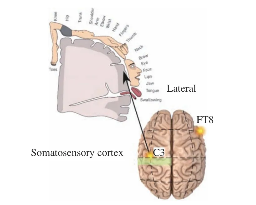
Fig.2 Illustration of electrodes C3 and FT8,which showed significant differences in the beta wavebands of all participants during increased temperature and humidity conditions
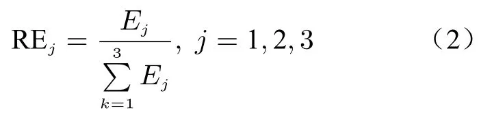
Furthermore,the energy of these three wavebands can be calculated as

where A(i) is the amplitude of the frequency wavebands at each sampling data i and Ejis the total energy of the theta,alpha,and beta activities of the same electrode(s).Then,the relative energy is as follows:
The analysis and comparison results revealed that the relative energies of alpha and beta activities increased in the selected electrodes for most participants,except for Participant 5,as shown in Fig.3.
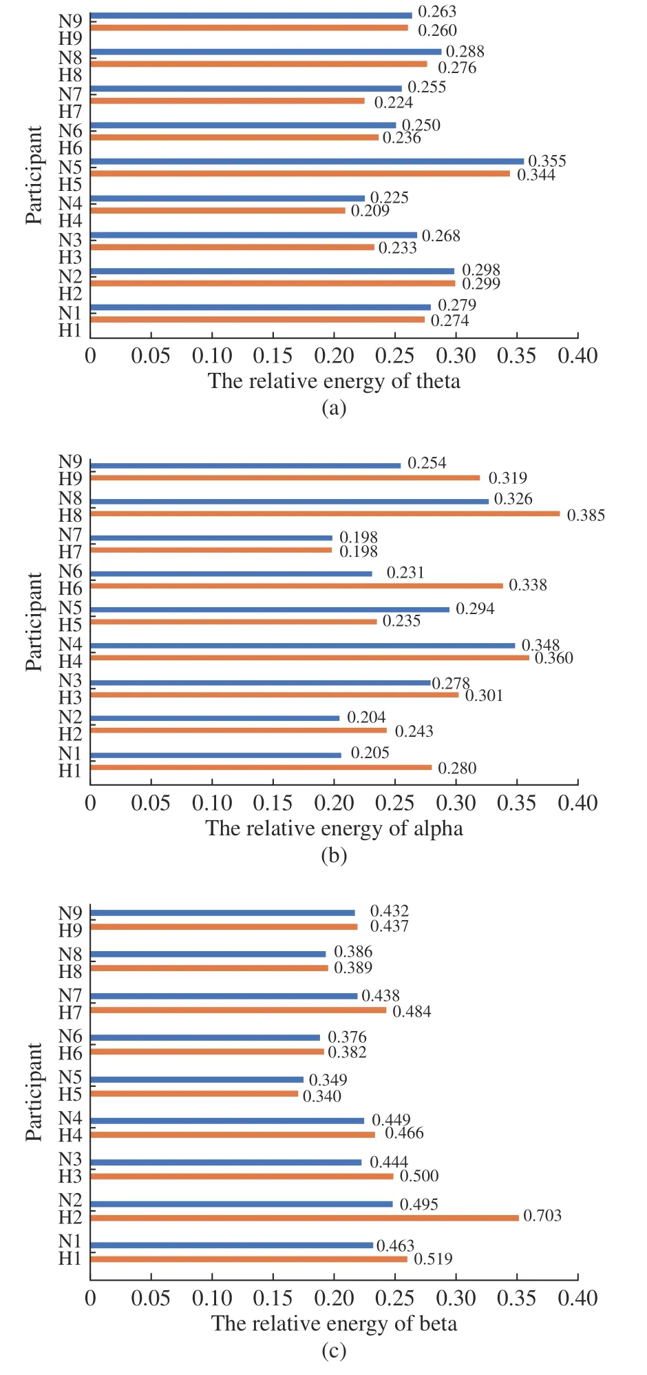
Fig.3 Relative energies of (a)theta,(b)alpha,and (c)beta activities under extreme and standard conditions
The relative energy of the theta waveband in AF4 declined under the extreme condition. In Fig.4,the value of θ/(α+β) can be adopted as an indicator of environmental change because it decreases when the temperature and humidity increase,except for Participant 5.The analysis of the ratio of θ and α+β showed that the change rate of θ activity was likely to be smaller than the total increase rate of α and β.
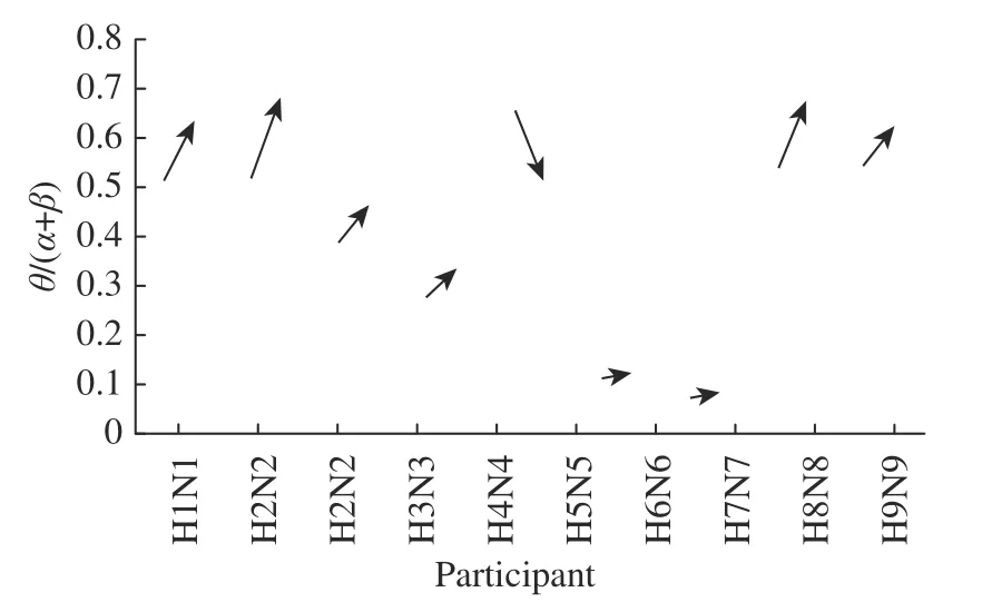
Fig.4 Changes in θ/(α+β) values in extreme and standard conditions
4 Discussion
4.1 Cardiovascular and Thermoregulatory Response
The cardiovascular response under different environmental conditions during exertion has been widely discussed and cerebrocortical activity also characterized. Fatigue or discomfort should be acknowledged as an integrated complex influenced by both central and peripheral nervous systems.Thus,this study analyzed EEG changes combined with the cardiovascular response in hot and humid environments.
Shortly before,our research group conducted a manned experiment in a hyperthermal and humid environment (35℃,80% RH),where PPG signals were collected and measured.The blood volume changes in the microvascular bed of tissues can be determined using plethysmograms.In our study,the correlation between the EEG activity and the PPG signal can be preliminarily identified. The Kolmogorov complexity (KC)was adopted to describe the PPG signal disorder.The KC process compresses the original string into a short description that requires little effort.Hence,a smaller KC index means that the information processing is relatively easier.The KC of the PPG signal of the 9 subjects under the hyperthermia and normal conditions is reported in Tab.3.The PPG signal complexity essentially decreased (p =0.006) when the environmental temperature increased.A decreasing KC is a reliable indicator that the PPG signal becomes more uniform due to human or environmental influences.The reason for the high KC complexity under the normal condition was related to the larger amplitude of the PPG signal that contained more intrinsic randomness or noise.Therefore,the impact of environmental temperature on PPG signals can be identified by the significant indicator of the KC complexity.

Tab.3 Kolmogorov complexity of PPG signal
4.2 Comparisons to Other Studies
While the association between temperature and EEG activity has been reported previously,they varied in the number of participants,variables measured,number of electrodes,and factors analyzed.
The findings of these studies demonstrated that environmental factors and fatigue (both physical and mental) were associated with significant changes in brain wave activity.For example,long driving duration caused an amplitude increase in the alpha band,while a hyperthermal environment was also identified as the major cause of strong alpha activity. Several studies reported that physical fatigue caused by exertion in heat reportedly led to an increased ratio of alpha activity to beta activity.However,the impact of temperature on brain wave activity was inconsistent among studies.Beta wave activity increased in the hot environment,but it showed a decreasing trend in hot and humid conditions. Furthermore,several studies analyzed low-frequency wavebands (delta and theta) and reported that a hyperthermal environment led to increased delta activity.In addition,it is unclear whether hypoxia suppresses brain activity.The most likely causes of contradictory results are differences in experimental procedures,types of EEG headsets and subject states.The change in cerebral activity is likely caused by multiple factors including environmental change,mental fatigue,or peripheral fatigue.For example,artifacts caused by dry headsets and unnecessary movement degrade the quality of EEG data.Our study used a dedicated EEG cap with 64 Ag-AgCl electrodes to assess the impact of temperature and humidity on brain wave activity.The results showed increased wave amplitudes of the theta,alpha,and beta wavebands in most of the participants.
4.3 Limitation
One limitation of this study is that the age range of the subjects is small since all the participants were undergraduates. However,the cerebral activity is probably impacted by the age factor.Youths and elders differ not only in the response of thermal comfort but also in the physical attributes in heat exposure.A similar study conducted with comprehensive population would produce more convincing results.
The second limitation of this study is that the duration of hyperthermal experiment is 10 min,which is tolerable for most people.However,the cerebral activity under heat exposure is probably change over time,the EEG signal in extreme hyperthermal condition should be deeply investigated for longer exposure in the future experiment.
5 Conclusions
This study examined the impact mechanism of a hyperthermal environment on human cerebral activity.The EEG signals of nine sedentary participants were collected for comparison under standard (25°C,30% RH) and extreme (35°C,80% RH) conditions in a climate chamber.Dedicated measurement with 64 electrodes was used to collect comprehensive EEG signals with high signal-to-noise ratios.The main results were as follows:
1) Most participants showed heat-induced increases in theta,alpha,and beta waves.Particularly,frontal AF7 showed the most pronounced most theta wave changes in seven participants.Eight participants showed significant differences in the alpha wave in the left frontal area (BA45 and BA8). Finally,the EEG activities of the beta waveband increased in electrodes C3 and FT8 in seven participants because of the hot and humid conditions.
2) Relative energies of alpha and beta activities increased in the extreme condition for most participants except for Participant 5,while the relative energy of the theta waveband declined in the extreme condition.Furthermore,the value of θ/(α+β) could be an indicator of heat exposure because it decreased in most participants when the temperature and humidity increased.
3) Correlations between the EEG activity and the PPG signal were preliminarily identified under extreme and standard conditions. The amplitude of the PPG signal dramatically declined due to the high temperature and humidity,while the EEG activity increased in most participants.
These results generally indicated that environmental temperature and humidity greatly impact human cerebral activity,which was reflected by reading from several electrodes.These significant findings could form a fundamental theory for the development of wearable equipment to monitor the status of industrial workers.The present study is the first step in addressing a challenging problem regarding early warning of health damage from multiple hazards.Although this study considered only the physical factor of heat stress,other hazards such as hypoxia,noise,and low illumination should be considered in future work.To improve the proposed framework in recognizing workers’ status and safety under different scenarios,future experiments should consider various working activities.
杂志排行

Journal of Beijing Institute of Technology的其它文章
- A Time-Frequency Associated MUSIC Algorithm Research on Human Target Detection by Through-Wall Radar
- High-Precision Vital Signs Detection Method Based on Spectrum Refinement and Extended DCMA
- The Wireless Power Transmission on the Wristto-Forehead Path Based on the Body Channel
- A Novel Flexible Antenna at Very High Frequency Band for On-Body Applications
- A Comparative Study of the Electrodes Gels’Electrical Properties in the Measurement Issues of Intrabody Communication
- A Modeling Method of the IBC System Based on the Composite Fading Channel