便携式静电纺丝装置在医学方面的应用
2021-12-08刘钟刘现峰王学艳张俊于淼相宏飞陈勇龙云泽
刘钟 刘现峰 王学艳 张俊 于淼 相宏飞 陈勇 龙云泽


摘要: 针对传统纳米纤维制备装置存在的运输困难、需要外部电源和灵活性差的现状,为解决在医学伤口敷料方面应用受限的问题,本文综述了不依赖外部电源的小型便携式静电纺丝装置组成和应用。通过对电纺纳米纤维的形态结构、聚合物组成、抗菌性能、止血效果、促进组织修复能力进行讨论,提出原位电纺的概念,即根据患者情况在伤口上直接快速制备纳米纤维敷料膜,用于肝脏止血、烧烫伤处理、脑膜封闭等。最后,讨论了便携式静电纺丝装置在个性化美容和伤口敷料等方面的研究挑战和发展趋势。本综述为便携式静电纺丝装置的原位应用提供参考。
关键词:静电纺丝; 聚合物纳米纤维; 儀器; 医学
中图分类号: TQ340.64; R318.08 文献标识码: A
基金项目: 国家重点研发计划(2019YFC0121402,2019YFC0121404,2019YFC0121405);国家自然科学基金(51973100,11904193);生物多糖纤维成形与生态纺织国家重点实验室(RZ2000003334);青岛市博士后应用研究项目(2020年)
静电纺丝是一种纳米纤维制造工艺,在高压电场作用下,聚合物溶液或熔体经喷射和延展,得到纳米级纤维材料,并广泛应用于能源环境[1-2]、医疗保健[3-4]和纳米器件开发[5-6]等领域。特别是在近年来以伤口护理为代表的新兴生物医学领域,纳米级纤维敷料形成的屏障不仅有助于保护伤口部位免受感染源的侵袭,而且透气性好,止血更快[7-8]。医用级静电纺丝三维材料,可使纳米纤维定向排列,从而引导细胞的定向生长与迁徙[9],还可通过加工工艺对材料调控,使其与生物体达到良好的相容性与可降解性[10],其具有较高的比表面积和孔隙率,适合药物递送和细胞增殖[11]。同时,还可制备支架材料[7-8,12-14],高度模拟体内细胞外基质(extracellular matrix,ECMs)结构,为软组织再生提供适宜条件。传统的伤口敷料是电纺网状材料,其生产封装好后择机使用,但制备和使用分开独立。近年来,为了制备应用于伤口的理想敷料,一系列便携式装置相继研发并面世,通过手持操作,可降解药液在伤口上被直接喷涂成纳米纤维,将制备和使用过程合二为一。这种在伤口上直接“原位”沉积纳米纤维的方法,操作简单,减少疼痛和出血量,并可根据患者的个人需求定制敷料[8-19]。因此,随着技术的不断发展,能够满足个性化伤口护理的便携式静电纺丝装置的需求日趋紧迫,而且该本文主要对便携式静电纺丝装置在医学上的应用进行研究,为便携式静电纺丝装置的原位应用提供理论依据,应用前景可期。
1静电纺丝的原理
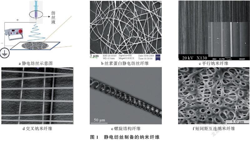
静电纺丝技术始于1934年,20世纪90年代以来,随着纳米技术的迅速发展,为制备微纳米纤维提供了一种简单易行的方法。静电纺丝制备的纳米纤维如图1所示。传统静电纺丝装置包括高压电源、纺丝喷头、接地金属收集器3部分(如图1a所示)。纤维直径从纳米级调整到微米级,直径分布受溶液粘度、导电率、挤出率、静电场、注射器直径、选择距离、温度和湿度的影响[15]。由于高压静电场作用,聚合物溶液(或熔体)克服表面张力形成喷流,并随着溶剂的蒸发或固化形成纤维,最终沉积在不同类型的收集器上,形成形态各异的纤维网(图1b~图1f),如纱线[16-20],或可直接组装成其他取向各异的纳米纤维[21-26]。
在纺丝过程中,聚合物溶液通过喷头进料,并施加高压,增强的电场拉伸液滴,电荷产生相互作用,当带电荷溶液相互间的静电斥力大于其表面张力时,一束射流从锥顶喷出,形成泰勒圆锥[27-28]。静电纺丝聚合物射流[27-29]运动规律十分复杂,静电纺丝射流路径如图2所示。射流在喷丝嘴的尖端可分为直线区、转换区和不稳定区,图2c展示了路径不稳定区的产生原理。首先形成一个带有细长包络圆锥体的线圈,这个细长的线圈形成一个较大的弯曲线圈,直径增加更快,线圈内部的灰色线表示形成“花环状”较大线圈的中心线。
2便携式静电纺丝装置的特点和优势
为实现纤维网中所需纤维的取向和形态的可控性[30-31],有必要对传统制备工艺的各个方面进行改良,例如喷头和收集器的自组装和改良,传统静电纺丝装置的改良如图3所示。Yang G等人[27]综述了一些代表性策略,例如使用不同的喷头[32-34](图3b)、无针头[35-38](图3c)、集电器[39](图3d)、平行电极[40](图3e)、环形收集器[41-42](图3f和图3g)、转盘收集器[43](图3h)、平行轨道[44](图3i)、水浴[45](图3j)、动态支撑系统[46](图3k)、近场电纺[47](图3l)、微流控[48](图3m)以及3D打印[49](图3n)。图案化收集器也可用于生产复杂的纤维几何形状[50-52],需要固定尖端和离心喷丝头才能形成定向纳米纤维组件[53-55]。上述对静电纺丝装置的简单改良,可用于定制静电纺丝网的结构和性能,并且用于药物输送、组织工程和诊断等生物医学领域[27]。组织修复是一个特殊的生物学过程,包括胶原代谢、伤口收缩、上皮形成及炎症。如果便携式静电纺丝设备对应的收集器是生物伤口的部位,仅依赖于某一部分(喷头或收集器)的改良是不可行的。
为适应生物收集体的特点,迫切要求对大型标准台式纺丝装置的组件进行改进,以提高便携性和可操作性。模拟野外伤口紧急处理工作条件下,可使用内置的锂电池和高电压转换器来供电,无需外部电源供应,提高便携性[56]。为解决高压单元和加热单元的静电干扰,可将超高热传导和高绝缘单元内置在头部装置中[57]。为精确沉积敷料,可使用定向罩辅助电纺,形成对伤口的定向性保护[58-59]。其它有效的改进措施还包括手持式喷丝头[60-64],使用带转换器的微型高压电源[64-68],以及使用替代电源(电池或发电机)[69-73]供电等。标准台式装置与便携式装置的优缺点如下:
1)标准台式。其优点为环境参数可控,高压可调,电压和流量数值精确,安全管理可靠。其缺点为运输困难,需要外部电源,灵活性差,价格昂贵。
2)便携式。其优点为便携性(小巧,轻便,手持),方向灵活,应用目标广泛,原位纺丝,手动控制纺丝/喷涂,电池供电,使用安全,价格适中,满足医学应用。其缺点为限流,高压不稳定,电池容量限制,固定尺寸。
便携式静电纺丝装置的优势,结合生物可降解聚合物、修复佐剂和组织替代品,使敷料具有抗菌、免疫调节、促细胞增殖和血管形成等多种功能。由于伤口类型较为复杂,如深度损伤[74]、表面积缺损[75]、烧烫伤[66,76]、內脏切除后的伤口[77-78]和硬脑膜损伤[79],因此定制化的伤口处理十分关键。聚合物功能敷料、原位纺丝和精确沉积都为快速处理伤口部位、促进伤口愈合提供了机会[80]。
3聚合物功能敷料
多种不同类型的便携式电纺装置已经制备,为抗菌敷料在伤口上的快速应用提供了机会。便携式静电纺丝材料的开发对伤口敷料的发展至关重要[81],理想的功能敷料应满足止血迅速、抑菌和生物相容性高、创面粘附力强、透气性好等特点,因此一些抗菌剂被掺入纳米纤维中,如中药制剂、金属和非金属、医用胶α氰基丙烯酸酯(Noctyl2cyanoacrylate,NOCA)等。静电纺纳米纤维在医学(伤口敷料)中的应用如表1所示。
目前,功能敷料的发展方向为简单、易用和多功能快速止血,多功能纳米纤维敷料的制备和性能实验如图4所示。Liu X F等人[89]使用便携式静电纺丝装置合成CuS复合纳米纤维(图4a和图4b),原位沉积在伤口上,在室外同时实现快速止血和抑菌,纳米纤维不仅能快速止血(<6 s),而且能缩短铜绿假单胞菌感染创面的愈合时间(图4c);Liu C L等人[90]通过原位沉积法制备了核壳结构的光动力纳米纤维,能有效杀灭耐甲氧西林金黄色葡萄球菌,向创面输送活性氧,增强光动力效果,可作为抗菌敷料与止血剂结合使用。
4便携式静电纺丝装置及其原位应用
便携式静电纺丝装置无需外部电源,内置电源可运行3 h以上[57],其体积小,重量轻,组件适合手持(单手在室内和室外)操作,可在紧急情况或偏远地区使用。便携式静电纺丝装置在操作过程中,内部高压经特殊处理,总功率不足1 W,近距离(2~12 cm)操作无伤害,可放心使用[81]。为了提高装置的便携性,实现原位电纺的要求,以下综述几种便携式静电纺丝装置及其原位应用。
4.1手持静电纺丝装置及其原位应用
Jiang K等人[62]首次设计一种气流定向原位静电纺丝装置,气流定向原位静电纺丝装置如图5所示。
由图5a可以看出,该装置主要由高压电源、注射泵、气泵、自制喷丝头手柄四部分组成。由图5b可以看出,在气流流速为12 L/min辅助下,可精确控制沉积范围和位置,该装置可作为书写“静电纺丝”等文字的笔。猪体内原位肝脏止血实验如图6所示。在麻醉条件下,从猪的腹腔中暴露一块肝脏,并用一对止血钳固定,部分肝脏被切开后,伤口可见明显出血,用无菌纱布和气流进一步清洁血液,然后用气流定向原位静电纺丝装置在20 s内迅速覆盖一层NOCA纤维膜,1 min后取下止血钳,3 h内未见渗血。
Dong R H等人[82]使用新鲜离体动物肝脏来建立肝切除术引起的出血模型,发现使用气流定向装置,不仅能将NOCA纤维精确沉积在伤口上,提高操作性能和安全性,而且显著减少NOCA用量近80%,显著降低毒副作用和炎性反应。不同于喷涂法(使雾化涂料在高压电场或气流引导下吸附于基底表面)与传统电纺法,气流定向原位静电纺丝法能够精确沉积,高效止血,对肝细胞损伤小,有效促进组织再生。用于体外止血实验的气流定向原位静电纺丝装置如图7所示。图7c包括暴露肝脏,固定肝叶,切除肝脏,沉积NOCA胶等。由图7c可以看出,喷头距离伤口3~4 cm,伤口在10 s内迅速被一层NOCA纤维膜覆盖,切开缝合线恢复肝血流后,未观察到血液或胆汁渗出。
Lü F Y等人[79]还研究了原位电纺NOCA纤维用于山羊硬脑膜修复,实验表明,这种方式纺成柔性、致密的膜不仅有助于快速缝合,而且能修复硬脑膜缺损,避免组织粘连,原位电纺NOCA纤维修复山羊硬脑膜实验如图8所示。
在活体器官上原位沉积纳米纤维可提高纳米纤维膜与器官的粘附力,从而加速止血效果。Zhang J等人[83]将原位静电纺丝和微创手术结合,通过腹腔镜可视,并直接沉积到活体器官上,原位静电纺丝和动物微创止血手术如图9所示。动物微创手术通过一根长的腹腔镜管(37 cm)进行(图9a),可最大限度减少施术者手抖动对伤口部位的影响。在腹腔镜下,进行长针静电纺丝装置微创手术止血实验(图9b),原位电纺法组(图9g)止血时间5 s,优于传统缝合止血组(图9e)止血时间3 min和涂抹NOCA组(图9f)止血时间14 s。纳米纤维膜的沉积面积可通过锥形会聚结构和电纺距离进行微调,不影响纤维直径。长针静电纺丝装置结合腹腔镜微创手术较传统手术止血快,术后炎症反应小,恢复快。
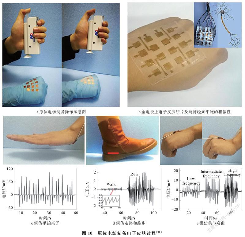
2018年,Wang X X等人[91]报道了一种原位电纺制备的电子皮肤,原位电纺制备电子皮肤过程如图10所示。研究采用聚偏氟乙烯(poly vinylidene fluoride,PVDF)纳米纤维的单电极压电纳米发电机传感器,插图展示了它与神经元细胞的相似性(图10b),它由原位电纺制备(图10a),可在单个单元上实现冷/热集成压力的稳态传感,压电信号表现为方波信号,热敏信号表现为脉冲信号,由这种纳米纤维构成的电子皮肤,可实现对温度和压力的高灵敏探测(图10c~图10e)。
针对肠道内容物复杂,伤口密封要求更高的特点,Zhou T T等人[84]将原位电纺应用于猪离体肠道伤口止血,发现能有效防止肠道积液,纳米纤维膜的平均直径约为0.5 μm,可延长90%而不断裂,该装置的便携性和可控沉积,进一步增加了原位电纺在伤口愈合中的应用潜力。
4.2电池驱动便携式静电纺丝装置及其原位应用
早期传统纺丝装置高压和加热组件需要220 V的工作电压,沉重的设备体积,外部电源供应,在便携性、可移植性方面存在短板。为了克服上述不足,已有研究报道了几款不依赖外部电源供应、电池驱动的便携式纺丝装置[85-86],简介如下:
手持式熔体电纺装置如图11所示,重量约450 g,长24 cm,厚6 cm,高13 cm。内置的高压单元和加热单元完全由可充电锂电池供电,可集成在设备内,内置锂电池可连续工作2 h以上(图11b)。该装置配有高效传热电绝缘单元,以解决加热单元与高压单元之间的静电干扰。它便于携带,单手操作,纤维可直接沉积在另一只手上(图11c)。利用该方法制备了直径为4~17 μm的聚己内酯(poly caprolactone, PCL)/Fe3O4磁性纳米复合纤维,可将电磁能转化为热能,用于磁热疗。
手持式电池驱动静电纺丝装置如图12所示。包括装置整机和一个用手指按压的注射器(如图12a所示)。采用两节碱性电池提供3 V直流电压,在高压变频转换器的作用下,3 V电压被放大到10 kV,转换器的正极与注射器上的针头相连,负极与用于操作装置的导电金属箔相连。该装置可连续工作15 h以上,针头与收集器工作距离在2~10 cm之间。实际质量约120 g,长5 cm,厚3 cm,高10.5 cm,不同方位外观如图12b所示。使用该装置将聚乙烯吡咯烷酮(polyvinyl pyrrolidone, PVP)、PVDF和PCL静电纺制备纳米纤维敷料[81],用于大鼠体表伤口止血实验(如图12c所示)。
抗菌剂和治疗剂可结合在一起,以获得多功能纳米纤维膜,便携式电纺装置制备抗菌敷料实验如图13所示。Dong R H等人[67]将载银修饰介孔二氧化硅颗粒与PCL电纺成纳米纤维膜(图13a),发现随着载银质量分数从3%增加到8%,平均纤维直径从389 nm增加到986 nm,纤维变得更加粗糙。载银质量分数5%的纳米纤维膜(平均纤维直径658 nm)作为理想样品,对金黄色葡萄球菌和大肠杆菌均具有良好的抗菌活性(图13c),并可减轻Wistar大鼠的炎症反应,促进伤口愈合(图13b和图13c)。
定向罩辅助电纺沉积如图14所示。为实现内脏如肝、肺、肾等器官的快速止血,利用定向罩辅助电纺沉积过程,通过在纺丝喷嘴上安装一个金属锥,形成定向罩(图14a),改变静电场的分布,从而减少喷流发射角,调节电纺纤维的沉积范围,以实现纤维的可控精密沉积[58-59]。调整电纺距离和金属锥的边长,可聚焦沉积面积(如图14b和图14c所示)。传统的气流辅助电纺需要一个附加的空气泵电源,而定向罩辅助电纺不需要空气泵电源,便携性更好。但是由于电压固定,限制了敷料的尺寸和物理性能,并且纺丝溶液的输送取决于手指对扳机施加的压力,在使用过程中,会导致纤维沉积不一致。
4.3发电机驱动便携式静电纺丝装置及其原位应用
没有外部电源,传统的静电纺丝装置不能工作,手持静电纺丝装置的电池寿命仍有待提升。Han W P等人[69]设计了一种基于手动Wimshurst发生器的自供电静电纺丝装置,使用Wimshurst发生器的纺丝装置如图15所示。
其中,图15a为实物样机,其高压部分被Wimshurst发生器所取代,通过转动手柄,使2个圆盘反向旋转,由于静电感应产生的正、负电荷分别储存在2个莱登瓶中。随着手柄的不断转动,电荷积累,然后在针和收集器之间产生高压。利用该装置成功的将聚苯乙烯(polystyrene,PS)、PVDF、PCL和聚乳酸(polylactic acid,PLA)等聚合物电纺成超薄纤维(图15c和图15e),2 min即可纺成几厘米高的PS纤维三维堆垛结构(图15b),PLA纤维可原位静电纺到皮肤上,快速成膜(图15f),初步表明这种装置可用于医疗美容领域。
Qin C C等人[87]通过更换锥形喷头和添加带温度控制器的加热枪,设计了基于Wimshurst发生器的自供电熔体静电纺丝装置,手动Wimshurst发生器自供电熔体静电纺丝装置如图16所示。
使用该装置成功制备了直径为15~45 μm的PLA和PCL超细纤维,纤维被直接熔融电纺到猪肝上(图16c和图16d),研究并优化了纺丝速度(1.5 r/s)和纺丝距离(8~10 cm)对纤维的影响。测得沉积纤维的温度(24~44 ℃)更接近人体温度,PCL纤维的粘附性可达1.2 N,提示该装置制备熔融电纺聚合物超细纤维适于伤口愈合。
除了电池供电和手动发电,Yan X等人[71]介绍了一种太阳能电池和手摇发电机驱动的便携式电纺装置,太阳能电池和手摇发电机驱动的便携式纺丝装置如图17所示。该装置可在户外使用,无需电源插头(图17a)。电源系统包括太阳能电池(5 V/140 mA)、手持式发电机(5 V/200 mA)、一组可充电电池和高压转换器(图17b)。该装置可放在阳光下通过太阳能电池发电,也可通过转动发电机手柄给电池充电,为固定距离的溶液或熔融静电纺丝提供持续电源。将喷丝头和注射器改为锥形金属喷嘴(图17c和图17d),聚合物颗粒可通过热枪加热成熔体,PCL、PLA和聚乳酸羟基乙酸共聚物(polylacticcoglycolic acid,PLGA)可通过该装置熔融电纺成超细纤维(图17e~图17g)。
5结束语
从生物医学的角度来看,静电纺纳米敷料是最有前途的纳米材料,它们的形态尺寸与细胞外基质结构类似,高孔隙率可促进细胞呼吸,柔韧性可促进皮肤3D重组再生,还可作为抗菌药物、生长因子、维生素的有效载体。其优点在于敷料不需要经常更换,减少了人体疼痛和恐惧感,对于受试者十分容易接受。在过去几年中,由电池或发电机供电的便携式静电纺丝装置取得了重大进展,性能和便携性得到了系统性的提高,这些工作在一定情况下促进了静电纺丝技术在医学领域的发展,未来还可考虑从以下方面进一步研究,开发对外部环境要求低、廉价、使止血药物在创面精确沉积的纺絲装置。评估各种功能材料适合手持静电纺丝装置和直接沉积在伤口部位,包括位于体表的伤口和体腔内的深部伤口,能够避免组织粘连,并可作为抗菌药物、生长因子的递送载体,长效、循环、可回收利用。多中心、大样本的临床前评估和体内实验,为定制个性化的美容和伤口敷料装置的广泛应用提供了理论依据。
參考文献:
[1]Cui J X, Li F H, Wang Y L, et al. Electrospun nanofiber membranes for wastewater treatment applications[J]. Separation and Purification Technology, 2020, 250: 117116117132.
[2]Wang M L, Wang K, Yang Y Y, et al. Electrospun environment remediation nanofibers using unspinnable liquids as the sheath fluids: A review[J]. Polymers, 2020, 12(1): 103116.
[3]Ghosal K, Agatemor C, Spitalsky Z, et al. Electrospinning tissue engineering and wound dressing scaffolds from polymertitanium dioxide nanocomposites[J]. Chemical Engineering Journal, 2019, 358: 12621278.
[4]Abrigo M, McArthur S L, Kingshott P. Electrospun nanofibers as dressings for chronic wound care: Advances, challenges, and future prospects[J]. Macromolecular Bioscience, 2014, 14(6): 772792.
[5]Kim F S, Ren G Q, Jenekhe S A. Onedimensional nanostructures of piconjugated molecular systems: Assembly, properties, and applications from photovoltaics, sensors, and nanophotonics to nanoelectronics[J]. Chemistry of Materials, 2011, 23(3): 682732.
[6]Xue J J, Wu T, Dai Y Q, et al. Electrospinning and electrospun nanofibers: Methods, materials, and applications[J]. Chemical Reviews, 2019, 119(8): 52985415.
[7]Zahedi P, Rezaeian I, RanaeiSiadat S O, et al. A review on wound dressings with an emphasis on electrospun nanofibrous polymeric bandages[J]. Polymers for Advanced Technologies, 2010, 21(2): 7795.
[8]Pereira R F, Barrias C C, Granja P L, et al. Advanced biofabrication strategies for skin regeneration and repair[J]. Nanomedicine, 2013, 8(4): 603621.
[9]Liu Y, Gao X, Wang Z P, et al. Controlled synthesis of bismuth oxychloridecarbon nanofiber hybrid materials as highly efficient electrodes for rockingchair capacitive deionization[J]. Chemical Engineering Journal, 2021, 403: 126326126337.
[10]Graca M F P, Miguel S P, Cabral C S D, et al. Hyaluronic acidbased wound dressings: A review[J]. Carbohydrate Polymers, 2020, 241: 116364116381.
[11]Topuz F, Uyar T. Electrospinning of cyclodextrin functional nanofibers for drug delivery applications[J]. Pharmaceutics, 2019, 11(1): 641.
[12]Dias J R, Granja P L, Bartolo P J. Advances in electrospun skin substitutes[J]. Progress in Materials Science, 2016, 84: 314334.
[13]Mele E. Electrospinning of natural polymers for advanced wound care: Towards responsive and adaptive dressings[J]. Journal of Materials Chemistry B, 2016, 4(28): 48014812.
[14]Chen S X, Liu B, Carlson M A, et al. Recent advances in electrospun nanofibers for wound healing[J]. Nanomedicine, 2017, 12(11): 13351352.
[15]Gao F F, Jiang M L, Liang W C, et al. Coelectrospun cellulose diacetategraftpoly(ethylene terephthalate) and collagen composite nanofibrous mats for cells culture[J]. Journal of Applied Polymer Science, 2020, 137(44): 4935049361.
[16]Levitt A, Seyedin S, Zhang J Z, et al. Bath electrospinning of continuous and scalable multifunctional mxeneinfiltrated nanoyarns[J]. Small, 2020, 16(26): 20021582002170.
[17]Yalcinkaya F, Komarek M. Polyvinyl butyral (PVB) nanofiber/nanoparticlecovered yarns for antibacterial textile surfaces[J]. International Journal of Molecular Sciences, 2019, 20(17): 43174328.
[18]Levitt A S, Vallett R, Dion G, et al. Effect of electrospinning processing variables on polyacrylonitrile nanoyarns[J]. Journal of Applied Polymer Science, 2018, 135(25): 4640446413.
[19]O′Connor R A, McGuinness G B. Electrospun nanofibre bundles and yarns for tissue engineering applications: A review[J]. Proceedings of the Institution of Mechanical Engineers Part HJournal of Engineering in Medicine, 2016, 230(11): 987998.
[20]Wu J L, Huang C, Liu W, et al. Cell infiltration and vascularization in porous nanoyarn scaffolds prepared by dynamic liquid electrospinning[J]. Journal of Biomedical Nanotechnology, 2014, 10(4): 603614.
[21]Bhardwaj N, Kundu S C. Electrospinning: A fascinating fiber fabrication technique[J]. Biotechnology Advances, 2010, 28(3): 325347.
[22]Sun B, Long Y Z, Zhang H D, et al. Advances in threedimensional nanofibrous macrostructures via electrospinning[J]. Progress in Polymer Science, 2014, 39(5): 862890.
[23]Yu M, Dong R H, Yan X, et al. Recent advances in needleless electrospinning of ultrathin fibers: From academia to industrial production[J]. Macromolecular Materials and Engineering, 2017, 302(7): 17000021700011.
[24]Han T, Reneker D H, Yarin A L. Buckling of jets in electrospinning[J]. Polymer, 2007, 48(20): 60646076.
[25]Angammana C J, Jayaram S H. Fundamentals of electrospinning and processing technologies[J]. Particulate Science and Technology, 2016, 34(1): 7282.
[26]Buchko C J, Chen L C, Shen Y, et al. Processing and microstructural characterization of porous biocompatible protein polymer thin films[J]. Polymer, 1999, 40(26): 73977407.
[27]Yang G, Li X L, He Y, et al. From nano to micro to macro: Electrospun hierarchically structured polymeric fibers for biomedical applications[J]. Progress in Polymer Science, 2018, 81: 80113.
[28]Reneker D H, Yarin A L. Electrospinning jets and polymer nanofibers[J]. Polymer, 2008, 49(10): 23872425.
[29]Xin Y, Reneker D H. Garland formation process in electrospinning[J]. Polymer, 2012, 53(16): 36293635.
[30]Sun B, Long Y Z, Chen Z J, et al. Recent advances in flexible and stretchable electronic devices via electrospinning[J]. Journal of Materials Chemistry C, 2014, 2(7): 12091219.
[31]Huang Z M, Zhang Y Z, Kotaki M, et al. A review on polymer nanofibers by electrospinning and their applications in nanocomposites[J]. Composites Science and Technology, 2003, 63(15): 22232253.
[32]Li D, McCann J T, Xia Y N. Use of electrospinning to directly fabricate hollow nanofibers with functionalized inner and outer surfaces[J]. Small, 2005, 1(1): 8386.
[33]Han D, Steckl A J. Triaxial electrospun nanofiber membranes for controlled dual release of functional molecules[J]. Acs Applied Materials & Interfaces, 2013, 5(16): 82418245.
[34]Wang N, Chen H Y, Lin L, et al. Multicomponent phase change microfibers prepared by temperature control multifluidic electrospinning[J]. Macromolecular Rapid Communications, 2010, 31(18): 16221627.
[35]Alamein M A, Liu Q, Stephens S, et al. Nanospiderwebs: Artificial 3d extracellular matrix from nanofibers by novel clinical grade electrospinning for stem cell delivery[J]. Advanced Healthcare Materials, 2013, 2(5): 702717.
[36]Yang R R, He J H, Xu L, et al. Bubbleelectrospinning for fabricating nanofibers[J]. Polymer, 2009, 50(24): 58465850.
[37]Niu H T, Lin T, Wang X G. Needleless electrospinning. I. A comparison of cylinder and disk nozzles[J]. Journal of Applied Polymer Science, 2009, 114(6): 35243530.
[38]Wang X, Niu H T, Wang X G, et al. Needleless electrospinning of uniform nanofibers using spiral coil spinnerets[J]. Journal of Nanomaterials, 2012, 2012: 785920785959.
[39]Hejazi F, Mirzadeh H, Contessi N, et al. Novel class of collector in electrospinning device for the fabrication of 3d nanofibrous structure for large defect loadbearing tissue engineering application[J]. Journal of Biomedical Materials Research Part A, 2017, 105(5): 15351548.
[40]Li D, Wang Y L, Xia Y N. Electrospinning nanofibers as uniaxially aligned arrays and layerbylayer stacked films[J]. Advanced Materials, 2004, 16(4): 361366.
[41]Dalton P D, Klee D, Moller M. Electrospinning with dual collection rings[J]. Polymer, 2005, 46(3): 611614.
[42]Katta P, Alessandro M, Ramsier R D, et al. Continuous electrospinning of aligned polymer nanofibers onto a wire drum collector[J]. Nano Letters, 2004, 4(11): 22152218.
[43]Zussman E, Theron A, Yarin A L. Formation of nanofiber crossbars in electrospinning[J]. Applied Physics Letters, 2003, 82(6): 973975.
[44]Beachley V, Katsanevakis E, Zhang N, et al. A novel method to precisely assemble loose nanofiber structures for regenerative medicine applications\[J\]. Advanced Healthcare Materials, 2013, 2(2): 343351.
[45]Smit E, Buttner U, Sanderson R D. Continuous yarns from electrospun fibers[J]. Polymer, 2005, 46(8): 24192423.
[46]Teo W E, Gopal R, Ramaseshan R, et al. A dynamic liquid support system for continuous electrospun yarn fabrication[J]. Polymer, 2007, 48(12): 34003405.
[47]Bisht G S, Canton G, Mirsepassi A, et al. Controlled continuous patterning of polymeric nanofibers on threedimensional substrates using lowvoltage nearfield electrospinning[J]. Nano Letters, 2011, 11(4): 18311837.
[48]Zhang X, Gao X H, Jiang L, et al. Flexible generation of gradient electrospinning nanofibers using a microfluidic assisted approach[J]. Langmuir, 2012, 28(26): 1002610032.
[49]Park S H, Kim T G, Kim H C, et al. Development of dual scale scaffolds via direct polymer melt deposition and electrospinning for applications in tissue regeneration[J]. Acta Biomaterialia, 2008, 4(5): 11981207.
[50]Zhang D M, Chang J. Patterning of electrospun fibers using electroconductive templates[J]. Advanced Materials, 2007, 19(21): 36643667.
[51]Xu H, Li H Y, Chang J. Controlled drug release from a polymer matrix by patterned electrospun nanofibers with controllable hydrophobicity[J]. Journal of Materials Chemistry B, 2013, 1(33): 41824188.
[52]Yu G F, Yan X, Yu M, et al. Patterned, highly stretchable and conductive nanofibrous pani/pvdf strain sensors based on electrospinning and in situ polymerization[J]. Nanoscale, 2016, 8(5): 29442950.
[53]Kameoka J, Orth R, Yang Y N, et al. A scanning tip electrospinning source for deposition of oriented nanofibres[J]. Nanotechnology, 2003, 14(10): 11241129.
[54]King W E, Bowlin G L. Nearfield electrospinning and melt electrowriting of biomedical polymersprogress and limitationsJ]. Polymers, 2021, 13(7): 10971120.
[55]Liu S L, Long Y Z, Zhang Z H, et al. Assembly of oriented ultrafine polymer fibers by centrifugal electrospinning[J]. Journal of Nanomaterials, 2013, 2013: 713275713284.
[56]Dong W H, Liu J X, Mou X J, et al. Performance of polyvinyl pyrrolidoneisatis root antibacterial wound dressings produced in situ by handheld electrospinner[J]. Colloids and Surfaces BBiointerfaces, 2020, 188: 110766110772.
[57]Zhao Y T, Zhang J, Gao Y, et al. Selfpowered portable melt electrospinning for in situ wound dressing[J]. Journal of Nanobiotechnology, 2020, 18(1): 111121.
[58]Luo W L, Qiu X, Zhang J, et al. In situ accurate deposition of electrospun medical glue fibers on kidney with auxiliary electrode method for fast hemostasis[J]. Materials Science & Engineering CMaterials for Biological Applications, 2019, 101: 380386.
[59]Luo W L, Zhang J, Qiu X, et al. Electricfieldmodified in situ precise deposition of electrospun medical glue fibers on the liver for rapid hemostasis[J]. Nanoscale Research Letters, 2018, 13: 278286.
[60]Sofokleous P, Stride E, Bonfield W, et al. Design, construction and performance of a portable handheld electrohydrodynamic multineedle spray gun for biomedical applications[J]. Materials Science and Engineering CMaterials for Biological Applications, 2013, 33(1): 213223.
[61]Lau W K, Sofokleous P, Day R, et al. A portable device for in situ deposition of bioproducts[J]. Bioinspired Biomimetic and Nanobiomaterials, 2014, 3(2): 94105.
[62]Jiang K, Long Y Z, Chen Z J, et al. Airflowdirected in situ electrospinning of a medical glue of cyanoacrylate for rapid hemostasis in liver resection[J]. Nanoscale, 2014, 6(14): 77927798.
[63]Song C, Wang X X, Zhang J, et al. Electric fieldassisted in situ precise deposition of electrospun γFe2O3/polyurethane nanofibers for magnetic hyperthermia[J]. Nanoscale Research Letters, 2018, 13: 273284.
[64]Brako F, Luo C J, Craig D Q M, et al. An inexpensive, portable device for pointofneed generation of silvernanoparticle doped cellulose acetate nanofibers for advanced wound dressing[J]. Macromolecular Materials and Engineering, 2018, 303(5): 17005861700592.
[65]Yue Y P, Gong X B, Jiao W L, et al. Insitu electrospinning of thymolloaded polyurethane fibrous membranes for waterproof, breathable, and antibacterial wound dressing application[J]. Journal of Colloid and Interface Science, 2021, 592: 310318.
[66]Haik J, Kornhaber R, Blal B, et al. The feasibility of a handheld electrospinning device for the application of nanofibrous wound dressings[J]. Advances in Wound Care, 2017, 6(5): 166174.
[67]Dong R H, Jia Y X, Qin C C, et al. In situ deposition of a personalized nanofibrous dressing via a handy electrospinning device for skin wound care[J]. Nanoscale, 2016, 8(6): 34823488.
[68]Chui C Y, Mouthuy P A, Ye H. Direct electrospinning of poly(vinyl butyral) onto human dermal fibroblasts using a portable device[J]. Biotechnology Letters, 2018, 40(4): 737744.
[69]Han W P, Huang Y Y, Yu M, et al. Selfpowered electrospinning apparatus based on a handoperated wimshurst generator[J]. Nanoscale, 2015, 7(13): 56035606.
[70]Li C J, Yin Y Y, Wang B, et al. Selfpowered electrospinning system driven by a triboelectric nanogenerator[J]. Acs Nano, 2017, 11(10): 1043910445.
[71]Yan X, Yu M, Zhang L H, et al. A portable electrospinning apparatus based on a small solar cell and a hand generator: Design, performance and application[J]. Nanoscale, 2016, 8(1): 209213.
[72]Yan X, Duan X P, Yu S X, et al. Portable melt electrospinning apparatus without an extra electricity supply[J]. Rsc Advances, 2017, 7(53): 3313233136.
[73]Duan X P, Yan X, Zhang B, et al. Simple piezoelectric ceramic generatorbased electrospinning apparatus[J]. Rsc Advances, 2016, 6(70): 6625266255.
[74]Mouthuy P A, Groszkowski L, Ye H. Performances of a portable electrospinning apparatus[J]. Biotechnology Letters, 2015, 37(5): 11071116.
[75]Azimi B, Maleki H, Zavagna L, et al. Biobased electrospun fibers for wound healing[J]. Journal of Functional Biomaterials, 2020, 11(3): 67103.
[76]Mogosanu G D, Grumezescu A M. Natural and synthetic polymers for wounds and burns dressing[J]. International Journal of Pharmaceutics, 2014, 463(2): 127136.
[77]Gao Y, Xiang H F, Wang X X, et al. A portable solution blow spinning device for minimally invasive surgery hemostasis[J]. Chemical Engineering Journal, 2020, 387: 124052124059.
[78]Li D J, Nie W, Chen L, et al. Fabrication of curcuminloaded mesoporous silica incorporated polyvinyl pyrrolidone nanofibers for rapid hemostasis and antibacterial treatment[J]. Rsc Advances, 2017, 7(13): 79737982.
[79]Lü F Y, Dong R H, Li Z J, et al. In situ precise electrospinning of medical glue fibers as nonsuture dural repair with high sealing capability and flexibility[J]. International Journal of Nanomedicine, 2016, 11: 42134220.
[80]Guo B L, Ma P X. Conducting polymers for tissue engineering[J]. Biomacromolecules, 2018, 19(6): 17641782.
[81]Yan X, Yu M, Ramakrishna S, et al. Advances in portable electrospinning devices for in situ delivery of personalized wound care[J]. Nanoscale, 2019, 11(41): 1916619178.
[82]Dong R H, Qin C C, Qiu X, et al. In situ precision electrospinning as an effective delivery technique for cyanoacrylate medical glue with high efficiency and low toxicity[J]. Nanoscale, 2015, 7(46): 1946819475.
[83]Zhang J, Zhao Y T, Hu P Y, et al. Laparoscopic electrospinning for in situ hemostasis in minimally invasive operation[J]. Chemical Engineering Journal, 2020, 395: 125089125096.
[84]Zhou T T, Wang Y Z, Lei F C, et al. Insitu electrospinning for intestinal hemostasis[J]. International Journal of Nanomedicine, 2020, 15: 38693875.
[85]Hu P Y, Zhao Y T, Zhang J, et al. In situ melt electrospun polycaprolactone/fe3o4 nanofibers for magnetic hyperthermia[J]. Materials Science & Engineering CMaterials for Biological Applications, 2020, 110: 1170811716.
[86]Xu S C, Qin C C, Yu M, et al. A batteryoperated portable handheld electrospinning apparatus[J]. Nanoscale, 2015, 7(29): 1235112355.
[87]Qin C C, Duan X P, Wang L, et al. Melt electrospinning of poly(lactic acid) and polycaprolactone microfibers by using a handoperated wimshurst generator[J]. Nanoscale, 2015, 7(40): 1661116615.
[88]Liu G S, Yan X, Yan F F, et al. In situ electrospinning iodinebased fibrous meshes for antibacterial wound dressing[J]. Nanoscale Research Letters, 2018, 13: 309316.
[89]Liu X F, Zhang J, Liu J J, et al. Bifunctional cus composite nanofibers via in situ electrospinning for outdoor rapid hemostasis and simultaneous ablating superbug[J]. Chemical Engineering Journal, 2020, 401: 126096126108.
[90]Liu C L, Yang J, Bai X H, et al. Dual antibacterial effect of in situ electrospun curcumin composite nanofibers to sterilize drugresistant bacteria[J]. Nanoscale Research Letters, 2021, 16(1): 5462.
[91]Wang X X, Song W Z, You M H, et al. Bionic singleelectrode electronic skin unit based on piezoelectric nanogenerator[J]. Acs Nano, 2018, 12(8): 85888596.
作者簡介: 刘钟(1984),男,博士,主要研究方向为纳米医学材料。
通信作者: 龙云泽(1977),男,教授,博士生导师,主要研究方向为功能纳米纤维的制备、性能、应用及产业化。Email: yunze.long@qdu.edu.cn
Application of Portable Electrospinning Devices in Medicine
LIU Zhong1, LIU Xianfeng1, WANG Xueyan1, ZHANG Jun1, YU Miao2, XIANG Hongfei3, CHEN Yong4, LONG Yunze1, 5
(1. College of Physics, Qingdao University, Qingdao 266071, China; 2. Qingdao Junada Technology Co. Ltd, Qingdao 266199, China;
3. Department of Orthopedics, The Affiliated Hospital of Qingdao University, Qingdao 266035, China;
4. Qingdao Yuren Medical Technology Co. Ltd, Qingdao 266111, China;
5. State Key Laboratory of BioFibers and EcoTextiles, Qingdao University, Qingdao 266071, China)
Abstract: For the current situation of difficult transportation, external power supply, and poor flexibility in traditional nanofiber preparation devices, in order to solve the problem of limited application in medical wound dressings, this paper summarizes the composition and medical application of small portable electrospinning devices independent of external power supply. By discussing the morphological structure, polymer compositions, antibacterial properties, hemostatic effect, and promoting tissue repair ability of electrospun nanofibers, the concept of insitu electrospinning is proposed, that is, the nanofibers dressings films are directly and quickly prepared on the wound according to the patient′s situation, especially in hemostasis of liver, burn and scald treatment, meningeal sealing, etc. Finally, the research challenge and development trend of portable electrospinning devices in individual cosmetic and wound dressings are considered for further study. This review provides a reference for the insitu application of portable electrospinning devices in medicine.
