Photocrosslinking-Immobilized Polymer Vesicles for Lowering Temperature Triggered Drug Release
2021-11-22YuyaoLiaoZhenFanJianzhongDu
Yuyao Liao , Zhen Fan , Jianzhong Du ,4,*
1 Department of Orthopedics, Shanghai Tenth People’s Hospital, Tongji University School of Medicine, Shanghai 200072, China.
2 Department of Polymeric Materials, School of Materials Science and Engineering, Tongji University, Shanghai 201804, China.
4 Key Laboratory of Advanced Civil Engineering Materials of Ministry of Education, Tongji University, Shanghai 201804, China.
Abstract:The stability of nanocarriers in physiological environments is of importance for biomedical applications.Among the existing crosslinking approaches for enhancing the structural integrity and stability, photocrosslinking has been considered to be an ideal crosslinking chemistry, as it is nontoxic and cost-effective, and does not require an additional crosslinker or generate by-products. Meanwhile, most current temperature-responsive nanocarriers are designed and synthesized for drug release by increasing temperature.However, heating may induce cell damage during triggered drug release. Therefore, lowering temperature-triggered nanocarriers need to be developed for drug delivery and safe drug release during therapeutic hypothermia. In this study,we prepared an amphiphilic block copolymer, poly(ethylene oxide)-block-poly[N-isopropyl acrylamide-stat-7-(2-methacryloyloxyethoxy)-4-methylcoumarin]-block-poly(acrylic acid)[PEO43-b-P(NIPAM71-stat-CMA8)-b-PAA13], by reversible addition fragmentation chain transfer (RAFT)polymerization. Successful synthesis of the polymer was verified by proton nuclear magnetic resonance (1H NMR)and size exclusion chromatography (SEC). The copolymers selfassembled into vesicles in aqueous solution, with the P(NIPAM-stat-CMA)block forming an inhomogeneous membrane and the PEO chains and PAA chains forming mixed coronas. The cavity of this vesicle could be utilized to load hydrophilic drugs. The CMA groups could undergo photocrosslinking and enhance the stability of vesicles in biological applications,and the PNIPAM moiety endowed the vesicle with temperature-responsive properties. Upon decreasing the temperature,the vesicles swelled and released the loaded drugs. The size distribution and morphology of the vesicles were characterized by dynamic light scattering (DLS), scanning electron microscopy (SEM), and transmission electron microscopy (TEM)experiments. After staining with phosphotungstic acid, the hollow morphology of the vesicles with a phase-separated inhomogeneous membrane was observed by TEM and SEM. The DLS results showed that the hydrodynamic diameter of the vesicles was 208 nm and the polydispersity was 0.075. The size of the vesicles observed by TEM was between 180 and 200 nm, which was in accordance with that measured by DLS. To verify the drug loading capacity and controlled release ability of the vesicle, a water-soluble antibiotic was encapsulated in the vesicles. The experimental results showed that the drug loading content was 10.4% relative to the vesicles and the drug loading efficiency was approximately 32.7%. For vesicles containing the same amount of antibiotics, the release rate at 25 °C was 35% higher than that at 37 °C after 12 h in aqueous solution. Overall, this photocrosslinked vesicle with temperature-responsive properties facilitates lowering temperature-triggered drug release during therapeutic hypothermia.
Key Words:Polymer vesicle; Temperature-sensitive; Photocrosslinking; Controlled drug release;Reversible addition fragmentation chain transfer (RAFT)polymerization
1 Introduction
The stability of nanocarriers in physiological environments is of great importance for biomedical applications1–10. To enhance structural integrity and stability, shell crosslinking (SCL),membrane crosslinking (MCL), or nuclear crosslinking (CCL)nanocarriers have been studied and developed11–15. However,most crosslinking approaches generate by-products and demand non-biocompatible crosslinkers. Photocrosslinking has been regarded as an ideal crosslinking chemistry, which is non-toxic,cost-effective, does not need extra crosslinker or produce any byproducts16. In addition, it has been demonstrated that the size and shape of the nanocarriers could remain unchanged during the photocrosslinking process7,13,17–20. Meanwhile, the photocrosslinking process could be simply regulated through adjusting the wavelength and intensity of light21,22, which makes photocrosslinking chemistry a good choice to develop stable nanocarriers for biomedical applications. Besides the desired stability property of nanocarrier, controlled cargo release capability is also of natural significance for targeted and efficient treatment.
Various strategies have been developed for addressing controlled drug release1,4,23–26. Among existing triggering approaches, the release of drugs through body temperature changes caused by external temperature stimulation has been considered to be one of the most practical methods in the clinic27.PNIPAM and its derivatives are commonly used as temperature responsive polymer segments that undergo a sharp transition in hydrophilic and hydrophobic properties at lower critical solution temperatures (LCST)28–30. For example, Narainet al.31reported a degradable and thermally responsive micelle for targeted drug delivery and controlled release. Swelling of the micelle core can be achieved by heating above the phase transition temperature of PNIPAM, thereby triggering drug release. Gaoet al.32developed an amphiphilic thermally-responsive ABA triblock copolymer that can self-assemble into micelles. The hydrophobic PMMA was squeezed into the core, while the hydrophilic P(NIPAM-co-PEGMEMA)formed a thermosensitive shell. When temperature was higher than the LCST (about 39 °C), the drug can be released without precipitation of copolymers. Currently,most temperature-responsive nanocarriers are designed and synthesized to release the drugs triggered by temperature rise to about 40 °C33,34. However, for clinical applications, some drug carriers and encapsulated drugs are injected subcutaneously or intravenously and heated at the required site to trigger drug release, which demands heating the subcutaneous layer to about 40 °C by electromagnetic waves (such as radio waves or microwaves)35,36. In addition, when the body temperature is heated above 42 °C, the cells in the heated area would be damaged37.
Meanwhile, some studies have shown that cooling on the skin for 15–30 min with ice will cause the skin surface temperature to be below 25 °C, and there will be no significant frostbite on the cooled site38,39. In order to ensure the safety of heatingtriggered drug release materials, it is necessary to accurately control the increase in body temperature. Otherwise, such materials will be difficult to apply to the clinic. Meanwhile,many scenarios in the clinic demand drug release under mild hypothermia environments40,41. Therefore, lowering temperature triggered nanocarriers need to be developed for drug delivery and safe release during therapeutic hypothermia.
Here, we synthesized a photocrosslinkable, temperatureresponsive block copolymer by reversible addition fragmentation chain transfer (RAFT), which self-assembled by solvent switch method to form photocrosslinked polymer vesicle with temperatureresponsive property (Scheme 1). During the self-assembly process of the copolymers into vesicles, the poly[N-isopropyl acrylamidestat-7-(2-methacryloyloxyethoxy)-4-methylcoumarin] [P(NIPAM-stat-CMA)] block forms the inhomogeneous membrane,whereas the poly(ethylene oxide)(PEO)chains and the poly(acrylic acid)(PAA)chains form the mixed coronas. The CMA group could be photocrosslinked by ultraviolet (UV)irradiation and enhance the stability of vesicles for biological applications. The PNIPAM moiety endows the vesicle with temperature-responsive capability by a sharp transition in hydrophilic and hydrophobic properties at LCST. The PAA chains are designed for further functionalization. The photocrosslinked vesicles could encapsulate drugs and release them upon lowering temperature.

Scheme 1 Schematic illustration of photocrosslinked temperature-responsive polymer vesicle which can load and lock drugs at normal body temperature, and release them upon decreasing the temperature.
2 Materials and methods
2.1 Materials
Polyethylene oxide monomethylether (PEO43;Mn = 1900)was purchased from Alfa Aesar. 2-(Dodecylthiocarbonothioylthio)-2-methylpropanoic acid (DDMAT)was synthesized according to the general method in the reported article42. 7-hydroxy-4-methylcoumarin (98%), 2-bromoethanol (95%), methacryloyl chloride (95%),N-isopropyl acrylamide (NIPAM, 98%),tertbutyl acrylate (tBA, 99%), trifluoroacetic acid (TFA, 99%),ciprofloxacin hydrochloride monohydrate (CIP, 98%),Azobisisobutyronitrile (AIBN, 99%)were obtained from Aladdin Chemistry, Co. Ltd. Dichloromethane (DCM, 99.5%),1,4-dioxane (99%),n-hexane (97%), tetrahydrofuran (THF,99%),N,N-dimethylformamide (DMF, 99.5%), and other solvents were purchased from Sinopharm Chemical Reagent Co., Ltd. (SCRC, Shanghai, China).
2.2 Methods
2.2.1 Synthesis of PEO-DDMAT
The synthetic methods of PEO-DDMAT refer to our previously published article43.
2.2.2 Synthesis of PEO43-b-P(NIPAM71-stat-CMA8)-b-PtBA13copolymer by RAFT polymerization
Macro chain transfer agent (macro-CTA)PEO-DDMAT (0.34 g, 0.15 mmol), NIPAM (1.36 g, 12.0 mmol)and CMA (0.500 g,1.65 mmol)were added in a 10 mL round bottom flask. Then dissolve them with 2 mL of 1,4-dioxane. Argon was bubbled through the mixture for 30 minutes for deoxidation. Radical initiator AIBN (3.7 mg, 0.020 mmol)was then added quickly,followed by deoxygenating with argon for 30 min. The reaction was carried out in an oil bath at 70 °C under argon balloon protection.The molar ratio of [PEO-DDMAT]/[NIPAM]/[CMA]/[AIBN] is 1 :80 : 11 : 0.15. The polymerization was carried out for 24 h, and the reaction was terminated upon exposure to air. Then addtertbutyl acrylate(0.290 g, 2.25 mmol)to the mixed solution, repeat the previous steps of deoxygenation and addition of AIBN, then place the flask in an oil bath at 70 °C for 24 h. After evaporating the solvent by rotary evaporation, the mixed solution was dissolved in DCM, then precipitated three times in n-hexane, and finally dried in a vacuum oven at 25 °C to give a light yellow powder. The1H NMR spectrum is shown in Fig. S1 (see Supporting Information). Yield: ~67%.
2.2.3 Synthesis of PEO43-b-P(NIPAM71-stat-CMA8)-b-PAA13copolymer
PEO43-b-P(NIPAM71-stat-CMA8)-b-PtBA13(1.00 g, 0.080 mmol)was added to a round bottom flask, dissolved in 5 mL of 1,4-dioxane, and TFA (0.300 g, 5.20 mmol)was added. The mixed solution was stirred at room temperature. After reacting overnight, the solvent and TFA were removed by a rotary evaporator. The solid polymer was then dried in a vacuum oven at 25 °C for 2 days. Dissolve it in DMF, dialyze against deionized water for 2 days to remove organic solvents and byproducts, and lyophilize to obtain white powder solid PEO43-b-P(NIPAM71-stat-CMA8)-b-PAA13. The1H NMR spectrum is shown in Fig. S2 (see Supporting Information).
2.2.4 Self-assembly of PEO43-b-P(NIPAM71-stat-CMA8)-b-PAA13into vesicles
Polymer vesicles were prepared by solvent switch method44.PEO43-b-P(NIPAM71-stat-CMA8)-b-PAA13 (8.0 mg)polymer was dissolved in THF at 40 °C with an initial polymer concentration of 4.0 mg∙mL−1. Then, the polymer solution was added dropwise to 8.0 mL of deionized water under heating(40 °C)to induce formation of polymer vesicles. Subsequently,the mixed solution was added to a dialysis tube (cutoffMn=3500), and THF was dialyzed off with a large amount of deionized water to obtain an aqueous polymer vesicle solution.
2.2.5 Photocrosslinking of polymer vesicles
The dialyzed polymer vesicles (0.5 mg∙mL−1)were placed under a UV spot curing system (8000 mW∙cm−2)at a wavelength of 365 nm, and the vesicle membrane was fixed by UV irradiation. The absorbance curve of the vesicle solution was measured with a UV-Vis spectrophotometer after irradiation with UV rays for a different period of time, and the degree of crosslinking of the polymer was calculated by the change in the UV absorption at 317 nm.
2.2.6 Preparation of CIP-loaded vesicles
CIP (1.6 mg)was added to the polymer vesicles solution (0.50 mg∙mL−1, 10 mL)and stirred at room temperature (25 °C)for more than 3 h. It was then heated to 40 °C to form CIP-loaded vesicles by stirring for 12 h. Unloaded free drug was removed by dialysis using a dialysis tube. The dialysis tube was immersed in 1000 mL of deionized water and dialyzed at 40 °C at a stirring rate of 300 r∙min−1. The deionized water was renewed 6 times in 3 h (0.5 h each). The calibration curve was established by measuring the absorbance of a series of known concentrations of CIP aqueous solution at a wavelength of 314 nm by a microplate reader (Fig. S3 (see Supporting Information)). The CIP loading in the post-dialysis vesicles can be calculated by comparing the absorbance of the solution at a wavelength of 314 nm with a calibration curve of a known concentration of CIP aqueous solution. In addition, since the vesicles also absorb UV radiation,the UV absorption rate of the pure vesicle solution is subtracted.Drug loading content (DLC)and drug loading efficiency (DLE)were calculated according to the following equations:

2.2.7 Drug release behavior of CIP-loaded vesicles
After dialysis to remove the free drug, the vesicle/CIP mixture was divided into two parts and transferred to two new dialysis tubes (cutoffMn= 3500)to assess drug release behavior at different temperatures. The drug release process was performed by dialyzing 5.0 mL of CIP-loaded vesicles in a dialysis tube against 50 mL of deionized waterin a beaker (100 mL)at 37 °C and 25 °C, respectively, and then stirring at the rate of 150 rpm.During the measurement, ensure that the amount of liquid in the beaker (outside the dialysis tube)is approximately 50 mL. At the required time interval, remove 100 μL of the solution in the beaker and add to the 96-well plate, measured by microplate reader (absorbance at 314 nm)and calculated according to the calibration curve. The cumulative release profile of CIP was obtained.
2.3 Characterization
2.3.11H NMR analysis
1H NMR spectrum were recorded at room temperature by Bruker AV 400 MHz spectrometers with chloroform-d(CDCl3)as the solvent and tetramethylsilane (TMS)as the standard. For polypeptide, a drop of trifluoroacetic acid-d(CF3COOD)was added to break the hydrogen bond.
2.3.2 Size exclusion chromatography (SEC)
Three MZ-Gel SDplus columns (pore size 103, 104and 105Å(1 Å = 0.1 nm), with molecular weight ranges within 1000–2000000, respectively)with a 10 μm bead size and an Agilent differential refractive index (RI)detector were used to measure number-averaged molecular weight (Mn)and polymer dispersity index (Ð). DMF was used as an eluent at 40 °C at a flow rate of 1.0 mL·min−1and polymethyl methacrylate standard samples were used to calibrate the samples.
2.3.3 Dynamic light scattering (DLS)
Nano-ZS 90 Nanosizer (Malvern Instruments Ltd.,Worcestershire, UK)was used to characterize the size distribution of copolymer micelles at a fixed scattering angle of 90°. Each measurement was tested for three times. Aisposable cuvettes were used to analyze the solutions. Cumulative analysis of the experimental correlation function was used to obtain the data, and the computed diffusion coefficients from the Stokes-Einstein equation was used to calculate the particle diameters.
2.3.4 Transmission electron microscopy (TEM)
JEOL JEM-2100F instrument at 200 kV equipped with a Gatan 894 Ultrascan 1000 CCD camera was used to take TEM images. To prepare TEM samples were prepare according to the following steps. Diluted micelle solution (5.0 μL)was dropped on carbon-coated copper grid and then dried overnight under ambient environment. Next, phosphotungstic acid solution(PTA; pH 7.0, 1%)was dropped on the hydrophobic film(parafilm), and then the grids were laid upside down on the top of the PTA solution for one minute. A filter paper was used to blot up the excess PTA solution slightly. After that, the grids were dried overnight in ambient environment.
2.3.5 UV-Vis spectroscopy
The photocrosslinking process of the vesicles (0.5 mg∙mL−1in DI water)was monitored by a UV759S UV-Vis spectrophotometer (Shanghai Precision & Scientific Instrument Co., Ltd.)with a scan speed of 300 nm∙min−1. The absorbance and transmittance spectra of the aqueous vesicles solutions were recorded in the range of 250–400 nm. DI water was used as the blank. All samples were analyzed using quartz cuvettes with a path length of 10 mm.
3 Results and discussion
3.1 Synthesis of PEO43-b-P(NIPAM71-stat-CMA8)-b-PAA13 block copolymer
This copolymer was synthesized in three steps (Fig. 1). First,PEO43-b-P(NIPAM71-stat-CMA8) block copolymer was synthesized using initiator AIBN, monomers NIPAM and CMA in an oxygen-free RAFT polymerization. Second, the monomertBA was added to the above reaction solution under the same conditions to synthesize PEO43-b-P(NIPAM71-stat-CMA8)-b-PtBA13copolymer. Finally, PEO43-b-P(NIPAM71-stat-CMA8)-b-PAA13copolymer was obtained by completely hydrolyzing thetert-butyl ester groups of PtBA with TFA.

Fig. 1 Synthetic route to PEO43-b-P(NIPAM71-stat-CMA8)-b-PAA13 block copolymer by RAFT polymerization.
The chemical structures of related intermediate and end products were confirmed by1H NMR (Fig. S1–Fig. S2 in the Supporting Information). The SEC traces of PEO43-b-P(NIPAM71-stat-CMA8)-b-PtBA13and PEO43-b-P(NIPAM71-stat-CMA8)-b-PAA13were provided in Fig. S4 (see Supporting Information). The above analyses confirmed that the block copolymers were successfully synthesized.
3.2 Self-assembling block copolymer into temperature-sensitive vesicles
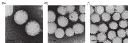
Polymer vesicles were prepared by solvent switch method44.The hydrophilic PEO and PAA chains form the coronas of the vesicle, while the P(NIPAM71-stat-CMA8)chains form the membrane in which PNIPAM is temperature-responsive and PCMA is crosslinkable. The size distribution and morphology of the vesicles were characterized by DLS, SEM and TEM. After stained with phosphotungstic acid, the hollow morphology of the collapsed vesicles with a phase-separated inhomogeneous membrane was observed from TEM and SEM images (Fig. 2 and Fig. S5 (see Supporting Information))45,46. The DLS results showed the hydrodynamic diameter (Dh)of vesicles is 208 nm and the polydispersity (PD)is 0.075 (Fig. 3). The morphology of the photocrosslinked vesicles at day 1 and day 7 was characterized by TEM with diameter between 180 and 200 nm(Fig. 2), which is in accordance with that measured by DLS.

Fig. 2 TEM images of photocrosslinked polymer vesicles at (a, b)day 1 and (c)day 7.

Fig. 3 Size distribution of vesicles as determined by DLS at 25 °C.
3.3 Photocrosslinking of block copolymer vesicles
In general, the PNIPAM segment is hydrophobic at 32 °C or higher and hydrophilic below 32 °C47. Therefore, all selfassembly processes are carried out at 40 °C, and the block copolymer can be self-assembled into vesicles by a solvent switch method. The dialyzed aqueous vesicle solution was diluted to 0.5 mg∙mL−1, and the CMA on the membrane was crosslinked under a UV spot curing system at a wavelength of 365 nm.After 120 seconds of UV irradiation, the degree of crosslinking of the vesicles was 88% (Fig. 4). As a result, the CMA crosslinking fixes the vesicle structure and enhances its stability in biological applications. When the temperature is decreased to 25 °C, the structure of the crosslinked vesicles will not change, but the permeability of the vesicle membrane will increase47. Thus, crosslinked and temperature-sensitive polymer vesicles can be used as drug carriers for controlled release under temperature changes.

Fig. 4 Photocrosslinking degrees of block polymer vesicles exposed to UV light at different time.
3.4 In vitro stability of photocrosslinked polymer vesicles
In vitrostability of photocrosslinked polymer vesicles were evaluated in the aspects of size and morphology. As shown in Fig. 2, no obvious change was observed of the morphology and diameter after one week. In addition, the hydrodynamic diameters and the size distribution of the photocrosslinked polymer vesicles in water and phosphate buffered saline (PBS;0.01 mol∙L−1at pH 7.4)at 25 °C were evaluated by DLS (Fig.5). From day 1 to day 7, the average hydrodynamic diameter of the vesicles in aqueous solution was between 203 and 216 nm with PD less than 0.25 during the whole process. Similarly, the average hydrodynamic diameter of the vesicles in PBS solution was between 197 and 215 nm with PD less than 0.25. The above experimental data indicated that the size and morphology of vesicles remained stable.

Fig. 5 Hydrodynamic diameter and polydispersity of photocrosslinked polymer vesicles in (a)water and(b)0.01 mol∙L−1 PBS at 25 °C.
3.5 Temperature-sensitive behavior of block copolymer vesicles
The temperature-responsive behavior of the uncrosslinked and crosslinked vesicles solution was evaluated by DLS. The temperature of uncrosslinked vesicles solution was gradually decreased from 45 to 10 °C. A longer equilibration time of 30 min was applied here to allow the temperature-sensitive PNIPAM chains to reach equilibrium. As shown in Fig. 6, the size of the uncrosslinked vesicles increased during cooling process when the temperature was above 25 °C. As the temperature was below 25 °C, the uncrosslinked vesicles started to disassemble with increased PD. The above results indicated that the uncrosslinked vesicles were not stable and would disassemble at low temperatures.

Fig. 6 Thermally controlled behavior of uncrosslinked polymer vesicles evaluated by temperature-dependent changes in size in aqueous solution. The temperature interval is 5 °C and the equilibration time is 30 min. During the temperature decrease process, the vesicles swelled and then dissociated below 25 °C.
Similarly, the temperature of the photocrosslinked vesicles solution was gradually increased from 5 to 45 °C and then decreased from 45 to 5 °C. The hydrodynamic diameter of the crosslinked vesicles during temperature variation was shown in Fig. 7. During the temperature-rising process, theDhof the crosslinked vesicles gradually decreased from 218 to 193 nm with PD less than 0.20. Then, theDhof the vesicles gradually increased from 191 to 223 nm with PD less than 0.20 during the cooling process. The above experimental data confirmed the temperature responsiveness and reversible size change during temperature variation of the crosslinked vesicles. The temperature rising and dropping processes were repeated three times with excellent repeatability.

Fig. 7 Thermally controlled behavior of photocrosslinked polymer vesicles evaluated by temperature-dependent changes in size in aqueous solution. The size of vesicles changed in the direction of(a)the temperature rising, and (b)the temperature decreasing. The temperature interval is 5 °C and the equilibration time is 30 min.
3.6 Drug release behavior of block copolymer vesicles
The cavity and membrane of vesicles can entrap hydrophilic and hydrophobic drugs, respectively. To verify the drug loading and controlled release capability, antibiotics were encapsulated by vesicles. CIP is a water-soluble antibiotic in the form of a hydrochloride, which was loaded into the polymer vesicles at 40 °C.
The temperature-regulated release profile of CIP in the vesicles is shown in Fig. 8. The drug loading content was estimated to be 10.4% relative to the vesicles. The drug loading efficiency was about 32.7%. The release experiments were carried out in deionized water at 37 and 25 °C, respectively. The experimental results showed that for the same CIP-containing vesicles, after 12 h, the CIP release rate in the surrounding of the deionized aqueous solution is nearly 35% higher at 25 °C than at 37 °C. In the first two hours, vesicles showed an initial burst of CIP release, reaching 68% and 52% at 25 and 37 °C,respectively. After that, the cumulative CIP release of vesicles continued to increase slowly at 25 °C, reaching 92% at 12 h. At 37 °C, the amount of CIP released continued to increase,stabilized after 2 h, and then the cumulative release of CIP eventually remained at about 57%. And the amount of CIP released during the whole cooling process was higher than at 37 °C. The release rate at different temperatures indicated that the drug release was significantly retarded at 37 °C due to its entrapment within the polymer vesicles, suggesting that the temperature-triggered vesicles swelling was the primary drug release mechanism.

Fig. 8 Cumulative release profile of CIP-loaded vesicles at 37 and 25 °C.
4 Conclusions
In summary, we have proposed a kind of temperaturesensitive polymer vesicles for lowering temperature triggered drug. TEM and SEM studies confirmed the structure of the vesicles, and DLS revealed their excellent stability against various temperatures after photocrosslinking. Moreover, for the same CIP-loaded vesicles, after 12 h, the CIP release rate was nearly 35% higher at 25 °C than at 37 °C. Overall, this photocrosslinked polymer vesicle with temperature-responsive properties has potential for future applications in the fields of lowering temperature triggered drug release for therapeutic hypothermia.
Supporting Information:1H NMR spectrum, calibration curve, SEC curve and SEM study (Fig. S1–Fig. S5)areavailable free of chargeviathe internet at http://www.whxb.pku.edu.cn.
