甲状腺乳头状癌中PDK1、ZEB1及P-cadherin的表达和临床意义
2021-05-07薛栋宋德坤王平安宋相孔夏修良
薛栋 宋德坤 王平安 宋相孔 夏修良

【摘要】 目的:探討甲状腺乳头状癌(papillary thyroid carcinoma,PTC)中3-磷酸肌醇依赖性蛋白激酶1(PDK1)、E盒结合锌指蛋白1 (ZEB1)和P-钙黏蛋白(P-cadherin)的表达情况和临床意义。方法:选取206例PTC和45例癌旁甲状腺标本,采用免疫组化检测PDK1、ZEB1和P-cadherin蛋白表达,并分析三个指标蛋白表达水平和临床病理各参数的关系。结果:PTC组织中PDK1、ZEB1阳性表达率分别为76.7%、72.3%,高于癌旁甲状腺组织的26.7%、28.9%(P<0.05)。P-cadherin在PTC组织中和癌旁甲状腺中的阳性表达率分别为22.3%、88.9%,两者比较差异有统计学意义(P<0.05)。PDK1、ZEB1及P-cadherin表达与TNM分期、腺体外侵及颈淋巴结转移有关(P<0.05);Spearman等级检验分析,PTC组织中PDK1和ZEB1蛋白表达呈正相关(rs=0.476,P=0.031),PDK1和P-cadherin蛋白表达呈负相关(rs=-0.419,P=0.029),ZEB1和P-cadherin蛋白表达呈显著负相关(rs=-0.586,P=0.019)。结论:PTC中PDK1、ZEB1及P-cadherin蛋白的异常表达与PTC侵袭和转移密切相关,PDK1、ZEB1可能对PTC的诊断和治疗具有重要作用。
【关键词】 甲状腺乳头状癌 3-磷酸肌醇依赖性蛋白激酶1 E盒结合锌指蛋白1 P-钙黏蛋白 免疫组织化学
[Abstract] Objective: To explore the expression and clinical significance of 3-phosphoinositide dependent protein kinase 1 (PDK1),zinc finger E-box-binding protein 1 (ZEB1) and P-cadherin protein in papillary thyroid carcinoma. Method: A total of 206 cases of PTC and 45 cases of adjacent thyroid specimens were selected, the expressions of PDK1, ZEB1 and P-cadherin were detected by immunohistochemistry, the relationship between three indicaters protein expression and clinicopathological parameters was analyzed. Result: The positive expression rates of PDK1 and ZEB1 protein in PTC were 76.7% and 72.3%, they were significantly higher than 26.7% and 28.9% in paracancerous normal thyroid tissues (P<0.05). The positive expression rates of P-cadherin in PTC and paracancerous normal thyroid tissues were 22.3%, 88.9%, the difference was statistically significant (P<0.05). The differences of the expression of PDK1, ZEB1 and P-cadherin protein in PTC of TNM stage, extrathyroidal invasion and lymph node metastasis were all significant (P<0.05). Spearman rank test showed significantly positive correlation was found between PDK1 and ZEB1 (rs=0.476, P=0.031), there was negative correlation between PDK1 and P-cadherin (rs=-0.419, P=0.029), the similar negative correlation was between ZEB1 and P-cadherin (rs=-0.586, P=0.019). Conclusion: The abnormal expression of PDK1, ZEB1 and P-cadherin protein in PTC is closely related to the invasion and metastasis of PTC, PDK1 and ZEB1 may have an important role in the diagnosis and theraphy of PTC.
[Key words] Papillary thyroid carcinoma PDK1 ZEB1 P-cadherin Immunohistochemistry
First-authors address: The Peoples Hospital of Binzhou City, Binzhou 256610, China
doi:10.3969/j.issn.1674-4985.2021.03.003
甲状腺乳头状癌(papillary thyroid carcinoma,PTC)是普外科工作实践过程中经常会接触到的一种甲状腺癌病理类型,占80%~85%[1]。最近几年PTC发病率呈明显上升态势,甚至有一部分微小乳头状癌病例早期就发生淋巴结转移[2]。磷脂酰肌醇3-激酶/蛋白激酶B(PI3K/Akt)是细胞分子网络中一个重要信号通路,它参与细胞的过度增殖、转分化和衰亡等过程。3-磷酸肌醇依赖性蛋白激酶1(3-phosphoinositide dependent protein kinase 1,PDK1)是一种重要的调节因子,激活PI3K/Akt信号,在转导信号过程中起着重要的作用[3]。PDK1主要包含两个分子结构域,分别为PH结构域(C端)和激酶结构域(N端),PDK1磷酸化Akt的苏氨酸(308位点)并激活PI3K/Akt信号,因此,PDK1与肿瘤细胞异常增殖、凋亡和侵袭转移都有关[4]。研究表明,急性髓细胞性白血病和乳腺癌中PDK1高表达,且与肿瘤不良预后有关[5-6]。上皮-间质转化(epithelial mesenchymal transition,EMT)是肿瘤侵袭转移过程中的重要事件。P-钙黏蛋白(P-cadherin)最初在1986年被发现,是EMT发生过程中的一个经典标志蛋白[7]。P-cadherin是一种细胞之间钙粘合蛋白,在维持正常细胞结构完整性、细胞形态、细胞增殖和转分化等过程中一直发挥重要生物学作用[8]。E盒结合锌指蛋白1(zinc finger E-box-binding protein 1,ZEB1)是一个重要的转录调控因子,促进肿瘤侵袭转移[9]。有研究表明,ZEB1在肺癌组织中高表达,且与病理分级及淋巴结转移明显有关,ZEB1还是一个不良预后因素[10]。目前关于PDK1、ZEB1和P-cadherin在PTC中研究报道较少。本文重点探讨PDK1、ZEB1和P-cadherin与PTC临床病理参数的相关性,为探索PTC侵袭转移的分子机制提供一个新的思路。现报道如下。
1 资料与方法
1.1 一般资料 收集2013年1月-2019年6月
滨州市人民医院206例PTC患者,男70例,女136例;年龄16~72岁,平均(44.1±6.4)岁;98例出现颈淋巴结转移,108例未出现颈淋巴结转移。纳入标准:符合甲状腺乳头状癌病理诊断标准;术中留取肿瘤组织或者癌旁组织。根据美国癌症联合委员会与国际抗癌联盟(AJCC)第八版进行TNM分期。排除标准:临床资料不完整;术前接受过放射治疗、化学药物治疗及免疫治疗等辅助治疗;合并其他组织器官癌变。选择同一时期45例癌旁正常甲状腺患者,男14例,女31例;年龄18~70岁,平均(42.2±5.2)岁。该研究已经滨州市人民医院伦理学委员会批准,患者知情同意并签署知情同意书。
1.2 方法 应用免疫组化方法(EnVision)检测PDK1、ZEB1及P-cadherin蛋白表达。鼠抗人PDK1单抗、鼠抗人ZEB1单抗和鼠抗人P-cadherin单抗采购于Santa Cruz公司,工作浓度为1∶200。EnVision试剂盒来源于Dako公司。阳性对照为已知阳性切片,阴性对照用PBS。
1.3 观察指标与判定标准 (1)比较PTC和癌旁甲状腺组织中PDK1、ZEB1和P-cadherin蛋白的表达情况;(2)分析PDK1、ZEB1和P-cadherin蛋白表達与PTC患者临床病理参数之间的关系;(3)分析PDK1、ZEB1和P-cadherin蛋白三者之间的相关性。阳性结果的判定:细胞出现黄色颗粒为阳性染色。PDK1蛋白定位在细胞质,ZEB1蛋白定位在细胞核,P-cadherin定位在细胞膜。先判定染色的强度:0、1、2、3分分别对应无色、淡黄、棕黄、棕褐色。然后计算阳性细胞百分数:0、1、2、3、4分分别对应≤5%、6%~25%、26%~50%、51%~75%和>75%以上。最后将前两者乘积≥4分为免疫反应阳性,而<4分为免疫反应阴性。
1.4 统计学处理 采用SPSS 22.0软件对所得数据进行统计分析,计量资料用(x±s)表示,组间比较采用t检验;计数资料以率(%)表示,比较采用字2检验。Spearman用于相关性分析。以P<0.05为差异有统计学意义。
2 结果
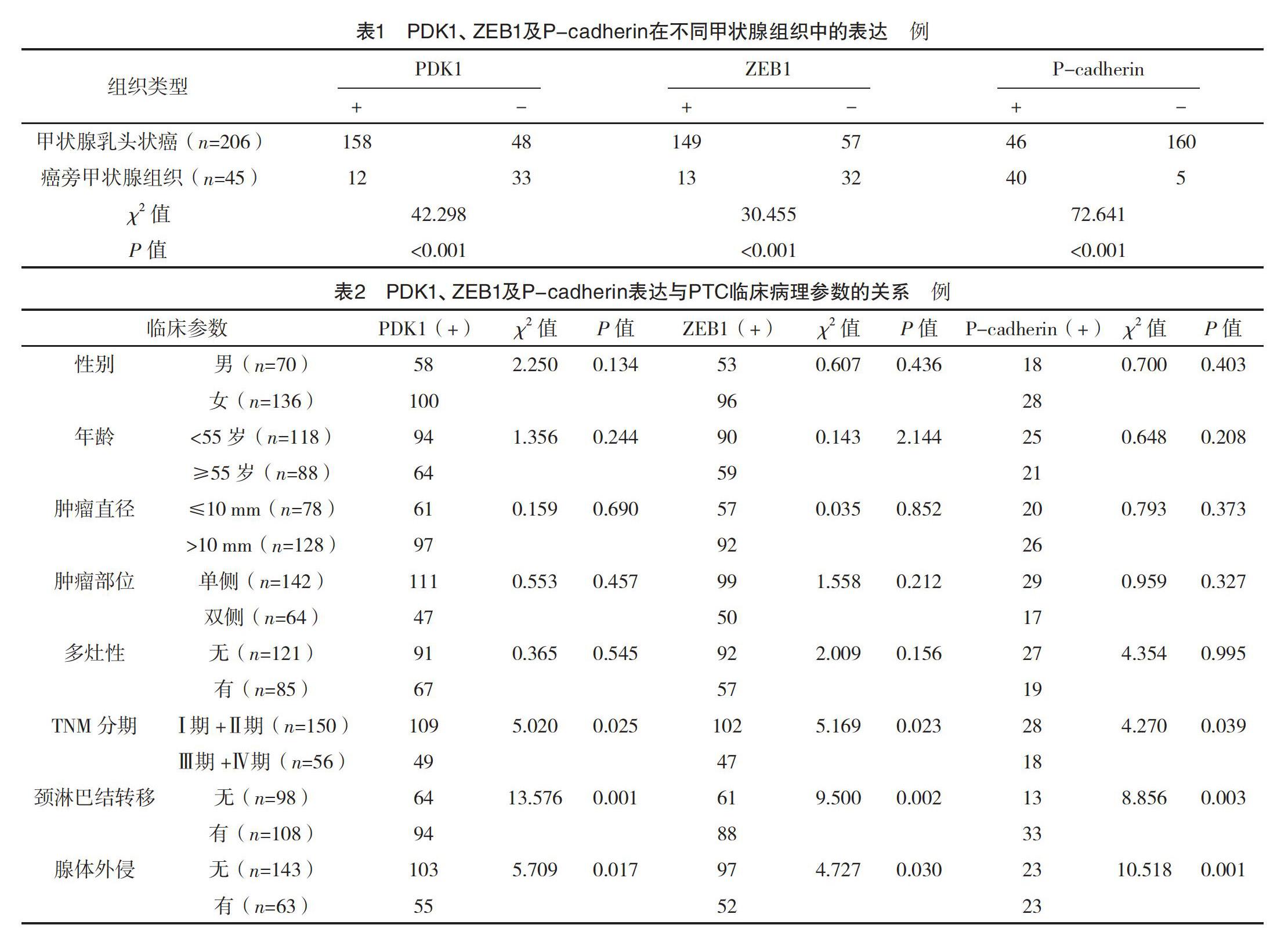
2.1 PTC和癌旁甲状腺中PDK1、ZEB1和P-cadherin蛋白的表达 细胞质内出现棕黄或棕褐色为PDK1,见图1;细胞核出现棕黄色为ZEB1蛋白,见图2;细胞膜出现黄染为P-cadherin,见图3。PTC组织中PDK1、ZEB1蛋白的阳性率分别为76.7%、72.3%,而癌旁正常甲状腺组织PDK1、ZEB1蛋白阳性率分别为26.7%、28.9%,PTC中PDK1、ZEB1的表达率均高于癌旁甲状腺组织(P<0.05)。PTC组织中P-cadherin阳性率为22.3%,而在癌旁甲状腺中为88.9%,两者比较差异有统计学意义(P<0.05)。见表1。
2.2 PDK1、ZEB1和P-cadherin表达与PTC患者临床病理参数间的关系 患者临床病理特征,如性别、年龄、肿瘤直径、肿瘤部位和多灶性与PDK1、ZEB1和P-cadherin阳性表达均无明显相关性(P>0.05);而PDK1、ZEB1和P-cadherin表达均与TNM分期、腺体外侵和颈淋巴结转移密切相关(P<0.05)。见表2。
2.3 PDK1、ZEB1和P-cadherin蛋白表达相关性 通过Spearman等级检验分析,PTC组织中PDK1与ZEB1蛋白表达呈正相关(rs=0.476,P=0.031),PDK1和P-cadherin蛋白表达呈负相关(rs=-0.419,P=0.029),ZEB1和P-cadherin蛋白表达水平呈显著负相关(rs=-0.586,P=0.019)。
3 討论
甲状腺癌在全球范围内均呈明显上升态势,目前居于我国女性恶性肿瘤的第四位[11]。近几年PTC病例越来越年轻化,且易发生引流区域的淋巴结转移,大约有70%的患者确诊时已发生淋巴结转移,因此关于侵袭转移的分子机制亟待解决。
PDK1可以通过多条途径调节蛋白激酶C的活性来激活Akt信号通路[12]。PDK1在喉癌组织中高表达,且其还与临床肿瘤分期、病理学分级及淋巴结转移有关,PDK1是喉癌患者生存预后的一个不良因素[13]。本研究结果显示,PTC组织中PDK1表达率高于癌旁组织(P<0.05),提示PDK1可能促进了PTC的发生发展。进一步还研究PDK1与PTC临床病理参数的关系,发现PTC组织中PDK1蛋白阳性表达与患者年龄、性别、肿瘤直径、肿瘤部位和多灶性没有密切关系,而与肿瘤的TNM分期、腺体外侵和颈淋巴结转移相关(P<0.05),表明PDK1在PTC细胞侵袭转移中起重要作用。
ZEB1作为一个转录调控因子,在恶性肿瘤侵袭转移中发挥重要的作用[14-15]。ZEB1在肺鳞癌中高表达,且和纵隔内淋巴结、远处器官转移均有关,干扰ZEB1基因表达后,肿瘤细胞的增殖和侵袭力均较前明显下降[16]。新近的研究表明,膀胱癌和食管癌组织中均发现了ZEB1蛋白表达,且与临床预后明显相关[17]。乳腺癌中ZEB1高表达的患者容易出现化疗耐受,且还与Bcl-xL、cyclin D1呈正相关[18]。本研究结果显示,ZEB1在PTC中有高表达,且ZEB1表达率与TNM分期、腺体外侵和颈淋巴结转移相关(P<0.05)。另外,PTC组织中PDK1与ZEB1存在正相关,推测PDK1可能通过调控ZEB1表达促进PTC侵袭转移。
实体恶性肿瘤在侵袭转移过程中经常发生着上皮-间质转化(EMT)现象。P-cadherin通过本身的钙黏附作用来保持细胞间的紧密连接,一旦表达减少后导致黏附力下降,肿瘤细胞就会容易侵袭和转移[19]。EMT在PTC细胞侵袭转移过程中发挥着重要作用[20]。有研究发现肝癌组织中出现P-cadherin低表达,并且35例肝癌完全没有表达,P-cadherin还与肿瘤分期明显有关,体外细胞实验发现抑制P-cadherin表达,促进了肝细胞癌细胞增殖[21]。本研究结果显示,PTC中P-cadherin表达明显低于癌旁甲状腺组织(P<0.05)。P-cadherin阳性表达与患者TNM分期、腺体外侵及颈淋巴结转移及有关(P<0.05)。进一步研究还发现,PDK1和P-cadherin存在负相关,P-cadherin和ZEB1存在负相关。PDK1、ZEB1和P-cadherin在PTC中有异常表达的现象,且三者有相关性,提示三者参与了肿瘤侵袭转移过程。
综上所述,PTC中PDK1、ZEB1高表达伴有P-cadherin低表达,且三个指标均与TNM分期、淋巴结转移和腺体外侵明显相关。但是关于PDK1促进PTC侵袭转移的分子机制尚不明确,仍需要深入研究。
参考文献
[1] Lundgren C I,Hall P,Dickman P W,et al.Clinically significant prognostic factors for differentiated thyroid carcinoma:a population-based,nested case-control study[J].Cancer,2006,106(3):524-531.
[2] Wei Sun,Xiabin Lan,Hao Zhang,et al.Risk Factors for Central Lymph Node Metastasis in CN0 Papillary Thyroid Carcinoma:A Systematic Review and Meta-Analysis[J].PLoS One,2015,10(10):e0139021.
[3] Gagliardi P A,Puliafito A,Primo L.PDK1:At the crossroad of cancer signaling pathways[J].Semin Cancer Biol,2018,48:27-35.
[4] Hossen M J,Kim S C,Yang S,et al.PDK1 disruptors and modulators:a patent review[J].Expert Opin Ther Pat,2015,25(5):513-537.
[5] Zabkiewicz J,Pearn L,Hills R K,et al.The PDK1 master kinase is over-expressed in acute myeloid leukemia and promotes PKC-mediated survival of leukemic blasts[J].Haematologica,2014,99(5):858-864.
[6] Du J,Yang M,Chen S,et al.PDK1 promotes tumor growth and metastasis in a spontaneous breast cancer model[J].Oncogene,2016,35(25):3314-3323.
[7] Nose A,Takeichi M.A novel cadherin cell adhesion molecule: its expression patterns associated with implantation and organogenesis of mouse embryos[J].J Cell Biol,1986,103(6 Pt 2):2649-2658.
[8] Vieira A F,Paredes J.P-cadherin and the journey to cancer metastasis[J].Mol Cancer,2015,14:178.
[9] Zhang P,Sun Y,Ma L.ZEB1:at the crossroads of epithelial-mesenchymal transition,metastasis and therapy resistance[J].Cell Cycle,2015,14(4):481-487.
[10] Dohadwala M,Yang S C,Luo J,et al.Cyclooxygenase-2-dependent regulation of E-cadherin: prostaglandin E2 induces transcriptional repressors ZEB1 and snail in non-small cell lung cancer[J].Cancer Research,2006,66(10):5338-5345.
[11] Wanqing Chen,Kexin Sun,Rongshou Zheng,et al.Cancer incidence and mortality in China 2014[J].Chinese Journal of Cancer Research,2018,30(1):1-12.
[12] Pearce L R,Komander D,Alessi D R.The nuts and bolts of AGC protein kinases[J].Nature Reviews Molecular Cell Biology,2010,11(1):9-22.
[13] Xiang G Z,Li X H,Cao L H,et al.Frequent overexpression of PDK1 in primary nasopharyngeal carcinoma is associated with poor prognosis[J].Pathology Research and Practice,2016,212(12):1102-1107.
[14] Larsen J E,Nathan V,Osborne J K,et al.ZEB1 drives epithelial-to-mesenchymal transition in lung cancer[J].J Clin Invest,2016,126(9):3219-3235.
[15] Chandra A,Jahangiri A,Chen W,et al.Clonal ZEB1-Driven Mesenchymal Transition Promotes Targetable Oncologic Antiangiogenic Therapy Resistance[J].Cancer Research,2020,80(7):1498-1511.
[16] Zhang J,Lu C,Zhang J,et al.Involvement of ZEB1 and E-cadherin in the invasion of lung squamous cell carcinoma[J].Mol Biol Rep,2013,40(2):949-956.
[17] Moussa R A,Khalil E Z I,Ali A I.Prognostic Role of Epithelial-Mesenchymal Transition Markers P-cadherin,β-Catenin,ZEB1,ZEB2 and p63 in Bladder Carcinoma[J].World Journal Oncology,2019,10(6):199-217.
[18] Zhang X,Zhang Z,Zhang Q,et al.ZEB1 confers chemotherapeutic resistance to breast cancer by activating ATM[J].Cell Death Dis,2018,9(2):57.
[19] Van Marck V,Stove C,Jacobs K,et al.P-cadherin in adhesion and invasion: opposite roles in colon and bladder carcinoma[J].Int J Cancer,2011,128(5):1031-1044.
[20] Mitchell B,Leone D,Yang S,et al.BRAF and epithelial-mesenchymal transition in papillary thyroid carcinoma-challenging the roles of Snail and P-cadherin?[J].American Journal of Translational Research,2016,8(11):5076-5086.
[21] Bauer R,Valletta D,Bauer K,et al.Downregulation of P-cadherin expression in hepatocellular carcinoma induces tumorigenicity[J].Int J Clin Exp Pathol,2014,7(9):6125-6132.
(收稿日期:2020-10-12) (本文編辑:程旭然)