Initial experience of single-incision plus one port left-side approach totally laparoscopic distal gastrectomy with uncut Roux-en-Y reconstruction
2020-09-23WeiZhouChangZhengDongYiFengZangYingXueXingGuoZhouYuWangYinLuDing
Wei Zhou, Chang-Zheng Dong, Yi-Feng Zang, Ying Xue, Xing-Guo Zhou, Yu Wang, Yin-Lu Ding
Abstract
Key words: Laparoscopy; Distal gastrectomy; Single-incision plus one port; Uncut Rouxen-Y gastrojejunostomy; Reduced port surgery; Gastric cancer
INTRODUCTION
Gastric cancer is the fifth most common cancer and the third leading cause of cancerrelated deaths among all malignant tumors in the world[1,2]. Although significant progress has been made in the diagnosis and comprehensive treatment of gastric cancer, the prognosis for patients with gastric cancer is still poor. Radical gastrectomy remains the first-choice treatment for patients with resectable gastric cancer. Laparoscopic techniques have recently been one of the main development directions for the surgical treatment of gastric cancer[3].
Laparoscopic distal gastrectomy has been widely accepted as a more effective approach than conventional open distal gastrectomy for early-stage gastric cancer according to several clinical studies[4-8]. The CLASS-01 randomized clinical trial by Yuet al[9]revealed that laparoscopic distal gastrectomy did not result in inferior 3-year disease-free survival compared with open distal gastrectomy among patients with a preoperative clinical stage indicating locally advanced gastric cancer[9]. As a type of reduced port surgery for patients with gastric cancer, single-incision laparoscopic surgery plus one port (SILS+1), in which fewer ports and shorter length of incisions are needed, has become increasingly popular in the past few years[10-14].
Digestive tract reconstruction after distal gastrectomy is still controversial because of the long-term postoperative complications that affect quality of life, such as Roux stasis syndrome and reflux gastritis. In 2005, Uyamaet al[15]first outlined a laparoscopic-assisted uncut Roux-en-Y digestive tract (uncut R-Y reconstruction) procedure following distal gastrectomy for the digestive tract. There is an increasing trend towards laparoscopic uncut R-Y reconstruction for distal gastrectomy due to its more satisfactory postoperative outcomes (lower incidence of bile reflux and Roux stasis syndrome) than conventional R-Y reconstruction[16-21].
It might be safe and feasible to treat gastric cancer, colon cancer and rectal cancer with SILS+1 by experienced surgeons[22-26]. SILS+1 provides minimal invasion without compromising the principles of tumor treatment. However, an analysis of an uncut RY reconstruction method after SILS+1 has not been carried out. To the best of our knowledge, there are no previous reports on the use of SILS+1 left-side approach (SILS+1/L) totally laparoscopic distal gastrectomy (TLDG) with uncut R-Y reconstruction.
The aim of this report is to present our initial experience in performing SILS+1/L TLDG with uncut R-Y reconstruction as well as the short-term postoperative outcomes and endoscopic findings.
MATERIALS AND METHODS
Patients and clinical data collection
Twenty-one patients who had undergone SILS+1/L TLDG with uncut R-Y anastomosis reconstruction to treat distal gastric cancer were enrolled in this study. The inclusion criteria were a clinical diagnosis of gastric cancer and a depth of invasion of gastric cancer ≤ T3. The exclusion criteria included preoperative radiotherapy and chemotherapy, tumor perforation, fused lymph nodes leading to difficult dissection and severe medical illness. All operations were carried out in The Second Hospital of Shandong University between March 2019 and February 2020. All operations were performed by the same team of doctors. The perioperative details, surgical details and pathological data of all the selected patients were collected retrospectively from cancer registry data and the electronic medical records. The pathological stage of each specimen was classified according to the American Joint Cancer Committee 8th Edition tumor, node, metastasis classification[27].
Food stasis, gastritis and bile reflux of the remnant stomach were evaluated by upper gastrointestinal radiography and endoscopy at 3 mo postoperatively. These findings were reviewed by two expert radiologist or two endoscopists. If the two reviewers did not reach an agreement, consultations and discussions on individual cases were held until the two reviewers reached a consensus.
This study was approved by the Ethics Committee of the Second Hospital of Shandong University. Written informed consent was obtained from each patient prior to enrollment in the present study.
Surgical procedures
The patient was placed in a reverse Trendelenburg position under general anesthesia. A 3.0-3.5 cm incision was made around the navel to place the SILS portTM(Hangtian Kadi Technology, Beijing, China) with four in-built trocars. Another 12-mm trocar was placed in the left upper quadrant as operative port for the passage of the surgical instruments such as the ultrasonic shear, the endoscopic linear stapler and the postoperative drainage tube (Figure 1). A 10 mm scope was inserted through the 10 mm in-built trocar at the extreme caudal position of the SILS port for the duration of the operation. Other surgical instruments were inserted through the other three inbuilt trocars of the SILS port as necessary.
The operator and assistant stood on the left and right side of the patient respectively, and the scope operator was located between the patient’s legs, which could be spread out. The monitor screen was placed above the head of the patient. The surgeon used the left flank 12 mm port and either the extreme head-side 12 mm port or the left-side 5 mm trocar in the SILS portTM. The assistant used the right-side 5 mm trocar in the SILS portTMto provide support.
The falciform ligament was suspended and fixed at the anterior abdominal wall, drawing the round ligament away with polypropylene monofilament. All of the twenty-one patients underwent laparoscopic-assisted distal gastrectomy with D2 lymphadenectomy, which was performed by the same operation team. After routine resection of the D2 group lymph nodes, the duodenum was cut 2 cm away from the pylorus, and the stomach was cut 5 cm from the upper edge of the tumor. Specimens were extracted from the umbilical port following a 3.0-3.5 cm vertical extension of the initial incision.
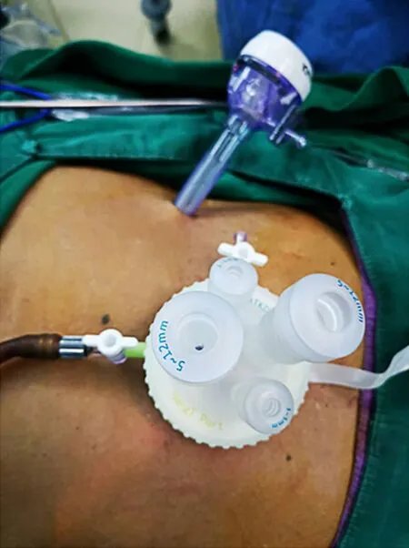
Figure 1 Settlement of port for single incision plus one port left-side approach.
The uncut R-Y reconstruction was performed intracorporeally. The transverse colon was retracted cephalad to expose the ligament of Treitz. First, the gastrojejunal (GJ) anastomosis site was marked on the jejunum 20 cm distal to the ligament of Treitz and on the posterior wall of the greater curvature of the residual stomach. The GJ anastomosis was accomplished through the 12 mm port in the left upper quadrant, using 60 mm endoscopic linear stapler (Ethicon, United States), Then the jejunojejunal anastomosis site was marked 5 cm distal to the ligament of Treitz and 40 cm distal to the GJ anastomosis site. The jejunojejunal anastomosis with a length of 4 cm was made through the 12 mm port in the left upper quadrant using a 60 mm endoscopic linear stapler. Both the common openings of the GJ anastomosis and the jejunojejunal anastomosis were closed using 3-0 V-LOCTMthread (Covidien, Mansfield, MA, United States). Finally, the biliopancreatic limb was closed 3 cm proximal to the GJ anastomosis using a 45 mm non-knife stapler (Ethicon, United States) through the port in the left upper quadrant. The completed uncut R-Y reconstruction is shown in Figure 2A-D. The appearance of the abdomen after surgery is shown in Figures 3 and 4.
Postoperative management
All patients received patient-controlled analgesia postoperatively. Nonsteroidal antiinflammatory drugs were used when the patient needed additional pain control. Water was provided after the first exhaust, followed by liquid diet and then soft diet. The patient was discharged when the diet was acceptable, and there was no adverse reaction.
Statistical analysis
Data exploration was used as appropriate. All statistical analyses were performed using SPSS for Windows (ver.22.0; SPSS, Inc., Chicago, IL, United States).
RESULTS
The patient population included 13 men and 8 women with a mean age of 48.2 years (ranged from 40 years to 70 years) and median BMI of 22.8 kg/m2. All patients had no previous history of abdominal surgery or preoperative chemoradiotherapy. None of the patients needed conversion to laparotomy or additional port insertion. The mean length of the proximal and distal margins was 4.2 and 6.4 cm, respectively. The number of retrieved lymph nodes ranged from 30-47 with a mean of 42. Six patients were in stage I and 15 patients were in stage II.
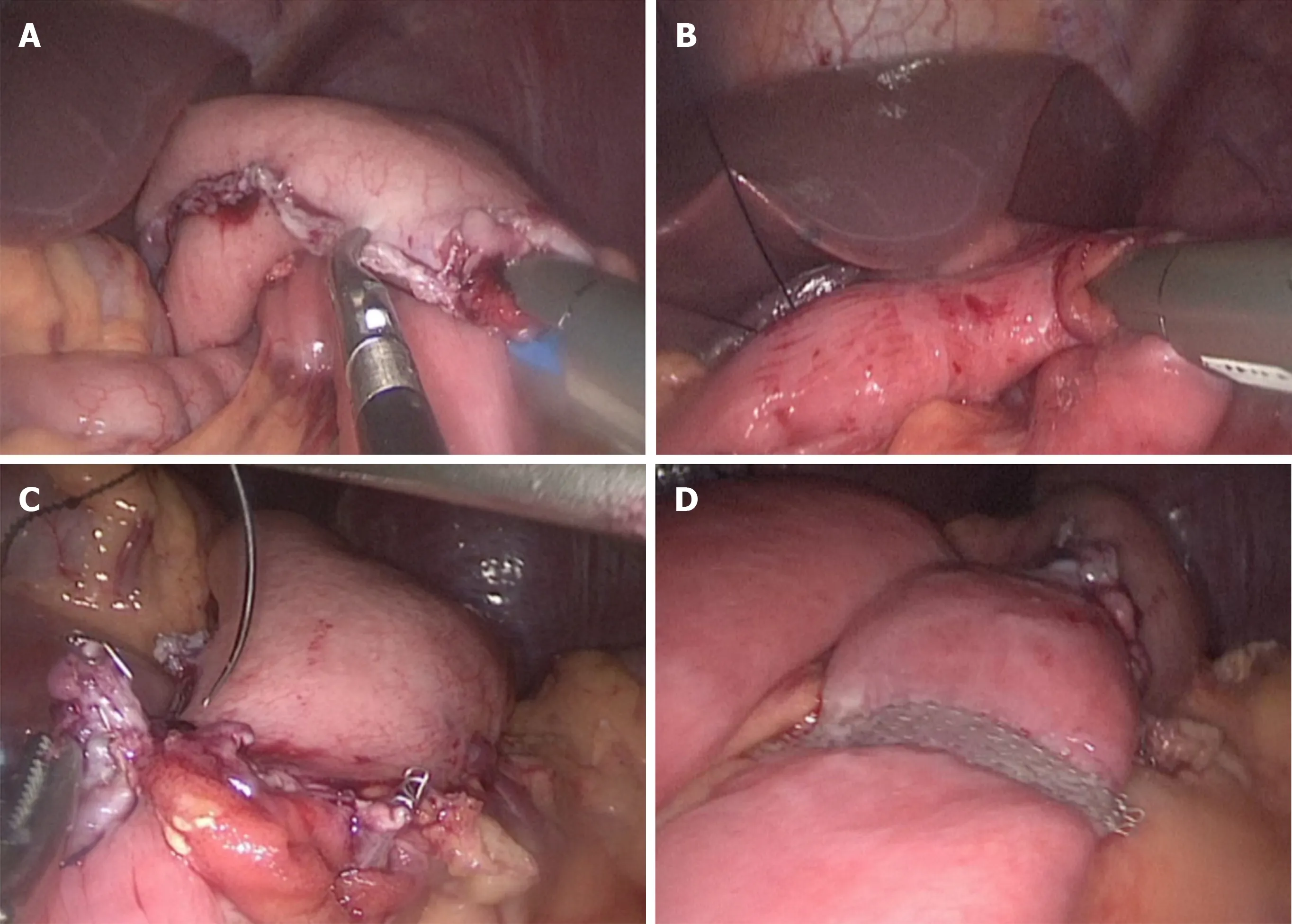
Figure 2 Procedures of reconstruction. A: The gastrojejunal anastomosis; B: The jejunojejunal anastomosis; C: Closing the gastrojejunal anastomosis; D: Uncut reconstruction.
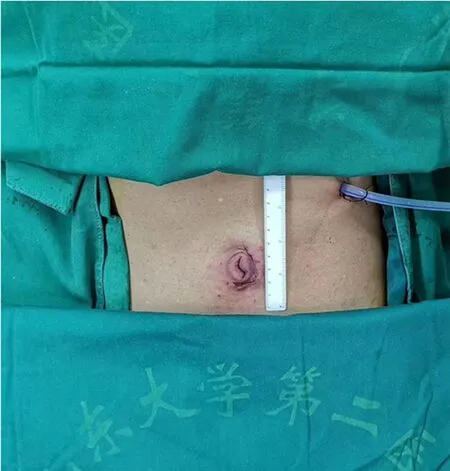
Figure 3 Appearance of the abdomen after surgery.
All patients underwent D2 radical gastrectomy with uncut R-Y reconstruction. No intraoperative complications occurred in any cases. The mean operating time was 146 min (ranged 130-180 min), and the estimated mean blood loss was 54 mL (ranged 20-110 mL). The mean duration to flatus and discharge was 2.3 d (ranged 1-3.5 d) and 7.3 d (ranged 6-9 d), respectively. One patient experienced mild postoperative pancreatic fistula ileus (grade A) and was managed without medication or surgical intervention. One patient experienced surgical site infection (wound at the navel incision) and was treated conservatively. The other complications such as postoperative pneumonia, postoperative small bowel ileus, bleeding, duodenal residual leakage, anastomotic complications and postoperative mortality did not occur in our study.

Figure 4 Appearance of the abdomen 3 mo after surgery.
All patients underwent upper gastrointestinal imaging examination using oral meglumine diatrizoate and gastroscopy examination 3 mo after surgery. At the 3-mo postoperative follow-up, no jejunal recanalization of the uncut staple line was observed by upper gastrointestinal imaging examination (Figure 5). Endoscopic examination showed that minimal food stasis was present in two patients (Figure 6A). No patient had moderate or severe food stasis. One patient had alkaline gastritis around the GJ anastomosis (Figure 6B). There was no moderate or severe alkaline gastritis, and no patients had bile reflux.
Surgical learning curve in SILS+1/L TLDG was reviewed. The data of operation time and blood loss were collected and plotted (Figures 7 and 8). With the case number increasing, operation time and blood loss gradually decreased, and the relatively stable plateau emerged. Experienced laparoscopic gastrectomy surgeons also need a long learning curve.
DISCUSSION
The digestive tract reconstruction procedure after distal gastrectomy is one of the most important factors that affects the quality of postoperative life. Although the reconstruction technique was a very hot spot issue in recent years, the optimal approach has not yet been proven by any trial or research yet. In most cases, B (Billroth)-I, B-II or R-Y are used[28,29]. The unique advantages and disadvantages of each method are determined by the location of the tumor, the progression of the cancer, the condition of the patient and the preference of the operator[30].
B-I gastroduodenal anastomosis is the most commonly used method for gastrointestinal reconstruction after distal gastrectomy[29]. However, this method requires enough remnant stomach to relieve anastomotic tension and has several disadvantages, including an increased incidence of food stasis, bile reflux, alkaline gastritis, dumping syndrome and reflux esophagitis[31-33]. Furthermore, the principle of tumor treatment should be fully considered. Once local recurrence occurs, a second opportunity for an operation may be lost.
B-II gastrojejunostomy is more suitable for extensive resection of advanced gastric antrum cancer and lymph node dissection. B-II type gastric jejunostomy is more likely to cause duodenal juice and bile reflux and residual gastritis. Roux-en-Y stomach jejunostomy can prevent these complications, may effectively control the blood sugar level of patients with type 2 diabetes and play a role in the treatment of diabetes. However, the additional anastomosis, the complexity of the procedure and the longer anastomosis time remain disadvantages of this technique. In addition, Roux stasis can occur in up to 30% of patients who undergo the R-Y procedure[34].
The uncut Roux-en-Y reconstruction preserves the advantages of Roux-en-Y gastrojejunostomy and builds a bridge that maintains neuromuscular continuity between the proximal jejunum and the Roux limb instead of cutting it off. Roux stasis is known to be an electrical and motor coordination problem that occurs after jejunal transection[35]. Uncut Roux-en-Y reconstruction may theoretically reduce the incidence of Roux retention syndrome[36].
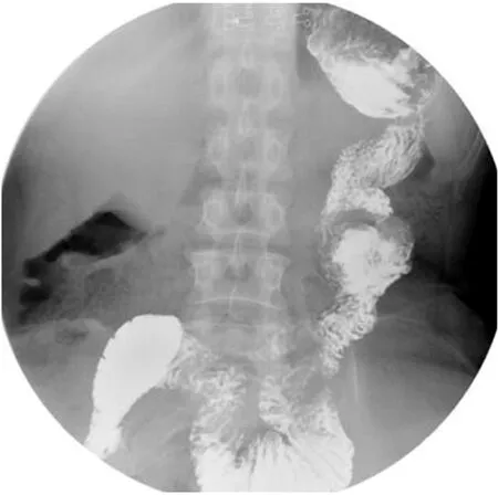
Figure 5 Upper gastrointestinal imaging examination 3 mo after surgery: No jejunal recanalization.
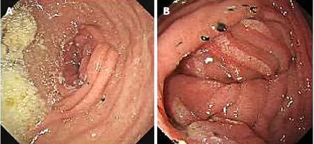
Figure 6 Endoscopic examination. A: Minimal food stasis; B: Alkaline gastritis around the gastrojejunal anastomosis.

Figure 7 Learning curve: Operation time plotted against case number.
Single-port and reduced-port laparoscopic radical gastrectomy as an innovative surgery for gastric cancer and has been gradually accepted and increasing attention has been paid to single-port and reduced-port laparoscopic radical gastrectomy due to its better cosmetic effect, less pain, lower incidence rate of surgical site infection and more advantages in enhanced recovery after surgery[37].

Figure 8 Learning curve: Blood loss plotted against case number.
Pure SILS has some technical limitations such as collision between instruments and inadequate countertraction. As a type of reduced port surgery, SILS+1 is more convenient than pure SILS. In the setting of SILS+1/L, the umbilical multichannel port is used to extend the lens, through which surgeons can manipulate forceps into the abdominal cavity for the operation with their left hand, and their right hand can manipulate energy instrument through the auxiliary port in the left upper quadrant. In Japan, surgeons are accustomed to adopting single-incision laparoscopic surgery plus one port in the right upper quadrant[38]. Therefore, the auxiliary operation port is set at 2 cm below the costal margin on the right side of the clavicle midline. The disadvantage of SILS+1 in the right upper quadrant is that the lymph node dissection in the subpyloric area is too close and the operation is inconvenient; the omentum is difficult to disperse without the help of an assistant. When standing on the right side, the energy instrument and lens in the surgeon’s right hand are passed through the multichannel puncture device, which is relatively close and prone to rear-end collisions. Conversely, the port in the upper left abdomen is the main operation hole in the case of SILS+1/L. The energy instrument and other surgical devices can enter the abdominal cavity through this hole and maintain a certain distance from the lens. Thus, this approach makes some surgical procedures, such as lymph node dissection and digestive tract reconstruction, easier without the need to replace instruments with the left and right hands.
In this study, no severe postoperative complications were found according to the Clavien-Dindo classification of surgical complications. Minimal to mild and limited gastritis were seen in 4.8% of the patients, and no patients had bile reflux. Food stasis was minimal or mild (9.5% of patients), and there was no moderate or severe food stasis. Moreover, no patients complained of nausea, vomiting or postprandial pain during the follow-up period. Our results are similar to the study by Yanget al[39]. This suggests that an uncut R-Y reconstruction after laparoscopic distal gastrectomy maintains the advantages of conventional R-Y in reducing the incidence of gastritis and bile acid reflux except for preventing roux stasis. In addition, we did not find any recanalization of the jejunum-jejunum anastomosis during the postoperative followup.
Standardized lymph node dissection and a sufficient number of lymph nodes for pathological examinations are the prerequisite and basis for radical surgery for gastric cancer. The number of lymph nodes recommended for pathological tumor, node, metastasis staging of gastric cancer in the 8th edition of American Joint Cancer Committee is ≥ 16[27]. In this study, the average number of retrieved lymph nodes was 42. This number meets the needs of judging tumor, node, metastasis stage in terms of oncology.
In conclusion, SILS+1/L with the uncut R-Y digestive reconstruction procedure in TLDG is secure and feasible. The uncut technique can be done by experienced surgeons in large medical center for patients with gastric cancer. Additional long-term follow-up is needed to evaluate the tumor effect compared to conventional laparoscopic radical gastrectomy.
ARTICLE HIGHLIGHTS
Research background
Single-incision laparoscopic surgery plus one port (SILS+1), in which fewer ports and shorter length of incisions are needed, has become increasingly popular in the past few years. However, the safety of SILS+1 left-side approach (SILS+1/L) totally laparoscopic distal gastrectomy (TLDG) with uncut Roux-en-Y reconstruction is not clear.
Research motivation
An analysis of an uncut R-Y reconstruction method after SILS+1 has not been carried out. To the best of our knowledge, there are no previous reports on the use of SILS+1/L TLDG with uncut R-Y reconstruction.
Research objectives
This study aimed to evaluate the safety and feasibility of SILS+1/L with the uncut R-Y digestive reconstruction procedure in TLDG. This report is to present our initial experience in performing SILS+1/L TLDG with uncut R-Y reconstruction as well as the short-term postoperative outcomes and endoscopic findings.
Research methods
The statistics of 21 patients who had undergone SILS+1/L TLDG with uncut R-Y anastomosis reconstruction to treat distal gastric cancer were collected. Data exploration was used as appropriate. All statistical analyses were performed using SPSS for Windows.
Research results
The mean operating time was 146 min (ranged 130-180 min), and the estimated mean blood loss was 54 mL (ranged 20-110 mL). The mean length of the proximal and distal margins was 4.2 and 6.4 cm, respectively. The number of retrieved lymph nodes ranged from 30-47 with a mean of 42. One patient experienced mild postoperative pancreatic fistula ileus, and one patient experienced surgical site infection. Other complications did not occur in our study.
Research conclusions
We found that SILS+1/L TLDG with uncut Roux-en-Y reconstruction is safe and effective and should be popularized.
Research perspectives
From this study, we can found that SILS+1/L TLDG with uncut R-Y reconstruction can be used not only for colon surgery but also for gastric surgery. In the future, the direction of the research is that an effective perioperative management program specific for gastric cancer is developed. The best method is to conduct a large-scale clinical trial to verify it.
杂志排行

World Journal of Gastroenterology的其它文章
- Current understanding of the metabolism of micronutrients in chronic alcoholic liver disease
- Establishment of a pattern recognition metabolomics model for the diagnosis of hepatocellular carcinoma
- Monocyte-to-lymphocyte ratio as a prognostic factor in peripheral whole blood samples of colorectal cancer patients
- Clinical and prognostic significance of CC chemokine receptor type 8 protein expression in gastrointestinal stromal tumors
- Feasibility and efficacy evaluation of metallic biliary stents eluting gemcitabine and cisplatin for extrahepatic cholangiocarcinoma
- Impact of interval between neoadjuvant chemoradiotherapy and surgery in rectal cancer patients