4,4′-二异硫氰基芪-2,2′-二磺酸对脓毒症小鼠心肌炎性反应的作用及机制
2020-04-02王艳夏琳蔡华忠熊御
王艳 夏琳 蔡华忠 熊御
[摘要] 目的 探討4,4′-二异硫氰基芪-2,2′-二磺酸(DIDS)对脓毒症小鼠心肌炎性反应的作用及机制。 方法 雄性BALB/c小鼠总计52只,采用随机数字表法分为4组,分别为空白对照组、脂多糖(LPS)组、DIDS低剂量组和DIDS高剂量,每组13只。预处理3 d,其中DIDS低剂量组、DIDS高剂量组每天分别腹腔注射7、14 mg/kg的DIDS,空白对照组、LPS组每天腹腔注射等量的磷酸缓冲盐溶液(PBS)。第3天预处理完成2 h后,空白对照组腹腔注射PBS,其余三组腹腔注射LPS 10 mg/kg建立小鼠脓毒症模型。在模型创建成功后,每组10只小鼠进行48 h生存状况观察,计算存活率。另外每组3只小鼠于4 h后处死,取心脏组织,采用real-time PCR法检测炎性因子[包括肿瘤坏死因子-α(TNF-α)、白细胞介素1β(IL-1β)和白细胞介素-6(IL-6)]及Notch信号通路相关基因(包括Notch1~4、Dll1、Jag1)mRNA表达,采用Western blot法检测Notch信号通路相关蛋白的表达。 结果 与LPS组比较,DIDS高剂量组可以明显提高小鼠存活率,差异有高度统计学意义(P < 0.01);与空白对照组比较,LPS组TNF-α、IL-1β和IL-6的 mRNA均显著升高,差异均有统计学意义(均P < 0.05);与LPS组比较,DIDS高剂量组TNF-α、IL-1β和IL-6的mRNA显著降低,差异均有统计学意义(均P < 0.05)。与LPS组比较,DIDS高剂量组Notch信号相关基因Notch1、Notch3、Dll 1及Jag 1的mRNA表达水平升高,Dll 1和Jag 1相关分子蛋白水平升高,差异均有统计学意义(均P < 0.05)。 结论 DIDS能够明显减轻脓毒症小鼠的心肌炎性反应,其机制可能与调控Notch信号通路有关。
[关键词] 4,4′-二异硫氰基芪-2,2′-二磺酸;炎性反应;脓毒症;Notch信号通路
[中图分类号] R967 [文献标识码] A [文章编号] 1673-7210(2020)01(c)-0004-05
Effect and mechanism of 4,4′-diisothiocyanostilbene-2,2′-disulfonic acid on myocardial inflammatory response in sepsis mice
WANG Yan XIA Lin CAI Huazhong XIONG Yuyun
1.Department of Pharmacy, Zhenjiang First People′s Hospital, Jiangsu Province, Zhenjiang 212001, China; 2.Department of Clinical Laboratory, Affiliated Hospital of Jiangsu University, Jiangsu Province, Zhenjiang 212001, China; 3.Department of Emergency, Affiliated Hospital of Jiangsu University, Jiangsu Province, Zhenjiang 212001, China
[Abstract] Objective To investigate the effect and mechanism of 4,4′-diisothiocyanostilbene-2,2′-disulfonic acid (DIDS) on myocardial inflammatory response in sepsis mice. Methods A total of 52 male BALB/c mice were divided into 4 groups by random number table method, namely blank control group, lipopolysaccharide (LPS) group, low dose DIDS group and high dose DIDS group, with 13 mice in each group. For 3 days of pretreatment, the low dose DIDS group and the high dose DIDS group were intraperitoneally injected with 7 and 14 mg/kg DIDS every day, respectively, while the blank control group and the LPS group were given the same amount of phosphate buffer salt solution (PBS) intraperitoneally every day. After 2 h of pretreatment on the third day, PBS was injected intraperitoneally into the blank control group, and LPS 10 mg/kg was injected intraperitoneally into the other three groups to establish a mouse sepsis model. After the model was created successfully, the survival status of 10 mice in each group were observed for 48 h, and the survival rates were calculated. In addition, 3 mice in each group were sacrificed after 4 h, and their heart tissues were taken. The mRNA expression of inflammatory factors [including tumor necrosis factor-α (TNF-α), interleukin-1β (IL-1β) and interleukin-6 (IL-6)] and genes related to Notch signaling pathway (including Notch1-4, Dll1 and Jag1) were detected by real-time PCR. Western blot was used to detect the expression of proteins related to the Notch signaling pathway. Results Compared with the LPS group, the survival rate of mice in the high dose DIDS group was significantly improved, with a highly statistically significant difference (P < 0.01). Compared with the blank control group, the mRNA levels of TNF-α,IL-1β and IL-6 in the LPS group were significantly increased, with statistically significant differences (all P < 0.05). Compared with the LPS group, the mRNA levels of TNF-α,IL-1β and IL-6 in the high dose DIDS group were significantly decreased, with statistically significant differences (all P < 0.05). Compared with the LPS group, mRNA expression levels of Notch1, Notch3, Dll 1 and Jag 1 of Notch signaling related genes in the high-dose DIDS group were increased, and protein levels of Dll 1 and Jag 1 were increased, with statistically significant differences (all P < 0.05). Conclusion DIDS can significantly reduce the myocardial inflammatory response in sepsis mice, and its mechanism may be related to the regulation of Notch signaling pathway.
[Key words] 4,4′-diisothiocyanostilbene-2,2′-disulfonic acid; Inflammation; Sepsis; Notch signaling pathway
脓毒症是指宿主对感染反应失调导致的威胁生命的器官功能障碍[1]。研究表明,约40%脓毒症患者出现心脏功能障碍(sepsis-induced cardiac dysfunction,SCD),死亡率高达70%~90%[2-3],主要原因有促炎因子的释放,线粒体功能失调以及由此引起的心肌收缩功能下降[4-7]。目前仍未有防治SCD的有效方法[8]。Notch信号通路是人体发育过程中高度保守的重要信号通路[9],参与了自身免疫和炎性反应相关的多种疾病[10-12],其相关基因的突变与多种类型心血管疾病有关[13-15]。4,4′-二异硫氰基芪-2,2′-二磺酸(4,4′-diisothiocyanostilbene-2,2′-disulfonic acid,DIDS)是一种非特异性氯离子通道阻断剂,具有抗凋亡、神经保护和抗炎作用[16-18]。已有文献报道,DIDS通过TLR4/NF-κB通路抑制脂多糖(lipopolysaccharide,LPS)诱导的RAW264.7细胞中促炎因子的释放。同时在LPS诱导的脓毒症小鼠模型中,DIDS可以减少小鼠的死亡率,保护肝肾等组织,下调促炎因子的表达[18]。目前尚未见DIDS抗脓毒症时心肌炎性反应的相关研究报道。为了进一步明确DIDS对抗小鼠脓毒症诱发的心肌炎性反应,本研究拟建立LPS诱导的小鼠脓毒症模型,探讨其抗心肌炎性反应的可能机制,以期为其后续深入研究奠定基础。
1 材料与方法
1.1 实验仪器
Multiskan GO多功能酶标仪(美国Thermo公司);ABI 7500 荧光定量PCR仪(ABI公司);BIO-RAD电泳转膜系统(美国BIO-RAD公司)。
1.2 药物与试剂
LPS(0111∶B4)、DIDS均购自Sigma-Aldrich公司;JAG1(2588T)和DLL1(2620T)抗体均购自SantaCruz公司,β-actin(abs132001)抗体购自absin公司;所有二抗均购自博士德生物科技公司;引物合成自金斯瑞生物科技有限公司;PrimeScriptTM RT-PCR试剂盒(RR037A)、SYBR Premix Ex TaqTM(RR420A)试剂盒均购自Takara公司;其他试剂均为国产分析纯。
1.3 实验动物
SPF级BALB/C小鼠52只,6~8周龄,雄性,体重18~20 g,由扬州大学比较医学中心提供[SCXK(苏)2012-0004]。小鼠实验遵循江苏大学实验动物伦理规范。小鼠自由饮食和饮水,室温20~23℃,湿度55%~65%,人工光暗周期12 h。
1.4 分组与干预方法
小鼠购入后适应性饲养3 d,采用随机数字表法分为空白对照组、LPS组、DIDS低剂量组、DIDS高剂量组,每组13只。DIDS低剂量组、DIDS高剂量组每天分别腹腔注射7、14 mg/kg的DIDS,空白对照组、LPS组每天腹腔注射与DIDS高剂量组等量的磷酸缓冲盐溶液(PBS)。第3天注射2 h后,空白对照组腹腔注射PBS,其余三组腹腔注射10 mg/kg的LPS,建立小鼠脓毒症模型。每组10只小鼠,进行生存状况观察研究。每组剩余的3只小鼠,在建立膿毒症模型后4 h,断颈处死后摘取心脏组织,立即置于液氮罐中-180℃保存。
1.5 观察指标
1.5.1 小鼠存活率比较 每隔4 h观察每组10只小鼠的死亡数,直到48 h为止,计算脓毒症小鼠的存活率
1.5.2 小鼠心脏组织中促炎因子的表达 提取心脏组织mRNA,对促炎因子肿瘤坏死因子-α(TNF-α)、白细胞介素1-β(IL-1β)和白细胞介素-6(IL-6)的mRNA水平进行检测。
1.5.3 脓毒症小鼠心脏中Notch相关基因mRNA表达 采用real-time PCR法。总RNA的提取利用Trizol试剂的方法进行。取1 μg RNA利用Takara PrimeScriptTM RT-PCR试剂盒的操作步骤进行逆转录反应合成cDNA。采用Takara公司SYBR Premix Ex TaqTM试剂盒进行real-time PCR操作。引物序列为:β-actin forward:5′-GAAGTCCCTCACCCTCCCAA-3′;β-actin reverse:5′-GGCATGGACGCGACCA-3′;TNF-α forward:5′-CATGGATCTCAAAGACAACC-3′;TNF-α reverse:5′-GGTATATGGGCTCATACCAG-3′;IL-1β forward:5′-GAAGTCAAGAGCAAAGTGG-3′;IL-1β reverse:5′-ACAGTCCAGCCCATACTTT-3′;IL-6 forward:5′-CTGATGCTGGTGACAACCAC-3′;IL-6 reverse:5′-TCCACGATTTCCCAGAGAAC-3′;Notch1 forward:5′-TCAGCGGGATCCACTGTGAG-3′;Notch1 reverse:5′-ACACAGGCAGGTGAACGAGTTG-3′;Notch2 forward:5′-TGCCAAGCTCAGTGGTGTTGTA-3′;Notch2 reverse:5′-TGCTAGGCTTTGTGGGATTC-AG-3′;Notch3 forward:5′-GGTTCCCAGTGAGCACCCTTAC-3′;Notch3 reverse:5′-GTGGATTCGGACCA-GTCTGAGAG-3′;Notch4 forward:5′-ACCTGCTCAA-CGGCTTCCA-3′;Notch4 reverse:5′-AGCTTCTGC-ACTCATCGATATCCTC-3′;Dll1 forward:5′-ACCT-TCTTTCGCGTATGCCTCAAG-3′;Dll1 reverse:5′-AGAGTCTGTATGGAGGGCTTC-3′;Jag1 forward:5′-C-AGGTCTTACCACCGAACA-3′;Jag1 reverse:5′-AC-AGGGAAGGCTCACACGG-3′。反应体系如下:SYBR?誖 Premix Ex Taq Ⅱ(Tli RNaseH Plus)(2×):12.5 μL;PCR Forward Primer(10 μmol/L):1 μL;PCR Reverse Primer(10 μmol/L):1 μL;反转录反应液(cDNA溶液):2 μL(500 ng);dH2O(灭菌蒸馏水):8.5 μL;共计25 μL。反应条件,步骤1:预变性95℃ 30 s;步骤2:95℃ 5 s,40个循环;60℃ 30 s。表达水平用其与内参β-actin的比值表示,以2-ΔΔCt法计算目标基因与内标基因荧光强度比值,相对定量比较各处理因素对目标基因表达的影响。
1.5.4 检测脓毒症小鼠心脏中Notch相关蛋白的表达 采用Western blot法。提取脓毒症小鼠心脏蛋白后,经SDS凝胶电泳,转膜,分别孵育Notch相关蛋白抗体后,将膜置于ECL发光液中,凝胶成像系统对条带进行观察和半定量分析。
1.6 统计学方法
采用SPSS 22.0对所得数据进行统计学分析,计量资料采用均数±标准差(x±s)表示,组间比较采用ANOVA方差分析,进一步两两比较采用t检验。以P < 0.05为差异有统计学意义。
2 结果
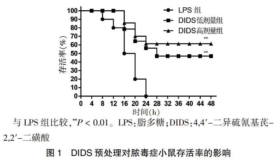
2.1 DIDS对抗LPS诱导的脓毒症小鼠存活率的降低
LPS腹腔注射24 h后,LPS组10只小鼠全部死亡。与LPS组比较,DIDS高剂量组可以明显提高小鼠存活率,差异有高度统计学意义(P < 0.01)。见图1。
与LPS组比较,**P < 0.01。LPS:脂多糖;DIDS:4,4′-二异硫氰基芪-2,2′-二磺酸
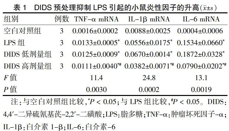
2.2 DIDS对抗LPS诱导的脓毒症小鼠心脏组织中促炎因子的升高
与空白对照组比较,LPS组TNF-α、IL-1β和IL-6的 mRNA均显著升高,差异均有统计学意义(均P < 0.05);与LPS组比较,DIDS高剂量组TNF-α、IL-1β和IL-6的mRNA显著降低,差异均有统计学意义(均P < 0.05)。见表1。
注:与空白对照组比较,*P < 0.05;与LPS组比较,#P < 0.05。DIDS:4,4′-二异硫氰基芪-2,2′-二磺酸;LPS:脂多糖;TNF-α:肿瘤坏死因子-α;IL-1β:白介素1-β;IL-6:白介素-6
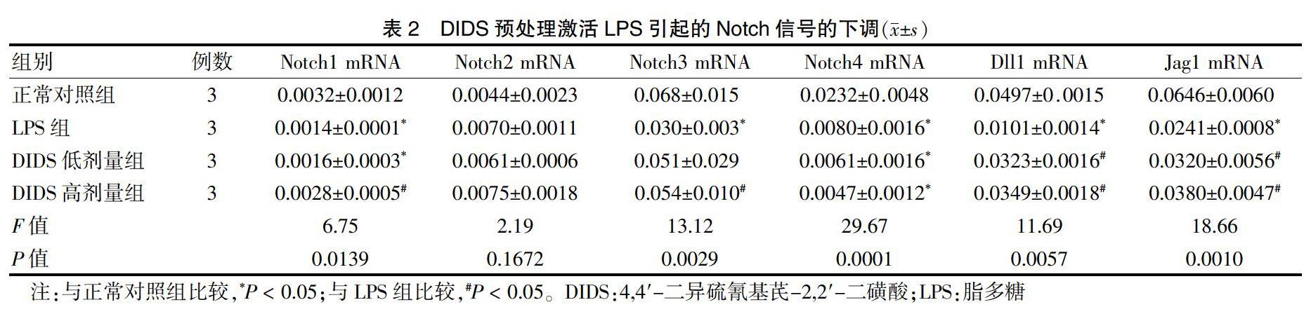
2.3 DIDS对抗LPS诱导的脓毒症小鼠心脏组织中Notch信号相关分子转录水平的降低
与空白对照组比较,LPS组脓毒症小鼠心脏组织Notch信号通路受体Notch1、Notch3、Notch4及配体Dll1、Jag1的mRNA显著降低,差异均有统计学意义(均P < 0.05)。与LPS组比较,DIDS高剂量组脓毒症小鼠心脏组织Notch信号通路受体Notch1、Notch3及配体Dll1及Jag1的mRNA显著升高,差异均有统计学意义(均P < 0.05)。见表2。
2.4 DIDS对抗LPS诱导的脓毒症小鼠心脏组织中Notch信号相关分子蛋白水平的降低
与空白对照组比较,LPS组Notch信号通路配体Dll1及Jag1显著降低,差异均有统计学意义(P < 0.05)。与LPS组比较,DIDS低剂量组、DIDS高剂量组脓毒症小鼠心脏组织Notch信号通路配体Dll1及Jag1显著升高,差异均有统计学意义(均P < 0.05)。见表2、图2。
注:与正常对照组比较,*P < 0.05;与LPS组比较,#P < 0.05。DIDS:4,4-二異硫氰基芪-2,2-二磺酸;LPS:脂多糖
LPS:脂多糖;DIDS:4,4′-二异硫氰基芪-2,2′-二磺酸
3 讨论
细胞因子被认为在脓毒症心肌损伤发病中具有重要作用。特别是TNF-α、IL-1β和IL-6等均参与了脓毒症心肌损伤的发病过程,在实验条件下,抑制这些细胞因子均可以有效缓解病程[19-20]。本研究发现,DIDS能够提高脓毒症小鼠存活率,可以抑制脓毒症小鼠心肌组织中促炎因子TNF-α、IL-1β、IL-6的mRNA升高,提示DIDS具有抗炎作用。
近年来,关于Notch信号转导通路在炎症中作用的研究日益受到关注。哺乳动物表达4个Notch受体(Notch1~4)及5个配体(Jag1、Jag2、Dll1、Dll3、Dll4)。研究发现,抑制Notch信号通路可以负调控多种促炎因子的表达[21],其机制可能与Notch可以与IL-6启动子区域结合,从而直接调控IL-6的表达有关[22]。本研究发现,DIDS高剂量组可以逆转脓毒症时心肌中Notch1、Notch3、Dll1及Jag1的mRNA和蛋白水平的降低,提示DIDS可能通过激活Notch信号通路,提高Notch受体1/3及配体Dll1、Jag1转录和翻译水平的表达,发挥抗炎作用,保护心脏。
综上所述,DIDS预处理可通过激活Notch信号通路,抑制LPS引起的小鼠心肌中促炎细胞因子的生成,从而减轻小鼠心脏炎性反应。
[参考文献]
[1] Singer M,Deutschman CS,Seymour CW,et al. The third international consensus definitions for sepsis and septic shock(sepsis-3) [J]. JAMA,2016,315(8):801-810.
[2] Vieillard-Baron A,Caille V,Charron C,et al. Actual incidence of global left ventricular hypokinesia in adult septic shock [J]. Crit Care Med,2008,36(6):1701-1706.
[3] Merx MW,Weber C. Sepsis and the heart [J]. Circulation,2007,116(7):793-802.
[4] Boyd JH,Mathur S,Wang Y,et al. Toll-like receptor stimulation in cardiornyoctes decreases contractility and initiates an NF-kappa B dependent inflammatory response [J]. Cardiovasc Res,2006,72(3):384-393.
[5] Avlas O,Fallach R,Shainberg A,et al. Toll-like receptor 4 stimulation initiates an inflammatory response that decreases cardiomyocytecontractility [J]. Antioxid Redox Signal,2011,15(7):1895-1909.
[6] Andrades M,Ritter C,de Oliveira MR,et al. Antioxidant treatment reverses organ failure in rat model of sepsis:role of antioxidant enzymes imbalance,neutrophil infiltration,and oxidative stress [J]. J Surg Res,2011,167(2):e307-e313.
[7] Dare AJ,Phillips AR,Hickey AJ,et al. A systematic review of experimental treatments for mitochondrial dysfunction in sepsis and multiple organ dysfunction syndrome [J]. Free Radic Biol Med,2009,47(11):1517-1525.
[8] Chen J,Wang B,Lai J,et al. Trimetazidine attenuates cardiac dysfunction in endotoxemia and sepsis by promoting neutrophil migration [J]. Front Immunol,2018,9:2015.
[9] Bray SJ. Notch signalling:a simple pathway becomes complex,Nature reviews [J]. Nat Rev Mol Cell Biol,2006,7(9):678-689.
[10] Park JS,Kim SH,Kim K,et al. Inhibition of Notch signalling ameliorates experimental inflammatory arthritis [J]. Ann Rheum Dis,2015,74(1):267-274.
[11] Zhang W,Xu W,Xiong S. Blockade of Notch1 signaling alleviates murine lupus via blunting macrophage activation and M2b polarization [J]. J Immunol,2010,184(11):6465-6478.
[12] Ito T,Allen RM,Carson WF 4th,et al. The critical role of Notch ligand Delta-like 1 in the pathogenesis of influenza A virus(H1N1)infection [J]. PLoS Pathog,2011, 7(11):e1002341.
[13] Kerstjens-Frederikse WS,van de Laar IM,Vos YJ,et al. Cardiovascular malformations caused by NOTCH1 mutations do not keep left:data on 428 probands with left-sided CHD and their families [J]. Genet Med,2016,18(9):914-923.
[14] Southgate L,Sukalo M,Karountzos ASV,et al. Haploinsufficiency of the NOTCH1 receptor as a cause of adams-oliver syndrome with variable cardiac anomalies [J]. Circ Cardiovasc Genet,2015,8(4):572-581.
[15] Meester JAN,Verstraeten A,Alaerts M,et al. Overlapping but distinct roles for NOTCH receptors in human cardiovascular disease [J]. Clin Genet,2019,95(1):85-94.
[16] Pamenter ME,Perkins GA,Gu XQ,et al. DIDS(4,4-diisothiocyanatostilbenedisulphonic acid)induces apoptotic cell death in a hippocampal neuronal cell line and is not neuroprotective against ischemic stress [J]. PLoS One,2013,8(4):e60804.
[17] Yao H,Felfly H,Wang J,et al. DIDS protects against neuronal injury by blocking Toll-like receptor 2 activated-mechanisms [J]. J Neurochem,2009,108(3):835-846.
[18] Xiang NL,Liu J,Liao YJ,et al. Abrogating ClC-3 inhibits LPS-induced inflammation via blocking the TLR4/NF-kappa B pathway [J]. Sci Rep,2016,6:27583.
[19] Maass DL,Hybki DP,White J,et al. The time course of cardiac NF-kappa B activation and TNF-alpha secretion by cardiac myocytes after burn injury:Contribution to burn-related cardiac contractile dysfunction [J]. Shock,2002,17(4):293-299.
[20] Maass DL,White J,Horton JW. IL-1 beta and IL-6 act synergistically with TNF-alpha to alter cardiac contractile function after burn trauma [J]. Shock,2002,18(4):360-366.
[21] Palaga T,Buranaruk C,Rengpipat S,et al. Notch signaling is activated by TLR stimulation and regulates macrophage functions [J]. Eur J Immunol,2008,38(1):174-183.
[22] Wongchana W,Palaga T. Direct regulation of interleukin-6 expression by Notch signaling in macrophages [J]. Cell Mol Immunol,2012,9(2):155-162.
(收稿日期:2019-09-05 本文编辑:顾家毓)
[基金项目] 江苏省镇江市重点研发计划--社会发展项目(SH2017042、SH2017025、SH2018027);江苏省青年医学重点人才培养项目(QNRC2016828)。
[作者简介] 王艳(1982-),女,碩士,副主任药师,主要从事药学及临床药学工作。
[通讯作者] 熊御云(1983-),女,博士,主要从事临床检验工作。