The appliances and prospects of aurum nanomaterials in biodiagnostics, imaging, drug delivery and combination therapy
2019-09-12DnYngFeiyngDengDechunLiuBoHeBingHeXingTngQingZhng
DnYng,FeiyngDeng,DechunLiu,BoHe,BingHe,XingTng,,QingZhng,,c,
a School of Pharmacy, Shenyang Pharmaceutical University, Shenyang 110016, China b Beijing Key Laboratory of Molecular Pharmaceutics and New Drug Delivery Systems, School of Pharmaceutical Sciences, Peking University, Beijing 100191, China c State Key Laboratory of Natural and Biomimetic Drugs, Peking University, Beijing 100191, China
Keywords:Gold nanomaterials Imaging and biodiagnostics Drug delivery Photothermal therapy Immunotherapy Combination therapy
ABSTRACT Aurum nanomaterials (ANM), combining the features of nanotechnology and metal elements, have demonstrated enormous potential and aroused great attention on biomedical applications over the past few decades. Particularly, their advantages, such as controllable particle size, flexible surface modification, higher drug loading, good stability and biocompatibility, especially unique optical properties, promote the development of ANM in biomedical field. In this review, we will discuss the advanced preparation process of ANM and summarize their recent applications as well as their prospects in diagnosis and therapy. Besides, multi-functional ANM-based theranostic nanosystems will be introduced in details,including radiotherapy (RT), photothermal therapy (PTT), photodynamic therapy (PDT), immunotherapy (IT), and so on.
1. Introduction
Gold nanomaterials, formed by aurum element, had been applied to biomedicine based on their unique physico-chemical properties since their first colloidal syntheses [1] . As a member of inorganic materials, the advantages of ANM promote their widespread use in biomedicine [2-7] : 1) Easy to synthesize.Various particle sizes can be achieved via adjusting preparation conditions. 2) Flexible surfaces modification. Gold cores can be modified for specific properties, such as improved stability, drug loading, active targeting, etc. 3) High biocompatibility. The cores are composed of non-toxic inert material and extremely stable. 4) Excellent drug carriers. The loaded components cover a wide range including small molecule chemicals, macromolecular proteins, nucleic acids, antibodies and so on. 5) Controlled drug release. Active substance release can be triggered by site-specific in vivo conditions . 6) Superior optical properties such as surface plasmon resonance (SPR),light scattering and photothermal heating, which can be utilized for tracking, imaging, diagnosis and therapy.
Since the first biomedical application of immungold labeling in 1971 [8] , ANM gradually plays an important role in preclinical studies. Nowadays, ANM is taken as one of the major tools for imaging and diagnostic techniques, such as surfaceenhanced Raman scattering (SERS) [9] , two-photon luminescence imaging (TPLI) [10] , X-ray computed tomography (CT)[11] and photoacoustic (PA) imaging [12] . The properties of gold promise the application of ANM. First, the strong SPR effect enhances optical signals, increases its laser penetration into tissues and also can be readily transformed into heat or acoustic waves. Second, ANM can be observed directly with electron microscope and treated as excellent contrast reagents due to its high electron density. Besides, similar to common nanomaterials, ANM is suitable as a carrier for drug delivery in small molecule chemical drugs as well as macromolecule protein,antibody and nucleic acid. Notably, with the two advantages of ANM, the combined treatment and theranostics develop rapidly now, including photothermal therapy, photodynamic therapy and immunotherapy. For example, Tian et al. reported the interventional treatment with gold nanoshells or clinical iodine-125 interstitial brachytherapy (IBT-125-I) against pancreatic cancer. After functional carriers were delivered to the tumor site via a special needle under ultrasonic guidance,gold nanoshells-based photothermal therapy as well as subsequent immune response showed a 25% higher survival rate of rats than IBT-125-I. Meanwhile, CT and optical imaging based on gold provided convenience in treatment [13] .
In clinic, safety and biocompatibility of drug systems are the issues of most concern, the same also applies to ANM.Currently, there are six ANM clinical trials [14] . AuroLase®,consisting of PEGylated gold-silica nanoshell, is under phase Ⅰtrial for thermal ablation of metastatic lung tumors, in which gold shell provides the thermal ablation based on their strong absorption of near-infrared light and PEG maintains particle stability and biocompatibility [15,16] . Throughout the research process, the better data indicates the enormous potential for clinical transformation.
Overall, ANM is one promising nanoplatform in both preclinical and clinical application. This review will provide insights into synthesis, preparation of gold drug delivery system, the application in biomedicine as well as combination therapy on the multi-function of ANM, and the further development in future.
2. Preparation of aurum nano drug delivery systems
ANM, with one-dimensional diameter ranging from a few to one hundred of nanometers, are readily prepared for desired core-shell structure shapes, such as nanospheres, nanorods,nanocages, nanostars, nanoshells, nanocubes and nanobowls,to achieve unique physical and chemical properties [6] . Among those, preparation of nanospheres and nanorods are mostly reported and will be further introduced as follows.
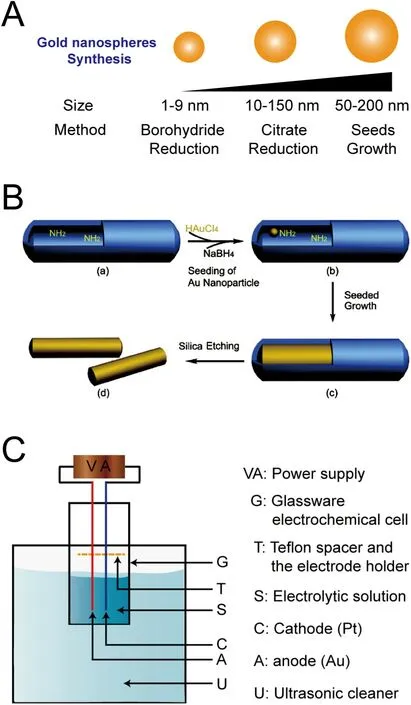
Fig. 1 -The synthesis methods of ANM cores. (A) The particle size distribution of gold nanospheres obtained through different approaches; construction of gold nanorods core with (B) template growth (Reproduced with permission from [33] ) Copyright 2011 American Chemical Society and (C) electrochemical process (Reproduced with permission from [36] ). Copyright 1999 American Chemical Society.
2.1. Synthesis of aurum core
The gold core provides the basic properties of metallic element and determines the genres of ANM based on its shape[17] . The methods of core synthesis vary from different ANM[18] . For gold nanospheres, reducing gold salt is the main synthetic approach [19] and particle sizes are controlled by regulating the ratio of reducing agent to gold ion. As shown in Fig. 1 A, Turkevich's citrate-mediated synthetic method (Citrate Reduction) [20,21] , is the most commonly used synthesis process and could produce monodisperse gold nanospheres with 10-150 nm diameter range. In which, citrate salt solution is always added into the boiled chloroaurate solution and the gold cores could be obtained under continuous stirring.Higher proportion of citrate salt leads to smaller particle size[22] , and the morphology of nanoparticles is also affected by the speed of mixing two aqueous solutions, the concentration of ions, temperature as well as pH value of reaction solution[23,24] . For preparation of gold nanoparticles in very small particle size (1-9 nm), the reaction of borohydride participation(Borohydride Reduction), also called Brust-Schiffrin method,is more suitable [25,26] . The reaction of core-forming is usually carried out in aqueous environment without heating due to the strong reducibility of borohydride [27] . Besides, a twostep protocol, spontaneous nucleation and isotropic growth(Seeds Growth), has addressed the need for more stable and larger gold nanoparticles (50-200 nm) by increasing Au deposition further on a small Au nucleus [28] . This process provides a novel approach to obtain stable, regular and uniform gold nanospheres with larger particle size, which are difficult to achieve with the first two methods [29] . These three kinds of core synthesis methods show investigators a wide range of selectivity according to the specific requirements during the construction of the round-shaped ANM.
Gold nanorods, which can be fabricated easily and diversely, are another important ANM due to their unique light properties in photothermal application. Seed-mediated growth, template growth as well as electrochemical process are the main synthesis methods, among which seed-mediated growth is especially reused on account of its long history and universal applicability [30,31] . Similar to the seeds growth in gold nanospheres preparation, the seed-mediated growth also starts with a small gold particle but continues to react in the solution containing surfactant, silver nitrate (AgNO 3 ), and cetyltrimethylammonium bromide (CTAB). The aspect ratio of nanorods is affected by the concentration of AgNO3, the diameters of the seeds, pH, etc. [32] . Template growth ( Fig. 1 B), also used in many other nanostructures preparations, is accomplished via Au deposition on the templates (porous aluminum,polycarbonate) in certain conditions. After selective dissolution of template, some polymeric stabilizers are added [33] and the gold nanorods can be dispersed in water or organic solvents with the help of external force (e.g. sonication, agitation)[34] . It is obvious that particle sizes of gold nanorods correspond to the pore diameters of the templates [35] . As for the electrochemical process ( Fig. 1 C) firstly established by Wang et al. [36] , they utilized two-electrodetype electrochemical cell with gold as anode while platinum as cathode to produce gold nanorods via electrolysis in a mixed solution containing CTAB,acetone and cyclohexane as well as other substances, in which CTAB also acted as stabilizer apart from one composition of the electrolyte. These three approaches, with other methods as complements [37] , can make up and cooperate with each other for synthesis of almost all types of gold nanorods.
2.2. Modification of functional shell
The gold nano core synthesized according to the methods mentioned above can keep good stability when stored at low temperature for several months, but they will aggregate when the weak protective layer (for example: citric acid, boride) is destroyed by external factor, such as high ionic strength solution, centrifugation, freeze and so on. In general, gold core only offers fundamental properties of gold nano-system while the satisfactory stability, solubility, biocompatibility and drug loading are determined by protective shells [19,31] .
2.2.1. Protective shell Although substances (e.g. CTAB, citric acid, hydroquinone)used in preparation of ANM contribute to system stability,poorly protective effect or toxicity (e.g. CTAB) to cells remain non-negligible [38,39] . To improve stability and biocompatibility, functional linkers have been designed for conjugation with gold core, which are based on groups like amine, carboxylate, thiolate, dithiocarbamate, selenide and phosphine to form Au-N, Au-COO-, Au-S, etc. [40,41] . It has been proved that Au-S bond is more stable and more easily formed (always only mixed at room temperature) than Au-N, Au-COO-or other bonds and it is considered the most popular and effective method [42] . To obtain these conjugates, thiolated poly (ethylene glycol), namely PEG-SH, is frequently employed as linkers in surface modification owing to its characteristics of hydrophilicity, neutral in charge and good biocompatibility. Besides, PEG overcomes the dilemma like nonspecific surface binding with biological molecules via electrostatic interactions and excess uptake by reticular endothelial system(RES) [43-45] . Meanwhile, Liu and Thierry found that facile surface modification did not affect the basic properties of gold nanoparticles except a slight increase of particle size [29] .These indicate PEG is an optimal candidate as protective layer.Notably, various stabilizers always demonstrate more advantages apart from stability. For example, protein (e.g. bovine serum albumin [46] ), surfactant (e.g. Tween [47] , polymerizable surfactant [48] ) and inorganic shell (e.g. silica [49] ) are applied in improving the water dispersity of ANM. Introduction of different charge stabilizers provides a controlled dispersion state and size distribution of gold nanoparticles via adjusting the ratio of the positively and negatively charged ligands [4] .Besides, Tween is confirmed to improve DNA modification [50] ,etc.
2.2.2. Drug loading shell Some drugs can form shells around in ANM and these will offer not only protective layer, but also a therapeutic effect.The drugs ( Fig. 2 ) are generally conjugated on cores in the following three ways: 1) by covalent bond directly; 2) by covalent bond with an intermediate linker; 3) by electrostatic binding,hydrophobic interaction, etc.
The formation of Au-S bond is the most popular and easiest method for drug loading in ANM. The active substance (chemical drugs, peptide, protein, etc.) containing or endowed with thiol group via modification can be conjugated on the surface of gold nanoparticles directly [51] . For instance, Prades et al.used cysteine to form Au-S bond with gold core, and modified the nanoparticles with the active peptide which has high affinity to transferrin receptor. Due to the enhanced bloodbrain barrier penetration ability of the metal carrier as well as active targeting properties, this system increased cerebric cellular uptake and reduced β-amyloid in Alzheimer's disease[52] . Li and co-workers completed a facile and effective protocol to attach thiolated DNA on gold core without the tedious salt addition process [47] . Sundaram et al. also constructed a co-delivered gold carrier by adding the thiol group at DNA 5'end and encapsulating neomycin inside the DNA, which realized controlled release by responding to temperature and affinity [53] .
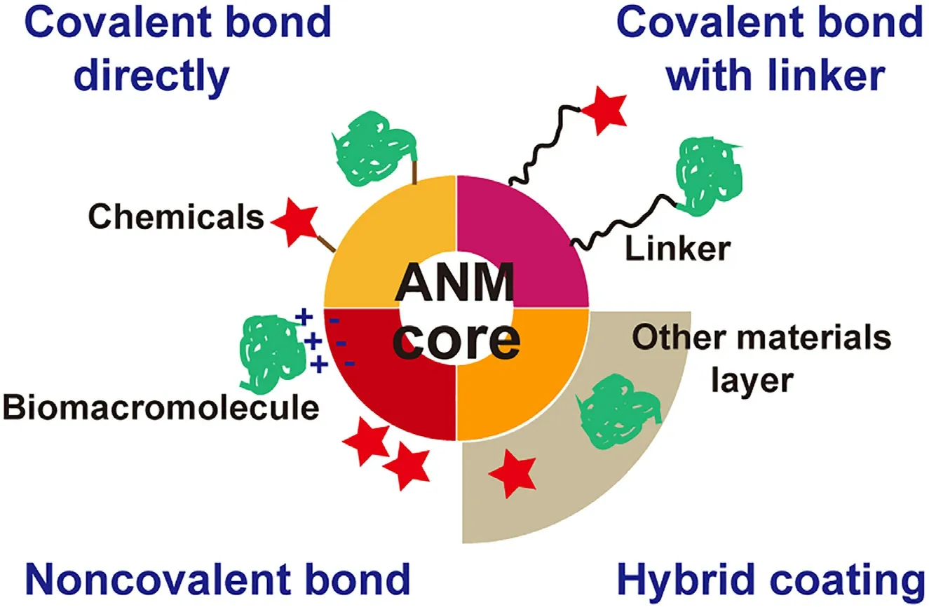
Fig. 2 -Schematic diagram of preparation of drug-loaded ANM platforms. Biomacromolecule includes peptides, proteins,lipids, carbohydrates, nucleic acids etc.
Apart from covalent bond, drugs can be also conjugated to gold core with by an intermediate linker, which holds two functional groups to connect with gold and active agents, respectively. Amino and carboxyl ends are two main groups for linking chemicals [54,55] , fluorescent dyes [56] , proteins[57] , ligands [5,58] etc. For instance, thiol-terminal PEG mixture (different molecular weight) was attached by protein on the carboxyl group at the other end and modified protein drugs efficiently and conveniently [29] . Similarly, paclitaxel was covalently linked to gold nanoparticles by DNA (aminoterminal) fragments and demonstrated 50-fold increase in solubility compared with bulk drug [59] .
Besides, drugs can also be physically adsorbed on the surface of gold cores to achieve rapid and effective drug release profile in vivo [60,61] . For example, Thomas and Klibanov combined the negative charge DNA with modified polyethylenimine (PEI) on the surface of gold nanoparticles and transfected monkey kidney (COS-7) cells. The result showed that the transfection efficacy was 12 times higher than using PEI vector alone [62] . Based on the hydrophobic interactions, Yu et al. loaded photodynamic therapy (PDT) drugs on the gold nanoparticles via noncovalent interaction and then coated PEG chain in the outermost layer, which ensured fast and complete drug release [63] . Antonio et al. prepared a multi-functional gold nanoparticles containing lactose, human galectin-3 and methotrexate (MTX) via hydrophobic interaction and steric hindrance [64] . Wang et al. also constructed a novel vaccine approach utilizing gold nanoparticles, which loaded recombinant trimetric A/Aichi/2/68 (H3N2),hemagglutinin (HA) and TLR5 agonist flagellin (FliC) with click chemistry and metal-chelating reactions [65] . Besides,new composite delivery systems were also developed with gold core entrapped in other materials, which provided the space for drugs and achieved a better biocompatibility, such as gold core-liposome system [66,67] , gold core-micelle system [68] , gold core-silica nanoparticles [69] , gold core-chitosan nanoparticles [70] , and so on.
3. Appliances of aurum based nano drug delivery systems
ANM can be constructed into an excellent platform for imaging, diagnosis and therapy on account of their changeable shape, tunable size, unique surface properties as well as good biocompatibility [71] . Their characteristics, like optical absorption of particular wavelength of light, abundant surface electrons, strong electron density and special appearance color,provide privilege for ANM as agents for imaging and diagnosis detection ( Fig. 3 ) [72] , while flexible modification, large surface area, good stability and low toxicity demonstrate feasibility for drug delivery [73] .
3.1. Imaging and diagnosis
3.1.1. Raman spectroscopy
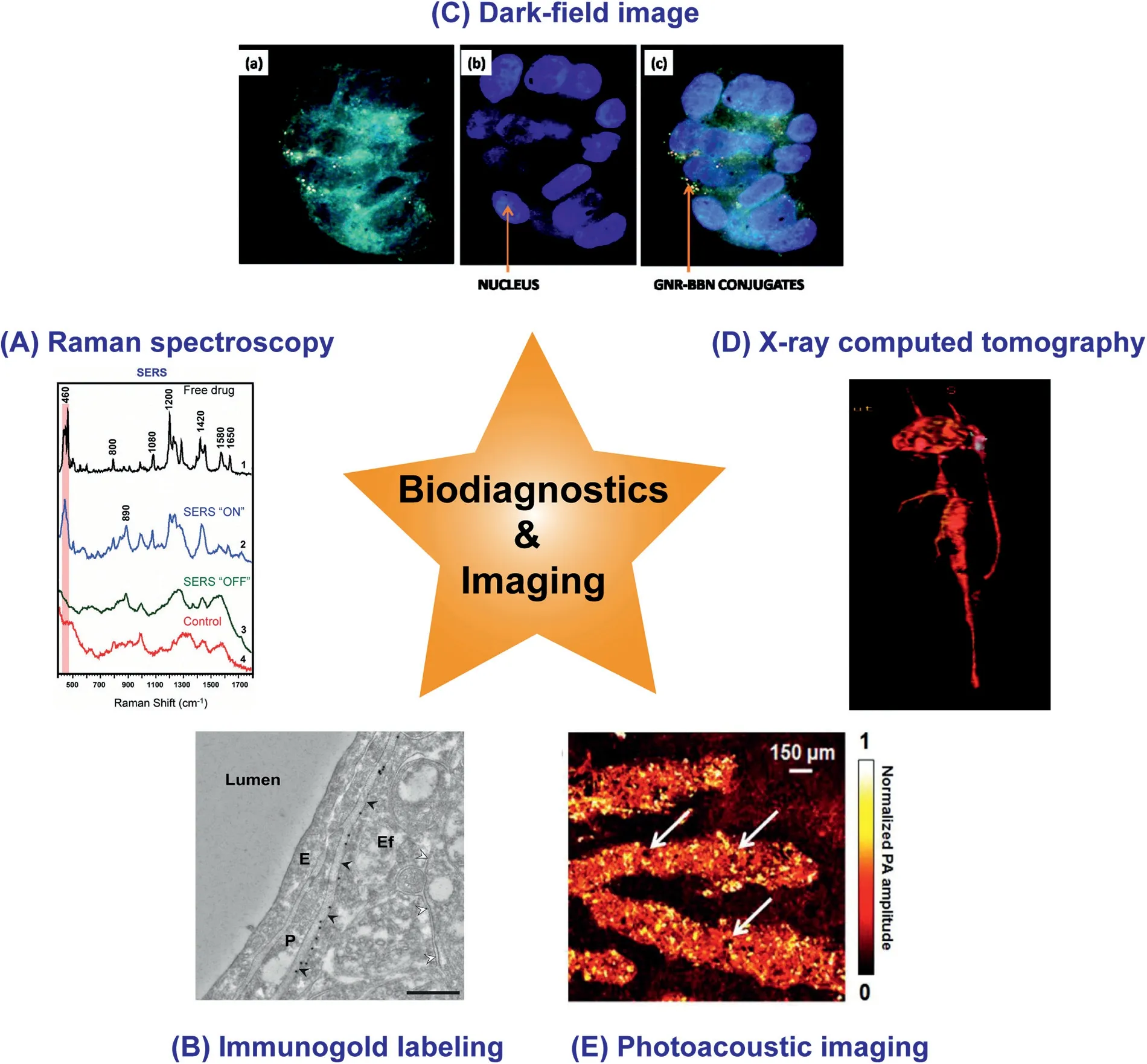
Fig. 3 -Applications of ANM in imaging and biodiagnostics. (A) Surface-enhanced Raman spectra, pink bond indicated the main viewing areas (Reproduced with permission from [75] ) Copyright 2013 American Chemical Society; (B) Immunogold labeling of perivascular astrocytic endfeet. The black dots were gold nanoparticles: E: endothelium, P: pericyte, Ef: endfoot process (Reproduced with permission from [82] ) Copyright 2017 Elsevier; (C) Dark-field image of breast tumor cells(Reproduced with permission from [86] ) Copyright 2009 American Chemical Society; (D) Three-dimensional in vivo CT angiogram image of the heart obtained after injection of PEG-coated GNPs (Reproduced with permission from [97] )Copyright 2007 American Chemical Society; (E) Photoacoustic images of latent fingerprints. Arrows indicated the pores(Reproduced with permission from [12] ) Copyright 2015 American Chemical Society. (For interpretation of the references to colour in this figure legend, the reader is referred to the web version of this article.)
Raman spectroscopy, discovered by Raman in 1928 as a supplement to the infrared spectroscopy, is a powerful tool to study molecular structure but always shows weak signals [6] .Plasmonic nanoparticles (completely made of or covered with plasmonic elements) can enhance Raman signs and thus are called surface-enhanced Raman scattering (SERS) [74] .ANM, as a main candidate for SERS due to its unique optical properties, has been applied in physical, chemical, biological analysis in the research of biomedical science. For instance,to improve the specificity and sensitivity of tumor diagnosis,a monoclonal anti-epidermal growth factor receptor (anti-EGFR) antibody was conjugated on the SERS-characterized gold nanorods, which showed higher-level Raman signals in cancer cells than normal ones [9] . Kang and co-workers designed a real-time doxorubicin (DOX) tracking system based on plasmonic-tunable Raman/fluorescence imaging spectroscopy ( Fig. 3 A). In brief, DOX (drug and fluorophore)and gold nanoparticles were conjugated by pH-sensitive hydrazine, after internalized into acidic endosomes, the hydrazone linker would be broken and DOX was released from gold particles. In this case, DOX got rid of quenching effect from gold nanoparticles and its fluorescence could be detected,while the Raman enhancement was weakened. Conversely,fluorescence disappeared while enhanced Raman spectrum could be detected when DOX was conjugated. This technique provided a viable and convenient idea to monitor the drug delivery dynamics in living cells through observing the change between fluorescence and Raman spectrum [75] .
3.1.2. Visual observation
The metallic nature (high optical density) of gold enables ANM to be observed by naked eyes with colorful appearance according to their shapes and sizes [76] . As the color of ANM would change when gold nanoparticles aggregates, it could be used for rapid test applications as lateral-flow immunoassays [77] .Lee et al. designed an intelligent ANM system based on color change along with distance-dependent optical properties. The original gold nanoparticles were purple in aggregation status in Hg2+ surroundings and turned red when the particles turned mono-dispersed when cysteine competed with Hg2+ .The color transformation could be observed naked eyes or an UV-vis spectrometer [78] . At present, the colorimetric assay has been proved a fast, highly selective and sensitive method applied in many researches [79] .
Immunogold staining technology is another useful tool in samples detection which was set up by Faulk and Taylor in 1971 and has been widely applied [8,80] . Orlov and co-workers conjugated antibodies of labeled RNA polymerase II to 0.8 nm gold nanoparticles. After endocytosis, this ANM could be targeted to the corresponding enzymes and detected by scanning electron microscopy (SEM), which provided a method for labeling cellular protein in situ [81] . Besides, the coupling technique of immunogold labeling and automatic gold detection software ( Fig. 3 B) [82] , serial multiplex immunogold labeling[83] as well as various labeling-detection methods promoted application of immunogold labeling into mature tools for tracing or detecting the distribution of samples.
3.1.3. Microscope detection
ANM show strong light scattering at their plasmonic wavelengths, which has been used for imaging and diagnostic with microscope. It was reported that gold nanoparticles with different shapes and particle sizes could be easily observed in a high contrast with common substance (e.g. cells, plasma) with a simple microscope at dark field condition [84,85] . Chanda et al. prepared a gastrin releasing protein receptor specific bombesin (BBN) peptide linked gold nanorods ( Fig. 3 C). For tracking nanocarriers internalized by receptor-specific pathway, dark-field images as well as TEM technology were used,based on the strong scattering light and high density of gold nanorods, respectively [86] .
Two-photon luminescence imaging (TPLI), which can not only enhance the laser penetration with longer wavelength,but also obtain a clear and remarkable sign with little damage to samples, has been used in vitro and in vivo in recent years.Gold nanorods, as well as other ANM, demonstrated outstanding ability for this technology owing to its near-infrared absorption wavelength and producing strong TPLI intensities [10,87] . Loumaigne and co-workers investigated the TPLI property of spherical gold particles (such as Brownian rotation) at single-particle aspect and they found that the average anisotropy could be influenced by either polarization or excitation wavelength [88] . Li and Gu used gold nanorods to conjugate with transferrin as a functional carrier to overcome tumors. They thought gold nanorods with high efficacy of TPLI were more effective and accurate in diagnosis than common fluorescein isothiocyanate. This finding could enhance microsurgery and a regulative apoptosis of cancer cells by adjusting laser energy [89] .
Different from TPLI, the images of confocal reflectance microscopy (CRM) are obtained with a lower power of light source so that photothermal effect could be avoided. Thus, CRM is another promising alternative to the detection of photo-signal excited by laser and widely used in studying the distribution of intracellular ANM [90,91] . In contrast to conventional fluorophores, the gold nanoparticles can be detected easily and the signals will not be quenched and disappeared, leading to more convenient and accurate without false-positive fluorescent leak. Yuan et al. set up a facile method via confocal laser scanning microscope (CLSM) to observe gold nanoparticles without fluorescence modification through fluorescent channels. When nanoparticles were excited by 633 nm laser,a sharp, stable reflected signal was obtained, which existed in real-time imaging and could be applied in co-localization or further quantitative analysis [92] .
3.1.4. Optical coherence tomography (OCT) and X-ray computed tomography (CT)
Optical coherence tomography (OCT) is a potential optical imaging technology based on light scattering and could be utilized for obtaining real-time and three-dimensional images with high resolution in situ [93] . In Yali Jia's research,it was proved that gold nanorods was a novel contrast agent for OCT imaging by methodology detection [94] . Prabhulkar and co-workers prepared gold nanorods conjugated with antiglucose transporter-1 antibodies as an indicating agent to evaluate ocular surface squamous neoplasia by OCT imaging and immunofluorescence in molecular histopathology [95] .
In X-ray computed tomography (CT), ANM is regarded as proper CT contrast agents for the large atomic weight to overcome the limits in clinical diagnosis like mild nephrotoxicity, short working time and weak contrast [11,96] . Kim and co-workers compared the CT imaging efficacy between gold nanoparticles (30 nm, PEG coating) and a clinical contrast agent, Ultravist. As result, gold nanoparticles showed 5.3-fold higher X-ray absorption coefficient in vitro and 24-fold longer of blood circulation time in vivo ( Fig. 3 D) [97] . Similar observation was reported in Park's comparison between hollow gold nanoparticles and Ultravist 300, which revealed that hollow gold nanoparticles had an equivalent HU value at the concentration less than one-fifth of Ultravist 300 [98] .
3.1.5. Photoacoustic (PA) imaging
Photoacoustic (PA) utilizes the acoustic waves induced by the thermal expansion of surrounding air from ANM rather than direct heat (e.g. photothermal effect) [99] . The ultrasound also deepen tissue penetration ability [100] . Song et al. utilized functional polymer modified gold nanoparticles to rapidly detect the latent fingerprints (LFP) with the co-action of PA and colorimetric visualization. This one-step strategy did not require silver staining to enhance signal and considered as a potential technique to identify chemicals of LFP residues, due to the high affinity with universal secretions and flexibility for different people ( Fig. 3 E) [12] . Yeager and coworkers constructed silica-coated gold nanorods as photoacoustic contrast agents for detection of temperature change of atherosclerotic plaques, which were also induced by the selective heating of plasmonic gold nanoparticles, suggesting that gold system was not only a diagnosis method but also a contrast agent according to their multiplicity [101] .
3.2. Application in therapy
Apart from imaging and diagnosis, ANM has also been widely applied to biomedical therapeutics because of the flexibility for various active agents, high drug loading and good biocompatibility. Different types of drugs, such as chemicals, peptides, proteins, lipids, carbohydrates or nucleic acid [102,103] are conjugated to ANM for various diseases treatments, including but not limited to anti-cancer,anti-viral, anti-microbial, anti-inflammatory therapeutic effect, which depend on the intrinsic activity of the drugs[73,104-106] .
Small molecule drugs, e.g. DOX [107] , paclitaxel [108] or other chemical drugs, which are most widely used in clinical and laboratory settings, could be easily assembled into the ANM as the protocol described in Fig. 2 and exhibit promising therapeutic efficacy. For example, ANM shows great potential in antibacterial drug delivery against pathogens, even to overcome resistant bacteria [109] . Li and co-workers prepared various tunable functional ANM with antimicrobial drugs and targeted ligands, which demonstrated effective against both Gram-negative and Gram-positive uropathogens, providing a novel antimicrobial strategy [110] . ANM are also fit for biomolecules, such as proteins and nucleic acid [111] . At present, a number of researches have reported gold nanoparticles as a common vector for gene effective delivery for repairing defective genes, regulating protein expression level,administrating therapeutically relevant processes owing to their high DNA loading ability, suitable biocompatibility. Generally, targeting nucleic acid is anchored onto ANM via covalent bonding and electrostatic absorption for its functional group and strong negative charge, respectively [112] . For instance, polyethyleneimine-g-bovine serum albumin (PEI-BSA)was synthesized as a non-viral gene vector and attached to ANM. After desired gene was adsorbed on the outermost of PEI-BSA-ANM via electrostatic interaction, the complex achieved high transfection efficacy [113] . As for protein delivery, a typical case was reported that ANM delivered silk fibroin(SF) delivery while SF also maintained the stability of ANM after lyophilization [114] .
3.2.1. Controlled release
Drug release profile is an important issue and requires careful consideration in drug delivery system design. By loaded onto ANM in different ways, drugs could release under the influence of endogenous or external factors, such as glutathione, pH, enzymes, temperature and light [115] .Glutathione-mediated release may serve as a strategy triggered by the large differences between intracellular glutathione (GSH) concentration (2-17 mM) [116] . Specifically, the drugs are separated from carriers through GSH reduction or exchange with the endogenous thiol substance. In a GSH sensitive system, Ce6, a hydrophobic photosensitizer, was conjugated covalently to surface of gold nanorods via disulfide bond and demonstrated great photothermal effect and singlet oxygen generation in A549 cells after endocytosis [117] .Besides, in some gene delivery systems, siRNA was released via being replaced by GSH on ANM, featuring an overall negative charge and thiol of GSH ( Fig. 4 A) [118] . For preparation of carriers, drugs could conjugate to ANM with ester bond,which might be fractured via hydrolysis or exert function by intracellular enzymes ( Fig. 4 B) [119] . Shieh et al. designed gold nanoparticles linked to paclitaxel via a phosphodiester bond which could be hydrolyzed by the high concentration of phosphodiesterase in tumor cells. In this way, free paclitaxel was successfully accumulated in the cell massively to obtain a strong anti-tumor effect [120] . pH-responsive is another principle for intelligent ANM. With the progress of intracellular endosome transport, the surrounding pH gradually decreases.Meanwhile, the acidic microenvironment of tumor also promoted the targeted release of the drugs in many researches( Fig. 4 C) [121,122] .
Different from drug released regulated by intracellular environment, external factors could also be utilized to induce drug release, such as light [123] , temperature [108,124] , osmotic pressure ( Fig. 5 A) [125] and so on. Based on the characteristic of gold nanoparticles, gold core can be ablated by near infrared irradiation (NIR) [126] . In Lee's study, DOX release depended on the power of NIR laser irradiation and tumor pH[127] . Wijaya et al. also utilized two kinds of gold nanorods as a carrier for delivering gene oligonucleotides. The two lengths of gold nanorods were sensitive to 800 nm and 1100 nm NIR, respectively, which promoted the selective release of gene oligonucleotides by using specific wavelength ( Fig. 5 B)[128] .
4. Theranostics
Theranostics, the superposition of therapy and diagnose, is one of the most promising application of ANM in biomedicine at present. It keeps developing with intense investigation currently and benefits from the fundamental optical, electronic characters and drug-loading properties [129] . According to reports, it has covered an extensive field like radiotherapy (RT),photothermal therapy (PTT), photodynamic therapy (PDT), immunotherapy (IT), etc.
4.1. Radiotherapy
ANM process high X-ray absorption as well as unique optical features and are an effective tool for radiotherapy with imaging function and therapeutic effect. They serve as a radiosensitizer based on three main mechanisms with ionizing radiation: increasing photoelectrons generation (physical); catalyzing the production of free radicals (chemical); promoting the damage via disturbing cell process (biological) [7] . Meanwhile,the key performances can also be summarized as the following five points, called as 5Rs: redistribution, repair, repopulation, reoxygenation and intrinsic radiosensitivity [130] . Zhang and co-workers prepared series of PEG-coated gold nanoparticles with different particle sizes and compared the in vitro and in vivo sensitization effects. All the nanoparticles showed good radioactive efficacy and caused rapid cell necrosis as well as subsequent apoptosis with less toxicity, among which 12.1 nm and 27.3 nm were the optimal choice [131] . With addition of antibody (trastuzumab) against HER-2, higher cytotoxicity was observed in vitro , so was a 46% reduction in tumor volume of MDA-MB-361 in mice compared with non-targeted nanoparticles with X-radiation [132] . Tsourkas et al. constructed goldloaded polymeric micelles, in which 1.9 nm gold nanoparticles and amphiphilic di-block copolymer were synthesized as core and shell, respectively. After intravenous injection, the system delineated the profile of tumor via CT and then exerted radiation therapeutic effect, exhibiting a 1.7-fold enhancement in mice survival time compared with simply radiation [133] .Dong et al. prepared folic acid-modified and DOX-loaded gold nanorods (FA-GSJNs-DOX), and utilized the triple mechanisms (chemoradiotherapy and CT imaging) to overcome the unresectable hepatocellular carcinoma ( Fig. 6 ). As a result, the multifunctional platform demonstrated remarkable synergistic anticancer effects in tumor regression with less systematic toxicity due to the active targeting simultaneously [134] .
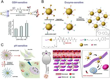
Fig. 4 -Common endogenous factors triggered the drug release from ANM, including (A) GSH (Reproduced with permission from [118] ) Copyright 2008 American Chemical Society, (B) enzyme (Reproduced with permission from [119] ) Copyright 2016 American Chemical Society, (C) pH (Reproduced with permission from [122] ) Copyright 2016 American Chemical Society.
4.2. Photothermal therapy
Photothermal therapy (PTT) is based on the induced heat.When suitable materials are exposed under laser, the phonons of plasmonic nanoparticles transfer by absorption light energy, which also induced temperature increase in nanoparticles as well as surroundings via conduction [135] . With strong light absorption properties, ANM can cause distinct temperature increase; Meanwhile, the high photo-stability and good biocompatibility guarantee ANM as promising hyperthermia agents [136,137] . Shen et al. prepared gold nanorods with siRNA on surface to achieve a synergy for antitumor combining gene silencing and photothermal therapy. The functional carrier provided siRNA endosomal escape for higher silencing efficacy and then promoted the inhibition of cancer cell proliferation further via gold nanorods heating activity [138] . The combination of photothermo-chemotherapy(photothermo-DOX) also showed obvious superiorities over either therapy alone on gold nanoshell [139] . Besides, Kim and co-workers designed a multifunctional carrier, in which a NIR responsive gold nanorods and a drug reservoirs mesoporous silica formed the core and shell of the nanoparticle, respectively, and then a thermoresponsive polymer was coated in the outermost. Temperature and pH controlled drug release.The therapeutic effect was enhanced via multi-modes treatments (chemo and hyperthermia) with precise detection of the X-ray from gold nanorods [140] . Generally speaking, when photosensitive materials are exposed with light, there also exists another phenomenon except temperature increase, which is the generation of reactive oxygen species (ROS) via energy conversion of surrounding oxygen molecules from light [141] .ROS is a byproduct substance in the cell oxidative metabolism,which might accelerate cell death [142] . This is the theory of photodynamic therapy (PDT). Based on the flexible nanoplatform of targeting gold nanorods, Liu et al. designed a mitochondrion-targeted and plasmonic properties from gold,which enhanced PDT therapy effect and reduced systemic toxicity ( Fig. 7 ) [143] . Besides, the optional conversion between PDT and PTT was also an interesting issue [144] and during the process of PTT, a small part of specific immune response also existed, completing by the way of immune cells capturing antigens from dry tumor cells [145] . These provided phototherapeutics of ANM more vigorous development and new insight.
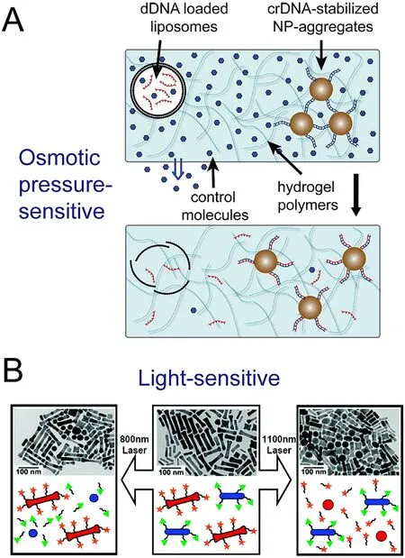
Fig. 5 -Drug release from ANM via regulating by (A) osmotic pressure (Reproduced with permission from [125] ) Copyright 2016 Elsevier; (B) light (Reproduced with permission from [128] ) Copyright 2009 American Chemical Society.
4.3. Immunological therapy
Immunological therapy (IT) is one of the most attractive and energetic strategies for cancer treatment in recent years,which works by activating or adjusting the body's immune response to overcome disease forcefully [146] . ANM are bioinert, easily-synthesized, capable to load antigen or immune adjuvant and can be internalized into immune system to regulate immune response accurately. In IT system, they deliver immune adjuvants, induce immune response, importantly, not only these, their diagnostic imaging property is further contributed them to become a new intelligent theranostics venues. Qu et al. designed an immunomodulatory cytosine-guanosine oligodeoxynucleotides (CpG ODNs) and DOX co-conjugated gold nanorods, which displayed high loading capability for both CpG and DOX and combined three therapy strategies (chemotherapy, hyperthermal therapy and immunotherapy) simultaneously, which showed significant advantage over each one alone ( Fig. 8 ) [147] . Cohen and coworkers reported a platform to track and delivery functional T-cells, on which T-cells could be observed precisely with assistance of gold nanoparticles via dark field microscopy,multiple modes CT scans and 3D volume-rendering images.Meantime, cytokine release in vitro and high tumor inhibition rate in vivo were maintained, indicating no negative effect on T cells [148] .
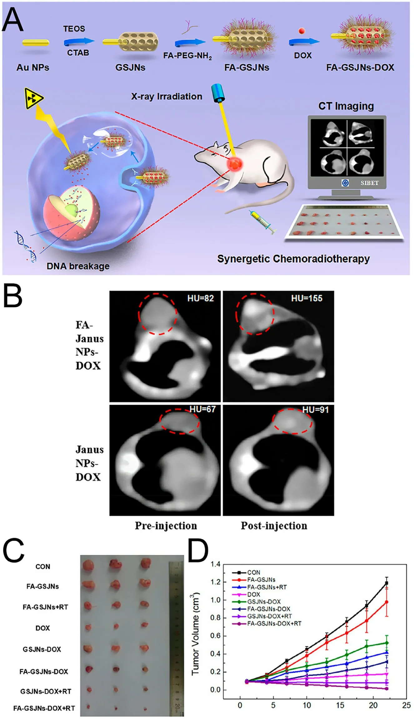
Fig. 6 -Radiotherapy. (A) Schematic illustration of multifunctional ANM for RT based on the combination of CT and chemoradiotherapy; (B) CT imaging of transverse section in SMMC-7721 tumor-bearing nude mice at 24 h post-injection with FA-GSJNs-DOX or GSJNs-DOX; Chemoradiotherapy effect in vivo was showed as (C) tumor photographs (D) tumor volume. (Reproduced with permission from [134] ) Copyright 2017 American Chemical Society.
5. Conclusion and outlook
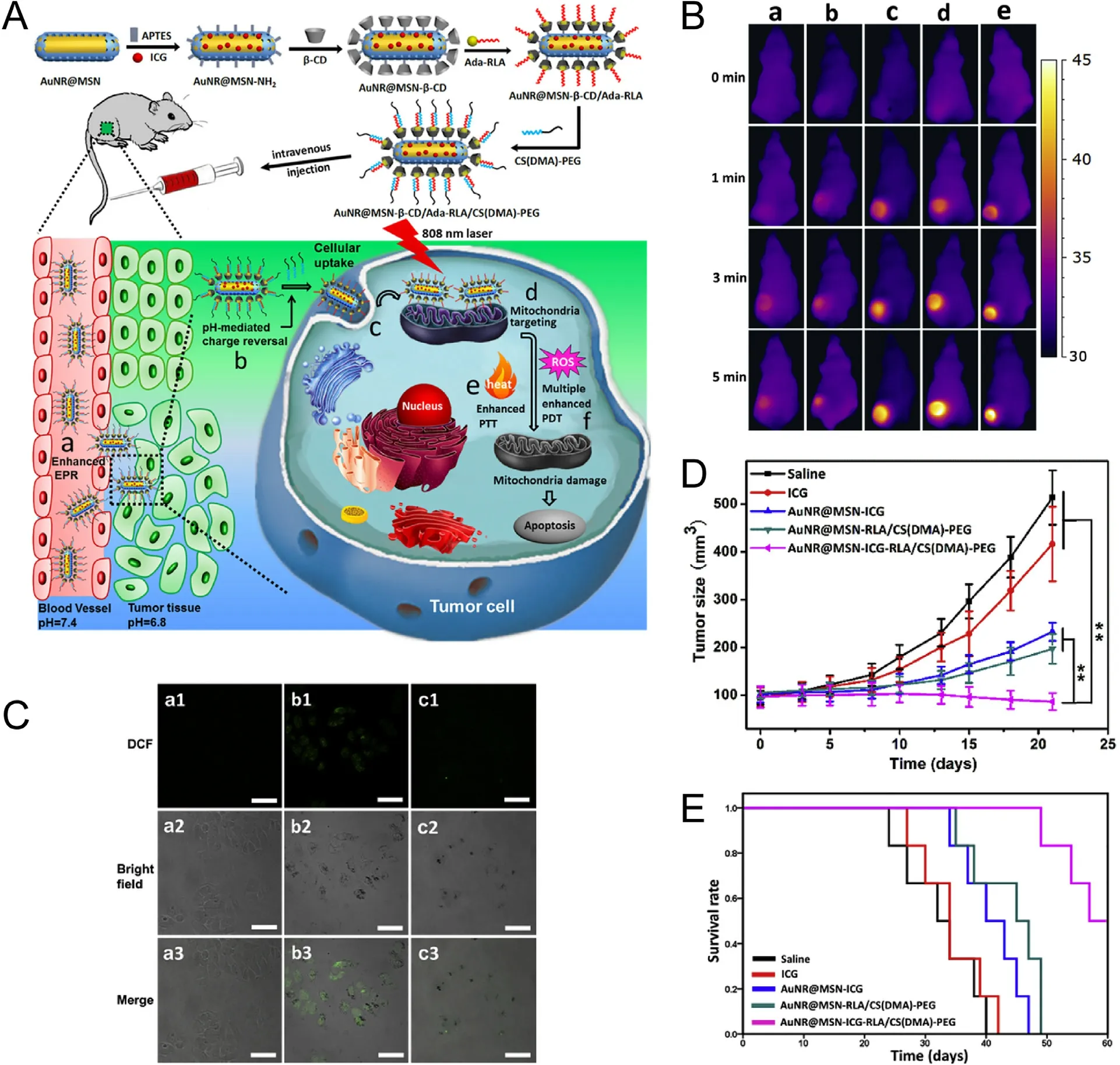
Fig. 7 -Photothermal/photodynamic therapy. (A) Schematic diagram of AuNR@MSN and in vivo work process:a-accumulation in tumor via EPR effect, b-release from shell due to the weak acid environment, c-internalization,D -mitochondrial targeting, e-PTT, f-PDT; (B) images of PTT in tumor bearing mice under 808 nm NIR laser irradiation 24 h after injection, a-saline, b-ICG, c- AuNR@MSN-ICG, D -AuNR@MSN-RLA/CS(DMA)-PEG, e-AuNR@MSN-ICG-RLA/CS(DMA)-PEG;(C) ROS producing detection, a-control, b-AuNR@MSN with irradiation, c-AuNR@MSN without irradiation; (D) Antitumor effect in vivo ; (E) Kaplan-Meier survival curves. (Reproduced with permission from [143] ) Copyright 2017 Elsevier.
A great deal of attention was focused on the ANM to biomedical area and has made palmary achievements. In biological imaging and diagnosis, ANM can well be performed based on their SPR characteristic, high electron density, strong scattering light as well as colorful appearance. In drug delivery, ANM can be considered as a qualified delivery system due to their suitability for diverse drugs, controlled drug release behavior and good biocompatibility. However, as the gold nanosystem always load drugs on their surface, the drug loading efficiency may be somewhat deficient. If sufficient target drug accumulation is to be obtained, a large amount of gold nanoparticles will be necessary, which may increase their toxic side effects on healthy tissues to some extent. Besides, in the metabolism and clearance in vivo , ANM cannot be removed from the body easily as there are poor corresponding enzyme as well as degradation pathway.
Thankfully, due to the development of nanotechnology,more and more gold nanomaterials have been synthesized,such as gold nanocages, gold nanoshells and other systems with large surface areas. At the same time, the hybrid system composed of organic and inorganic materials, including gold-micelle, gold-liposome, are also promising. These ANM are helpful in increasing drug loading and reducing biotoxicity. Specifically, theranostics, utilizing the abilities of multi therapeutic methods in one nanocarrier simultaneously will be going to be the most potential nanopatforms based on integrating the advantages of ANM in the future.In which, multi-strategies of combination therapy and precision treatment with the aid of imaging and biodiagnostics will greatly promote the therapeutic effect and the metabolism also will be improved due to the accelerated cleaning with the help of laser light. In summary, flexible multi-functional theranostics system is the focus of next research on gold nanomaterials.
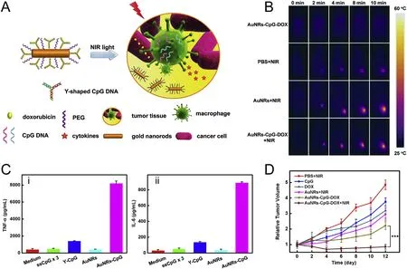
Fig. 8 -Immunological therapy. (A) Schematic illustration of multifunctional nanoplatform with cooperation of gold nanorods, DOX and CpG ODNs; (B) images of PTT in tumor bearing mice under 808 nm NIR laser irradiation(808 nm,1.5 W/cm 2 , 10 min); (C) Cytokine release from RAW264.7 cells; (D) Antitumor effect in vivo . (Reproduced with permission from [147] ) Copyright 2014 Elsevier.
Conflicts of interest
The authors declare that there are no conflicts of interest.
Acknowledgments
Supported by the National Basic Research Program of China( 2015CB932100 ).
杂志排行
Asian Journal of Pharmacentical Sciences的其它文章
- Current developments in drug delivery with thermosensitive liposomes
- The application of nitric oxide delivery in nanoparticle-based tumor targeting drug delivery and treatment
- Delivery of docetaxel using pH-sensitive liposomes based on D - α-tocopheryl poly(2-ethyl-2-oxazoline)succinate: Comparison with PEGylated liposomes
- Amino functionalized chiral mesoporous silica nanoparticles for improved loading and release of poorly water-soluble drug
- Optimizing pH-sensitive and time-dependent polymer formula of colonic pH-responsive pellets to achieve precise drug release
- A hybrid genipin-crosslinked dual-sensitive hydrogel/nanostructured lipid carrier ocular drug delivery platform
