MEBT/MEBO对慢性难愈合创面组织中CK10表达水平的影响
2019-04-09舒清峰陈端凯唐乾利单云龙岑小宁卓臣义
舒清峰 陈端凯 唐乾利 单云龙 唐 强 岑小宁 卓臣义 冯 时
作者单位:533000 广西 百色,右江民族医学院/桂西高发病重点实验室
近年来,大量临床实践及基础研究显示,皮肤再生医疗技术 (moist exposed burn therapy/moist exposed burn ointment,MEBT/MEBO) 在慢性难愈合创面的治疗中取得了卓越的成效[1-3],且具体的作用机制成为了临床研究的热点。如徐荣祥等的研究显示,MEBT/MEBO可激活创面组织内的潜能再生细胞,并将其转化为细胞角蛋白 (cytokeratin,CK)19阳性表达的干细胞,再在原位不断增殖分化为正常表皮细胞,最终实现缺损创面的再上皮化和组织结构重塑[4-8]。随着研究的不断深入,部分研究学者发现,CK10是表皮细胞分化成熟的标记分子之一,其表达水平的差异可体现创面的修复水平[9]。为此,笔者于本研究中动态监测了经不同方法处理后大鼠皮肤及创面组织内CK10表达水平的变化情况,探讨了MEBT/MEBO对表皮干细胞增殖分化的影响,以揭示MEBT/MEBO促进慢性难愈合创面修复的部分作用机制。
1 实验材料
1.1 实验动物
12周龄SPF级健康Wistar雄性大鼠90只[长沙市天勤生物技术有限公司提供,许可证号SCXK (湘) 2014⁃0011],体重 (220 ±20) g,实验过程中严格按照SPF级饲养环境饲养,湿度保持在60% ±10%,室温保持在 (25±2)℃,通风通气良好。本研究经右江民族医学院动物伦理委员会批准。
1.2 主要试剂
湿润烧伤膏 (moist exposed burn ointment,MEBO):汕头市美宝制药有限公司生产;重组牛碱性成纤维细胞生长因子 (recombinant bo⁃vine basic fibroblast growth factor,rb⁃bFGF) 凝胶:珠海亿胜生物制药有限公司生产;水合氯醛:泰兴市豪申化工贸易有限公司生产;醋酸氢化可的松注射液:上海通用药业股份有限公司生产;一抗稀释液、二抗稀释液:上海碧云天生物技术有限公司生产; Anti⁃CK10⁃Antibody:Abcam公司生产;辣根过氧化物酶标记山羊抗兔 IgG (100μL)、 β⁃actin 抗体 (100μL): 北京中杉金桥生物技术有限公司生产;PVDF膜:Millpore公司生产。
2 方法
2.1 实验分组与模型制备
将大鼠置于SPF级动物实验室饲养1周并确定大鼠健康后,随机分为空白组、急创组、慢创组、rb⁃bFGF组与MEBO组,每组18只,均选取脊柱两侧背部皮肤作为实验区域,其中空白组大鼠仅对其背部皮肤进行面积约3.0 cm×3.0 cm的备皮处理;急创组大鼠于7%水合氯醛腹腔注射 (4 mL/kg)麻醉及备皮处理 (方法同空白组)后,使用直径为1.5 cm的圆形印章进行标记,并在无菌环境下延标记线去除皮肤组织至筋膜层,建立急性皮肤组织缺损创面;慢创组、rb⁃bFGF组与 MEBO组大鼠于麻醉、备皮及去除皮肤组织 (方法均同急创组)后,立即注射醋酸氢化可的松注射液建立慢性难愈合创面模型[10]。
2.2 局部处理
模型制备成功后,空白组大鼠备皮处皮肤立即予以5%碘伏常规消毒3次,并依次覆盖生理盐水纱布及清洁敷料,胶带固定,每天换药2次;急创组与慢创组大鼠创面立即予以5%碘伏常规消毒3次,并依次覆盖生理盐水纱布及清洁敷料,胶带固定,每天换药2次;rb⁃bFGF组大鼠创面立即予以5%碘伏常规消毒3次,并均匀涂抹 rb⁃bFGF凝胶 (60 U/cm2)后依次覆盖生理盐水纱布及清洁敷料,胶带固定,每天换药2次;MEBO组大鼠创面立即予以5%碘伏常规消毒3次,并均匀涂抹MEBO(0.2 g/cm2)后依次覆盖生理盐水纱布及清洁敷料,胶带固定,每天换药2次。
2.3 标本采集
治疗第3、7、14天,每组随机选取6只大鼠于腹腔注射麻醉后,去除创面痂皮并于距离创面边缘0.5 cm处切取创面组织 (空白组大鼠取相同部位正常皮肤组织)均分为两份,其中一份立即置于装有10%甲醛的EP管中固定,并于标本全部采集后行石蜡包埋保存;另一份立即置入EP管内暂时存放在液氮中,并于标本全部采集后保存于-80℃冰箱内。
2.4 组织学检查
将石蜡包埋的组织切片后进行HE染色,并于电子显微镜下观察组织内炎性细胞与成纤维细胞的分布、新生血管的排列及皮肤附属器的形成等情况。
2.5 Western blotting法检测CK10表达水平
将冷冻组织置于放在冰上的装有组织裂解液的EP管中充分裂解后提取总蛋白,检测蛋白浓度,并使用蛋白缓冲液及组织裂解液的配比液高温变性后分装保存于-20℃冰箱中;将变性后的蛋白逐孔注入SDS⁃PAGE凝胶中,在100 V恒压下电泳至Marker条带均匀分开,并根据CK10分子量大小裁剪相应PVDF膜于250 mA恒流下行60~90 min转膜处理;转膜完成后,洗膜3次,在常温下孵育一抗、二抗,并于抗体孵育后进行发光曝光处理,最后使用Image J软件对蛋白灰度进行分析。每组大鼠标本重复检测3次以上,最后取均值进行对比分析。
2.6 统计学处理
采用SPSS 22.0统计软件对所得数据进行统计学分析,其中计量资料以均数±标准差(±s)表示,多个样本均数比较采用单因素方差分析 (one⁃way ANOVA), 并根据方差齐性检验结果采用LSD法或Games⁃Howell法进行组间对比,均以P<0.05为差异具有统计学意义。
3 结果
3.1 组织学检查结果
治疗第3天,空白组大鼠皮肤组织与正常皮肤组织无明显差异;急创组大鼠创面组织内可见大量炎性细胞及少量成纤维细胞与新生毛细血管,组织水肿明显;慢创组、rb⁃bFGF组及MEBO组大鼠创面组织内可见大量炎性细胞及少量成纤维细胞,局部可见红细胞溢出血管壁外,组织水肿较急创组更为明显。
治疗第14天,空白组大鼠皮肤组织与正常皮肤组织无明显差异;急创组、rb⁃bFGF组及MEBO组大鼠创面基本愈合,组织内可见排列整齐的新生毛细血管、完整的毛囊及皮脂腺等皮肤附属器,组织结构接近正常皮肤组织;慢创组大鼠创面明显缩小,组织内可见少量炎性细胞及大量成纤维细胞,部分成纤维细胞排列紊乱 (图1)。
3.2 CK10表达水平
治疗第3、7、14天,空白组大鼠皮肤组织内CK10表达水平无明显变化,P>0.05,差异无统计学意义;急创组、慢创组、rb⁃bFGF组及MEBO组大鼠创面组织内CK10表达水平均呈逐渐升高的趋势,P均<0.05,差异具有统计学意义 (表1,图2-3)。

图1 5组大鼠皮肤或创面组织HE染色典型图 (×200)Fig.1 The typical HE staining results of rat skin or wound tissues in the five groups(×200)
治疗第3天,5组大鼠皮肤或创面组织内CK10表达水平对比,空白组>急创组=慢创组=rb⁃bFGF组 =MEBO组,组间两两对比,除空白组分别与急创组、慢创组、rb⁃bFGF组及MEBO组对比,P均<0.05,差异具有统计学意义外,其余各组组间两两对比,P均>0.05,差异无统计学意义。治疗第7天,5组大鼠皮肤或创面组织内CK10表达水平对比,空白组>急创组=rb⁃bFGF组=MEBO组>慢创组,组间两两对比,除急创组、rb⁃bFGF组及MEBO组两两对比,P均>0.05,差异无统计学意义外,其余各组组间两两对比,P均<0.05,差异具有统计学意义。治疗第14天,5组大鼠皮肤或创面组织内CK10表达水平对比,空白组=急创组=rb⁃bFGF组=MEBO组>慢创组,组间两两对比,除空白组、急创组、rb⁃bFGF组及MEBO组分别与慢创组对比,P均<0.05,差异具有统计学意义外,其余各组组间两两对比,P均>0.05,差异无统计学意义 (表1,图2-3)。
表1 5组大鼠皮肤或创面组织内CK10表达水平对比 (±s)Table 1 Comparison of CK10 expression levels in rat skin or wound tissues among the five groups(±s)
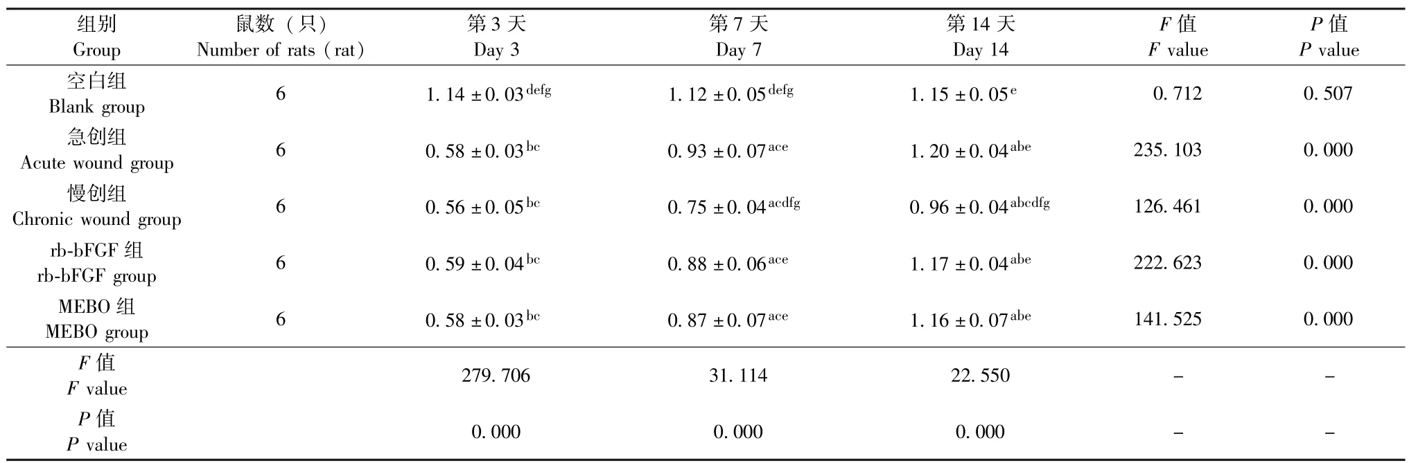
表1 5组大鼠皮肤或创面组织内CK10表达水平对比 (±s)Table 1 Comparison of CK10 expression levels in rat skin or wound tissues among the five groups(±s)
注:5组大鼠皮肤或创面组织内CK10表达水平组内对比,其中与第3天对比,aP<0.05,差异具有统计学意义;与第7天对比,bP<0.05,差异具有统计学意义。5组大鼠皮肤或创面组织内CK10表达水平组间对比,其中与空白组对比,cP<0.01,差异具有统计学意义;与急创组对比,dP<0.05,差异具有统计学意义;与慢创组对比,eP<0.05,差异具有统计学意义;与rb⁃bFGF组对比,fP<0.05,差异具有统计学意义;与MEBO组对比,gP<0.05,差异具有统计学意义Note: The expression levels of CK10 in rat skin or wound tissues were compared within each of the five groups,in which statistically significant differ⁃ences were observed in comparisons respectively with that on day 3 (aP <0.05) and with that on day 7 (bP <0.05).The between⁃group comparisons of CK10 expression levels were done among the five groups,in which statistically significant differences were observed in comparisons with the blank group(cP <0.01),with the acute wound group(dP <0.05),with the chronic wound group(eP <0.05),with the rb⁃bFGF group(fP <0.05) and with the MEBO group(gP<0.05)
组别Group鼠数 (只)Number of rats (rat)第3天Day 3第7天Day 7第14天Day 14 F值F value P值P value空白组Blank group 6 1.14±0.03defg 1.12±0.05defg 1.15±0.05e 0.712 0.507急创组Acute wound group 6 0.58±0.03bc 0.93±0.07ace 1.20±0.04abe 235.103 0.000慢创组Chronic wound group 6 0.56±0.05bc 0.75±0.04acdfg 0.96±0.04abcdfg 126.461 0.000 rb⁃bFGF 组rb⁃bFGF group 6 0.59±0.04bc 0.88±0.06ace 1.17±0.04abe 222.623 0.000 MEBO组MEBO group 6 0.58±0.03bc 0.87±0.07ace 1.16±0.07abe 141.525 0.000 F值F value 279.706 31.114 22.550- -P值P value 0.000 0.000 0.000- -

图2 Western blotting法检测5组大鼠皮肤或创面组织内CK10表达蛋白条带图Fig.2 CK10⁃expressed protein bands in rat skin or wound tissues in the five groups tested by Western blot⁃ting method

图3 5组大鼠皮肤或创面组织内CK10表达水平柱状图Fig.3 Histogram of CK10 expression levels in rat skin or wound tissues in the five groups
4 讨论
表皮干细胞具有强大的增殖、分化能力,其在皮肤受到创伤时可被激活,并逐步分化为表皮及皮肤附属器等组织结构,最终实现缺损皮肤的再生修复,在创面修复过程中发挥着至关重要的作用[11-12]。研究显示,表皮干细胞所生存的外界环境对表皮干细胞的增殖、分化影响巨大[13-15],如创面涂抹 MEBO 后,表皮干细胞可因所处的外界环境发生改变而提高其增殖、分化能力,从而加速创面修复[16]。亦有研究显示,CK10是表皮细胞增殖分化的标记性分子,其表达水平可随着表皮干细胞的不断分化、成熟而改变[9,17]。 因此,笔者为探讨 MEBT/MEBO对表皮干细胞影响的部分作用机制,最终实现加快慢性难愈合创面修复的目的,于本研究中对比观察了不同时间点经不同方法处理后大鼠皮肤及创面组织中CK10表达水平的变化情况。
研究结果显示,治疗第3、7、14天,空白组大鼠皮肤组织内CK10的表达水平无明显变化,P>0.05,差异无统计学意义,且镜下观组织形态与正常皮肤组织形态也无明显差异,符合其表皮未予任何处理的特征;而急创组、慢创组、rb⁃bFGF组及MEBO组大鼠创面组织内CK10的表达水平均逐渐升高,P均<0.05,差异具有统计学意义,且镜下观组织形态也有明显改变。治疗第3天,空白组大鼠皮肤组织内CK10的表达水平均较其他各组高,且P均<0.05,差异具有统计学意义,而急创组、慢创组、rb⁃bFGF组及MEBO组的组织形态及CK10表达水平并无明显差异,P均>0.05,差异无统计学意义,表明正常表皮内角化蛋白较为丰富,皮肤缺损后大量表皮细胞缺失,致使创面组织内CK10的表达水平也随之降低。治疗第7天,空白组大鼠皮肤组织内CK10的表达水平仍为最高,而急创组、慢创组、rb⁃bFGF组及MEBO组大鼠创面组织内CK10表达水平呈现升高的趋势,与第3天对比,P均<0.05,差异具有统计学意义,与基底层细胞不表达CK10,随着基底层干细胞不断增殖、分化为基底上层细胞,CK10的表达水平也不断升高[18]的研究结果一致;且急创组、rb⁃bFGF组及MEBO组的增长幅度均较慢创组明显,P均<0.05,差异具有统计学意义,说明慢创组大鼠创面修复速度较急创组慢,而rb⁃bFGF与MEBO对慢性难愈合创面均具有治疗作用。治疗第14天,除慢创组外,急创组、rb⁃bFGF组和MEBO组大鼠创面组织形态及CK10表达水平均接近空白组,P均>0.05,差异无统计学意义,表明除慢创组外,急创组、rb⁃bFGF组和 MEBO组大鼠创面已修复至接近正常皮肤组织,且组间对比,P均>0.05,差异无统计学意义,即MEBT/MEBO治疗慢性难愈合创面的疗效不亚于 rb⁃bFGF 凝胶。
综上所述,CK10表达水平的变化规律可表明表皮细胞增殖、分化的状况,MEBT/MEBO治疗慢性难愈合创面的疗效不亚于rb⁃bFGF凝胶,且通过对表皮干细胞外环境的影响促进表皮干细胞的增殖、分化可能是其加快创面修复的作用机制,仍需进一步深入研究考证。
In recent years,a large number of clinical practice and basic studies have shown that moist exposed burn therapy/moist exposed burn ointment(MEBT/MEBO) can realize significant curative effects in the treatment of chronic non⁃healing wounds[1-3],and its action mechanism has become a focus of clinical research.As demonstrated in Rongxiang Xu,et al.,MEBT/MEBO can activate the potential re⁃generative cells in wound tissues and transform them into stem cells with positive expression of cytokeratin 19,and such stem cells cancontinuously proliferate and differentiate into normal epidermal cells in situ and eventually the damaged wounds can realize re⁃epithelial⁃ization and remodeling of tissue structure[4-8].With the advances of research,some researchers found that CK10 is one of markers of epi⁃dermal cell differentiation and maturation,and its expression level may reflect the wound repair condition[9].To this end,this study dy⁃namically monitored the changes of CK10 expression in rat skin and wound tissues managed with different approaches,and explored the influence of MEBT/MEBO on the proliferation and differentiation of epidermal stem cells,with the aim of revealing partially the action mechanism of MEBT/MEBO in promoting the repair of chronic non⁃healing wounds.
1.Experimental material
1.1.Experimental animals
Ninety SPF healthy Wistar male rats(provided by Changsha Tianqin Biotechnology Co.,Ltd.,License number SCXK (Xiang)2014⁃0011),aged 12 weeks old and weighing (220 ± 20) g,were selected as subjects.During the experiment course,the subjects were kept strictly in the SPF feeding environment,with good ventilation,humidity at 60% ±10%and room temperature at(25±2)℃.This study was approved by the Animal Ethics Committee of Youjiang Med⁃ical University for Nationalities.
1.2.Main agents
Moist exposed burn ointment(MEBO) is manufactured by Shantou MEBO Pharmaceutical Co.,Ltd.,recombinant bovine bas⁃ic fibroblast growth factor (rb⁃bFGF) gel by Zhuhai Essex Bio⁃Pharmaceutical company Limited,chloral hydrate by Taixing Haoshen Chemical Trading Co.,Ltd.,hydrocortisone acetate injection by Shanghai General Pharmaceutical Co.,Ltd.,primary antibody dilu⁃tion buffer and secondary antibody dilution buffer are from Shanghai Beyotime Biotechnology Co.,Ltd.,and Anti⁃CK10⁃Antibody from Abcam.Horseradish peroxidase⁃labeled goat anti⁃rabbit IgG (100μL)and β⁃actin antibody (100μL) are manufactured by Beijing ZSGB Biotechnology Co.,Ltd.,and PVDF membrane by Millpore.
2.Methods
2.1.Grouping and modelling
The subjects were fed in the SPF animal laboratory for one week and,after being identified as healthy,were randomly divided into blank group,acute wound group,chronic wound group,rb⁃bFGF group and MEBO group,18 rats each group.The back skin of bilat⁃eral spine was selected as the experimental area.In the blank group,the skin at an area of about 3.0 cm×3.0 cm was prepared on the back of rats.However,in the acute wound group,after intraperitone⁃al injection of 7%chloral hydrate (4 mL/kg) and skin preparation(same method as in the blank group),mark the prepared skin with acircular seal of about 1.5 cm in diameter,remove skin tissues to the fascial layer along the marked line under sterile condition to establish the skin defect wound.In the chronic wound group,rb⁃bFGF group and MEBO group,Hydrocortisone acetate injection was injected im⁃mediately after the same procedures of anesthetizing,preparing skin and removing skin tissues as in the acute wound group to establish chronic non⁃healing wound models[10].
2.2.Local management of wounds
After successful model establishment,the prepared skin in the blank group was immediately given the routine disinfection with 5%iodophor for three times,followed by the successive covering of nor⁃mal saline soaked gauze and clean dressing,and the fixation with ad⁃hesive tape.The dressing was changed twice a day.The wounds in the acute wound group and chronic wound group were disinfected im⁃mediately with 5%iodophor for three times,and also covered with normal saline soaked gauze and clean dressing in turn,followed by the fixation with adhesive tape.The dressing change was performed twice a day.After the immediate routine disinfection with 5% io⁃dophor for three times,the wounds in the rb⁃bFGF group and MEBO group were respectively managed with rb⁃bFGF gel(60 U/cm2) and MEBO (0.2 g/cm2) before the successive covering of normal saline soaked gauze and clean dressing,and the fixation of adhesive tape,and the dressing was also changed twice a day in the two groups.
2.3.Sample collection
On day 3,7 and 14 of treatment,randomly select 6 rats in each group to undergo intraperitoneal anesthesia,and then remove the wound scab and take out wound tissues at 0.5 cm away from the wound edges(take out the normal skin tissues at the same location in the blank group).Divide the wound tissues into two samples,of which one was fixed immediately in EP tube containing 10%formal⁃dehyde and then embedded in paraffin after complete sample collec⁃tion,and the other was immediately put into EP tube and temporarily stored in liquid nitrogen,and then stored in a refrigerator of-80℃after complete sample collection.
2.4.Histological examination
Do HE staining after the slicing of paraffin⁃embedded tissues,and observe,under electron microscope,the distribution of inflamma⁃tory cells and fibroblasts,the arrangement of new blood vessels and the formation of skin appendages in tissues.
2.5.Test the expression level of CK10 by Western blotting method
Fully decompose the frozen tissues in the EP tube containing tis⁃sue lysate on ice to extract the total protein,and detect the concentra⁃tion of the total protein.Subpackage and store the protein into a re⁃frigerator of-20℃ after high⁃temperature degeneration by using the composition of protein buffer and tissue lysate.Pour the degenerated protein into SDS⁃PAGE gel hole by hole,and then proceed with the electrophoresis at the constant voltage of 100 V till the Marker bandswere spread uniformly.Cut PVDF membrane into suitable size in ac⁃cordance with CK10 molecular weight,and transfer the protein onto a membrane for 60-90 min under the constant current of 250 mA.Af⁃ter this,the membrane was washed for three times,incubated with the primary antibody and secondary antibody at room temperature,followed by exposure development.And finally the protein shades of gray was analyzed using Image J.The samples in each group were de⁃tected repeatedly for more than three times and the obtained mean val⁃ues were compared and analyzed.
2.6.Statistical analysis
Statistical software SPSS 22.0 was used for statistical analysis,in which measurement data was expressed as mean±standard devia⁃tion(±s),the means of multiple samples were compared with the one⁃way ANOVA,and between⁃group comparisons were done with the LSD method or Games⁃Howell method according to the variance homo⁃geneity.P<0.05 was considered statistically significant.
3.Results
3.1.Results of histological examination
On day 3 of treatment,there was no obvious difference between the skin tissues in the blank group and the normal rat skin tissues,while in the acute wound group,there was a large number of inflam⁃matory cells and a few fibroblasts and newly⁃born capillaries in wound tissues,and the tissues were markedly edematous.In the chronic wound group,rb⁃bFGF group and MEBO group,besides a large number of inflammatory cells and a few fibroblasts in the wound tissues,some red blood cells overflowing vascular wall can be seen in some locations and the tissue edema was more severe than the acute wound group.
On day 14 of treatment,the skin tissues in the blank group had no obvious difference from the normal skin tissues.The wounds in the acute wound group,rb⁃bFGF group and MEBO group were basically healed,well⁃arranged new capillaries and intact skin appendages in⁃cluding hair follicle and sebaceous glands were observable in tissues,and the tissue structure was similar to normal skin tissues.The wounds in the chronic wound group decreased obviously in size,few inflammatory cells and a large number of fibroblasts can be seen in tissues,and some fibroblasts were arranged disorderly (Fig.1).
3.2.Expression level of CK10
On day 3,7 and 14 of treatment,no significant change was ob⁃served on the expression level of CK10 in rat skin tissues in the blank group,and there was no statistically significant difference,P>0.05.In contrast,the expression of CK10 in rat skin tissues in the acute wound group,chronic wound group,rb⁃bFGF group and MEBO group all showed an increasing tendency,and the differences were statisti⁃cally significant,allP<0.05 (Table 1,Fig.2-3).
On day 3 of treatment,the comparison of CK expression levels among the five groups showed a pattern of the blank group>the acute wound group=chronic wound group=rb⁃bFGF group=MEBO group,
and the between⁃group pairwise comparisons all showed no statistically significant difference(P>0.05) except the comparisons of the blank group with the other four groups (P<0.05).On day 7 of treatment,the comparison of CK10 expression levels among the five groups showed a pattern of the blank group > the acute wound group = rb⁃bFGF group =MEBO group > chronic wound group,and the between⁃group pairwise comparisons all showed statistically significant differences(P<0.05) except the pairwise comparisons between acute wound group,rb⁃bFGF group and MEBO group (allP>0.05).On day 14 of treatment,the comparison of CK10 expression levels among the five groups showed a pattern of the blank group=the acute wound group=rb⁃bFGF group =MEBO group >chronic wound group,and the between⁃group pairwise comparisons all showed no statistically significant difference(P>0.05) except the comparisons of the chronic wound group with the other four groups(allP<0.05) (Table 1,Fig.2-3).
4.Discussion
Epidermal stem cells have strong ability of proliferation and dif⁃ferentiation,which can be activated when skin is injured to gradually differentiate into tissue structures of epidermis and skin appendages,and eventually realize the regenerative repair of injured skin,playing an important role in wound repair[11-12].Studies showed that the liv⁃ing environment of epidermal stem cells exerts huge impacts on the proliferation and differentiation of epidermal stem cells[13-15].For ex⁃ample,after the application of MEBO,with the change of the external environment of epidermal stem cells,their ability of proliferation and differentiation will be enhanced,facilitating the wound repair[16].Moreover,some studies confirmed that CK10 is the molecule marker of epidermal cell proliferation and differentiation,and its expression level may change with the continuous differentiation and maturation of epi⁃dermal stem cells[9,17].Therefore,with the aim of investigating the ac⁃tion mechanism of MEBT/MEBO on epidermal stem cells to realize the accelerated repair of chronic non⁃healing wounds,this study compared the changes of CK10 expression levels in rat skin and wound tissues at different time points after managed with different treatment approaches.
And the results showed that on day 3,7 and 14 of treatment,no significant change was observed on the expression level of CK10 in rat skin tissues in the blank group,and there was no statistically signifi⁃cant difference,P>0.05.Microscope observation found that the tis⁃sue morphology has no significant difference from the normal skin tis⁃sues,being in line with the features of non⁃managed epidermis.In contrast,the expression of CK10 in rat wound tissues in the acute wound group,chronic wound group,rb⁃bFGF group and MEBO group all showed an increasing tendency,and the differences were statistical⁃ly significant(allP<0.05).The tissue morphology under microscopeobservation also presented marked changes.On day 3 of treatment,the expression level of CK10 in rat skin tissues in the blank group was higher than that in the other four groups and the differences were statis⁃tically significant,allP<0.05.While no statistically significant differences was observed in the tissue morphology and CK expression levels among the acute wound group,chronic wound group,rb⁃bFGF group and MEBO group (allP>0.05),which indicated that though the cytokeratin in normal epidermis was very abundant,with the dam⁃age of skin,plenty of epidermal cells will lose,subsequently resulting in the decrease of CK10 expression in wound tissues.On day 7 of treatment,the expression level of CK10 in the blank group was still highest,meanwhile the CK10 expression levels in wound tissues in the other four groups presented an increasing tendency,and there were sta⁃tistically significant differences as compared with that on day 3 of treat⁃ment(allP<0.05),which was consistent with the study results that basal layer cells don’t express CK10 and the expression level of CK10 will increase continuously with the constant proliferation and differenti⁃ation of basal layer stem cells into cells above the basal layer[18].How⁃ever,the increase extents in the acute wound group,rb⁃bFGF group and MEBO group were more significant than that in the chronic wound group (allP<0.05),which indicated that the wound repair speed in the chronic wound group was slower than the acute wound group,and rb⁃bFGF gel and MEBO have the function of treating chronic non⁃healing wounds.On day 14 of treatment,except the chronic wound group,the tissue morphology and CK10 expression levels in the acute wound group,rb⁃bFGF group and MEBO group were close to that in the blank group,and the differences were not statistically significant(allP>0.05),which demonstrated that except the chronic wound group,the rat wounds in the acute wound group,rb⁃bFGF group and MEBO group have basi⁃cally restored to the status of normal skin tissues,and their between⁃group comparisons showed no statistically significant differences(allP>0.05),which indicated the curative effect of MEBT/MEBO in treating chronic non⁃healing wounds was not inferior to that of rb⁃bFGF gel.
To sum up,the change law of CK10 expression levels can reflect the condition of epidermal cell proliferation and differentiation,and the clinical efficacy of MEBT/MEBO in the treatment of chronic non⁃healing wounds is not inferior to that of rb⁃bFGF gel.Promoting the proliferation and differentiation of epidermal stem cells by changing the external living environment of epidermal stem cells may be the action mechanism of MEBT/MEBO in facilitating the restoration of chronic non⁃healing wounds,which,however,needs to be further studied in the future.
(收稿日期: 2018⁃12⁃28)
