川崎病休克综合征临床特征及外周血细胞因子水平分析
2017-12-04李艳蝶郭莉邹丽霞滕丽萍郑嵘君徐益萍卢美萍
李艳蝶 郭莉 邹丽霞 滕丽萍 郑嵘君 徐益萍 卢美萍
川崎病休克综合征临床特征及外周血细胞因子水平分析
李艳蝶 郭莉 邹丽霞 滕丽萍 郑嵘君 徐益萍 卢美萍
目的 分析川崎病休克综合征(KDSS)的临床特征及外周血细胞因子水平,为该病的诊治提供依据。方法 选择KDSS患儿17例(KDSS组),并选择同期住院且年龄、性别匹配的川崎病(KD)患儿43例(KD组)。采用流式细胞术检测两组患儿外周血细胞因子IL-2、IL-4、IL-6、IL-10、TNF-α和IFN-γ水平,并比较两组患儿确诊前的临床特征、常规实验室指标、肝肾功能及凝血功能指标、疗效及预后情况。结果 与KD组比较,KDSS组发热时间和住院时间显著延长,外周血WBC、中性粒细胞比例、C反应蛋白、ESR、肌酐、血尿素氮、D-二聚体,IL-6、IL-10、TNF-α、IFN-γ水平,冠状动脉损伤、丙种球蛋白(IVIG)抵抗、进重症监护室及其他脏器损伤的发生率均显著升高,而Hb、血钠、血钾、白蛋白均显著下降,差异均有统计学意义(均P<0.05)。两组均未发现死亡病例。结论细胞因子IL-6、IL-10、TNF-α和IFN-γ水平的升高、IVIG抵抗导致持续炎症可能参与KDSS的发生,KDSS患儿发热时间和住院时间显著延长,病情更重,更易发生冠状动脉及其他脏器损伤。
川崎病休克综合征 临床特征 细胞因子
川崎病(kawasaki disease,KD)是儿童较为常见的急性发热性血管炎,以5岁以下儿童多见[1]。该病主要累及中等血管,尤其是冠状动脉,15%~25%未治疗的患儿可能导致冠状动脉损伤(coronary artery lesions,CALs)[2],是儿童获得性心脏病的重要病因[3]。近年来,Kanegaye等[4]将KD急性期伴有低血压、休克等血流动力学改变者定义为川崎病休克综合征(kawasaki disease shock syndrome,KDSS),由于其发病机制尚不明确,而且KDSS有可能发生在KD典型症状出现前,有时与脓毒性休克很难鉴别,给诊断和治疗带来一定困难。因此,本研究通过分析KDSS的临床特征及外周血细胞因子水平,以进一步了解该病的发病机制,为早期临床诊治提供依据。
1 对象和方法
1.1 对象 选择2014年1月至2016年12月在本院住院的KDSS患儿17例(KDSS组),男11例,女6例;年龄 2~102(42.29±29.89)个月。并选择同期住院且年龄、性别匹配的KD患儿43例(KD组),男19例,女24例;年龄 3~89(28.82±21.50)个月。KD 诊断标准根据美国心脏病学会标准[5]。KDSS诊断标准参照Kanegaye等[4]提出的定义:KD患者伴有血流动力学障碍包括低血压和休克,儿童休克与年龄密切有关,年龄<1个月,收缩压<60mmHg;年龄 1~12个月,收缩压<70mmHg;年龄1~10 岁,收缩压<70+(年龄×2)mmHg;年龄>10 岁,收缩压<90mmHg。其他临床症状包括心率快、精神状态变化、毛细血管再灌注时间延长(>3s)、皮肤花白、少尿。KDSS的低血压标准还包括收缩压从基线水平下降≥20%,或者临床出现低灌注表现[4]。CALs根据日本卫生部标准:年龄<5岁,最大绝对内径>3mm;年龄≥5岁,最大绝对内径>4mm,或者动脉内径是临近段或者不规则段的1.5倍[6]。左心室功能障碍指射血分数<54%[7-8]。丙种球蛋白(IVIG)抵抗是指丙球完全注射(2g/kg)后48h内再次发热[9]。所有患儿均常规使用IVIG及阿司匹林治疗。本研究获得医院伦理委员会批准和患儿家属知情同意。
1.2 方法
1.2.1 外周血细胞因子 IL-2、IL-4、IL-6、IL-10、TNF-ɑ、IFN-γ 水平检测
1.2.1.1 试剂和仪器 流式细胞微球芯片技术(CBA)捕获人T辅助细胞1/2细胞因子试剂盒Ⅱ和FACSCalibur流式细胞仪均购自美国BD公司。
1.2.1.2 样品制备及流式细胞测定 取被检者外周血2ml,EDTA抗凝,1 000r/min离心10min,分离血浆备用。CBA技术测定:在每支流式细胞荧光分选技术样品管中加入 IL-2、IL-4、IL-6、IL-10、TNF 和 IFN-γ 混合捕获微球和待测血浆各50μl,PE抗体50μl,振荡混匀,20~25℃避光放置3h。每管中加入0.5ml PBS缓冲液,1 000r/min离心5min,弃上清液后,加0.3ml PBS缓冲液,振荡混匀,待测。同法制备标准品上样管。24h内流式细胞仪测定。测试条件:激光器电源15mV,电流6.20A,样本电压5.90V。根据仪器获取的数据,用CBA分析软件自动绘制标准曲线,并将样本数据与标准曲线拟合,自动计算出血浆中各种细胞因子含量。外周血细胞因子正常参考范围为1.0~5 000.0pg/ml。
1.2.2 观察指标 患儿确诊前的临床特征:发热时间,结膜炎、口唇充血、颈部淋巴结肿大、手足硬肿或脱皮、皮疹等发生率。常规实验室指标:外周血WBC、中性粒细胞比例、Hb、PLT、ESR、C 反应蛋白(CRP)、血钠、血钾。肝肾功能及凝血功能:白蛋白、ALT、AST、肌酐、血尿素氮、纤维蛋白原和D-二聚体。外周血细胞因子:IL-2、IL-4、IL-6、IL-10、TNF-ɑ、IFN-γ。患儿疗效及预后比较:住院时间,冠状动脉损伤、IVIG抵抗、进ICU以及其他脏器损伤等发生率,有无死亡病例。
1.3 统计学处理 采用SPSS 20.0统计软件。符合正态分布的计量资料以表示,组间比较采用两独立样本t检验;不符合正态分布的计量资料以M(Q1~Q)3表示,组间比较采用Mann-Whitney U检验。计数资料以百分率表示,组间比较采用χ2检验。P<0.05为差异有统计学意义。
2 结果
2.1 两组患儿确诊前的临床特征比较 与KD组比较,KDSS组确诊前发热时间显著延长,差异有统计学意义(P<0.01);而两组结膜炎、口唇充血、颈部淋巴结肿大、手足硬肿或脱皮、皮疹的发生率比较差异均无统计学意义(均 P >0.05),见表 1。

表1 两组患儿确诊前的临床特征比较
2.2 两组患儿常规实验室指标比较 与KD组比较,KDSS组外周血WBC、中性粒细胞比例、ESR、CRP均显著升高,而Hb、血钠、血钾均下降,差异均有统计学意义(均P<0.05);而两组PLT比较差异无统计学意义(P>0.05),见表 2。
2.3 两组患儿肝肾功能及凝血功能比较 与KD组比较,KDSS组白蛋白显著下降,肌酐浓度、血尿素氮、D-二聚体显著升高,差异均有统计学意义(均P<0.05);而两组ALT、AST和纤维蛋白原比较差异均无统计学意义(均 P >0.05),见表 3。
2.4 两组患儿外周血细胞因子水平比较 与KD组比较,KDSS组外周血IL-6、IL-10、TNF-ɑ和IFN-γ水平显著升高,差异均有统计学意义(均P<0.01);而两组IL-2、IL-4水平比较差异均无统计学意义(均P>0.05),见表4。
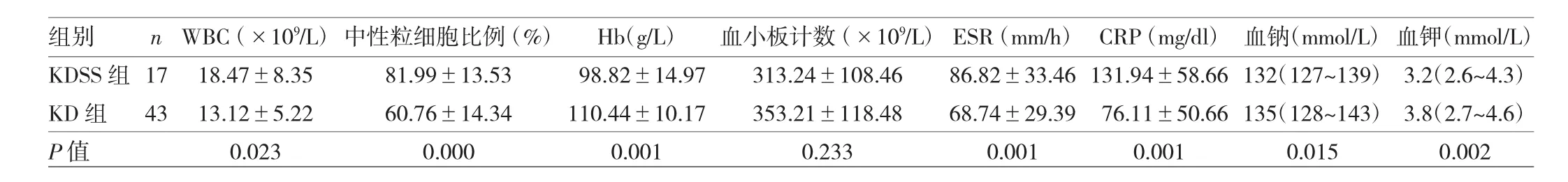
表2 两组患儿常规实验室指标比较

表3 两组患儿肝肾功能及凝血功能比较

表4 两组患儿外周血细胞因子水平比较
2.5 两组患儿疗效及预后比较 与KD组比较,KDSS组患儿住院时间显著延长,CALs、IVIG抵抗、进ICU以及其他脏器损伤的发生率显著升高,差异均有统计学意义(均P<0.05)。KDSS组所有患者均使用激素治疗,两组患儿均病情好转出院,未见死亡病例,见表5。

表5 两组患儿疗效及预后比较
3 讨论
KDSS是KD的严重形式,早期多以低血压、休克症状为主,症状不典型易误诊。Kanegaye等[4]报道女性患儿较多,本研究KDSS组男性多于女性,可能与样本量有关。KDSS常伴有外周血WBC、中性粒细胞比例、CRP、ESR升高、低钠血症[10]。本研究也得到相同的结果。
KDSS确切病因不明确,有学者认为细胞因子在KDSS发病中起关键作用,细胞因子失调导致心肌缺血、毛细血管渗漏和IVIG抵抗是导致KDSS的重要机制[11-12]。KD活动期免疫系统被激活,促进细胞因子产生,如 IL-6、IL-8、IL-27 和 TNF-ɑ 水平升高[13]。本研究中 KDSS 组外周血 IL-6、IL-10、TNF-ɑ 和 IFN-γ 水平显著高于KD组。干酪乳杆菌诱导的心肌病变可能与细胞因子水平(IL-1、TNF-α 和 IL-6)增加相关[14]。KD 患儿外周血IL-6、IL-10和IFN-γ水平升高与CALs密切相关,也是IVIG抵抗的标志[15-17]。IL-6过度升高与IVIG抵抗密切相关[18]。Wang等[15]研究表明KD患儿经IVIG治疗后血清IL-6和IL-10水平下降缓慢与IVIG抵抗相关。实验室数据对KDSS的诊断也具有指导意义,如WBC升高、CRP增高、ESR增快、贫血。这些指标与细胞因子水平升高密切相关。KD患儿IVIG治疗前,WBC、CRP与血清IL-6水平呈正相关[19-20]。Kuo等[21]报道KDSS患儿IL-6升高诱导铁调素下调,与贫血有关。
低蛋白血症、低钠血症、低钾血症、多器官损伤与细胞因子的高水平密切相关[22-23],与本研究结果相符。促炎细胞因子过度产生导致发热、冠状动脉壁损伤和多脏器损伤[24]。IL-2会增加内皮通透性,白蛋白可通过血管壁外渗到血管外导致低白蛋白血症[22]。KDSS患儿低钠血症的病理生理机制包括脱水、抗利尿激素分泌异常和急性肾损伤[23]。低钠血症会增加血管渗漏,加重心肌受损,是导致休克的原因之一[23]。IL-6促进多器官损害和衰竭,如胃肠道并发症[25-27]。
KDSS临床表现与脓毒症休克相似,均有发热和皮疹,难以鉴别[28]。KDSS常伴有贫血、心瓣膜炎,在脓毒症休克中很少出现[29],可作为鉴别的依据。两者治疗方案不同,脓毒症休克患者需要合适的抗生素治疗,IVIG对KDSS的治疗是必需的,还包括液体复苏、血管加压药来维持血压,因此及时确诊对患者预后非常重要。本研究样本量小,具有一定的局限性,可能需要更多的研究来证实。
综上所述,典型或不典型KD患儿出现细胞因子IL-6、IL-10、TNF-ɑ和 IFN-γ 水平持续升高、IVIG 疗效不佳、低白蛋白血症、低钠血症和贫血时,儿科医生应警惕休克、多器官损伤,冠状动脉扩张。及时再次给予IVIG治疗、皮质类固醇或生物制剂抗炎治疗减少并发症的发生。此外,在休克患儿抗感染治疗效果不佳,应考虑KDSS的可能性,及时给予IVIG治疗。KDSS患儿常表现为发热持续时间和住院时间显著延长,冠状动脉损伤及其他脏器损伤的风险增加,病情较重。笔者期待越来越多的儿科医生认识KDSS,并能及时治疗,从而减少并发症的发生。
[1] Nakamura Y,Yashiro M,Uehara R,et al.Epidemiologic features of Kawasaki disease in Japan:results of the 2007-2008 nationwide survey[J].J Epidemiol,2010,20(4):302-307.doi:10.2188/jea.JE 20090180.
[2] Singh S,Sharma D,Bhattad S,et al.Recent advances in Kawasaki disease-proceedings of the 3rd Kawasaki disease summit,Chandigarh,2014[J].Indian J Pediatr,2016,83(1):47-52.doi:10.1007/s12098-014-1456-x.
[3] Kumar R K,Tandon R.Rheumatic fever&rheumatic heart disease:the last 50 years[J].Indian J Med Res,2013,137(4):643-658.
[4] Kanegaye J T,Wilder M S,Molkara D,et al.Recognition of a Kawasaki disease shock syndrome[J].Pediatrics,2009,123(5):e783-e789.doi:10.1542/peds.2008-1871.
[5] McCrindle B W,Rowley A H,Newburger J W,et al.Diagnosis,treatment,and long-term management of Kawasaki disease:a scientific statement for health professionals from the American Heart Association[J].Circulation,2017,135(17):e927-e999.doi:10.1161/CIR.00000 00000000484.
[6] Newburger J W,Takahashi M,Gerber M A,et al.Diagnosis,treatment,and long-term management of Kawasaki disease:a statementforhealth professionals from the committee on rheumatic fever,endocarditis and Kawasaki disease,council on cardiovascular disease in the young,American Heart Association[J].Circulation,2004,110(17):2747-2771.doi:10.116 1/01.CIR.0000145143.19711.78.
[7] Adler AC,KodavatigantiR.Kawasakidisease with giant coronary aneurysms requiring a ventricular assist device to separate from extracorporeal membrane oxygenation:coronary issues can be a pediatric problem too![J].AACase Rep,2016,7(4):77-80.doi:10.1213/XAA.0000000000000349.
[8] Protogerou A,Zampeli E,Fragiadaki K,et al.A pilot study of endothelial dysfunction and aortic stiffness after interleukin-6 receptor inhibition in rheumatoid arthritis[J].Atherosclerosis,2011,219(2):734-736.doi:10.1016/j.atherosclerosis.2011.09.015.
[9] Saneeymehri S,Baker K,So T Y.Overview of pharmacological treatment options for pediatric patients with refractory Kawasaki disease[J].J Pediatr Pharmacol Ther,2015,20(3):163-177.doi:10.5863/1551-6776-20.3.163.
[10] Dominguez S R,Friedman K,Seewald R,et al.Kawasaki disease in a pediatric intensive care unit:a case-control study[J].Pediatrics,2008,122(4):e786-e790.doi:10.1542/peds.2008-1275.
[11] Gatterre P,Oualha M,Dupic L,et al.Kawasaki disease:an unexpected etiology of shock and multiple organ dysfunction syndrome[J].Intensive Care Med,2012,38(5):872-878.doi:10.1007/s00134-012-2473-8.
[12] Chen P S,Chi H,Huang F Y,et al.Clinical manifestations of Kawasaki disease shock syndrome:a case-control study[J].J Microbiol Immunol Infect,2015,48(1):43-50.doi:org/10.1016/j.jmii.2013.06.005.
[13] Si F,Wu Y,Gao F,et al.Relationship between IL-27 and coronary arterial lesions in children with Kawasaki disease[J].Clin Exp Med,2017.doi:10.1007/s10238-017-0451-8.[Epub ahead of print]
[14] Okitsu-Negishi S,Nakano I,Suzuki K,et al.The induction of cardioangitis by Lactobacillus casei cell wall in mice.I.The cytokine production from murine macrophages by Lactobacillus casei cell wall extract[J].Clin Immunol Immunopathol,1996,78(1):30-40.
[15] Wang Y,Wang W,Gong F,et al.Evaluation of intravenous immunoglobulin resistance and coronary artery lesions in relation to Th1/Th2 cytokine profiles in patients with Kawasaki disease[J].Arthritis Rheum,2013,65(3):805-814.doi:10.1002/art.37815.
[16] Weng K P,Hsieh K S,Hwang Y T,et al.IL-10 polymorphisms are associated with coronary artery lesions in acute stage of Kawasaki disease[J].Circ J,2010,74(5):983-989.doi.org/10.1253/circj.CJ-09-0801.
[17] Guo M M,Tseng W N,Ko C H,et al.Th17-and Treg-related cytokine and mRNA expression are associated with acute and resolving Kawasaki disease[J].Allergy,2015,70(3):310-318.doi:10.1111/all.12558.
[18] Watanabe M,Uchida K,Nakagaki K,et al.High avidity cytokine autoantibodies in health and disease:pathogenesis and mechanisms[J].Cytokine Growth Factor Rev,2010,21(4):263-273.doi:10.1016/j.cytogfr.2010.03.003.
[19] Peng Q,Wu Q,Chen C H,et al.Value of serum soluble interleukin-2R,interleukin-6 and C-reactive protein in the early diagnosis of Kawasaki disease[J].Zhongguo Dang Dai Er Ke Za Zhi,2006,8(3):208-210.
[20] Honda H,Hirano T,Ueda M,et al.Associations among apolipoproteins,oxidized high-density lipoprotein and cardiovascular events in patients on hemodialysis[J].PLoS One,2017,12(5):e177980.doi:10.1371/journal.pone.0177980.
[21] Kuo H C,Yang Y L,Chuang J H,et al.Inflammation-induced hepcidin is associated with the development of anemia and coronary artery lesions in Kawasaki disease[J].J Clin Immunol,2012,32(4):746-752.doi:10.1007/s10875-012-9668-1.
[22] Park S,Eun L Y,Kim J H.Relationship between serum sodium level and coronary artery abnormality in Kawasaki disease[J].Korean J Pediatr,2017,60(2):38-44.doi:10.3345/kjp.2017.60.2.38.
[23] Gitiaux C,Kossorotoff M,Bergounioux J,et al.Cerebral vasculitis in severe Kawasaki disease:early detection by magnetic resonance imaging and good outcome after intensive treatment[J].Dev Med Child Neurol,2012,54(12):1160-1163.doi:10.1111/dmcn.12002.
[24] Schuster J E,Palac H L,Innocentini N,et al.Hyponatremia is a feature of Kawasaki disease Shock Syndrome:A Case-Control Study[J].Journal of the Pediatric Infectious Diseases Society,2017.doi:10.1093/jpids/piw081.[Epub ahead of print]
[25] Gamez-Gonzalez L B,Murata C,Munoz-Ramirez M,et al.Clinical manifestations associated with Kawasaki disease shock syndrome in Mexican children[J].Eur J Pediatr,2013,172(3):337-342.doi:10.1007/s00431-012-1879-1.
[26] Gatterre P,Oualha M,Dupic L,et al.Kawasaki disease:an unexpected etiology of shock and multiple organ dysfunction syndrome[J].Intensive Care Med,2012,38(5):872-878.doi:10.1007/s00134-012-2473-8.
[27] Singh R,Verma A,Aljabari S,et al.Urinary biomarkers as indicator of chronic inflammation and endothelial dysfunction in obese adolescents[J].BMC Obes,2017,4:11-19.doi:10.1186/s40608-017-0148-2.
[28] Sinhabahu VP,Suntharesan J,Wijesekara D S.Kawasaki shock syndrome in a 12-year-old girl mimicking septic shock[J].Case Rep Infect Dis,2016,2016(4949036):1-3.doi:10.1155/2016/4949036.
[29] Lin Y,Cheng M,Lo M,et al.Early differentiation of Kawasaki disease shock syndrome and toxic shock syndrome in a pediatric intensive care unit[J].Pediatr Infect Dis J,2015,34(11):1163-1167.doi:10.1097/INF.0000000000000852.
(本文由浙江省医学会风湿病学分会推荐)
Clinical characteristics and serum cytokine levels in patients with Kawasaki disease shock syndrome
LI Yandie,GUO Li,ZOU Lixia,et al.
Department of Rheumatology Immunology&Allergy,Children's Hospital,Zhejiang University School of Medicine,Hangzhou 310003,China
Objective To investigate the clinical characteristics and serum cytokine levels in children with Kawasaki disease shock syndrome (KDSS). Methods Seventeen KDSS children and 43 age-and gender-matched patients with Kawasaki disease (KD)admitted during January 2014 to December 2016 were included in this study.Serum levels of interleukin-2(IL-2),IL-4,IL-6,IL-10,tumor necrosis factor-α(TNF-α),and interferon gamma(IFN-γ)were measured by flow cytometry in two groups.The clinical characteristics,serum cytokine levels and routine laboratory parameters,the liver,kidney and blood coagulation functions and disease outcomes were compared between two groups. Results Compared with KD group,the duration of fever and length of hospital stay in KDSS group were significantly prolonged,the WBC,neutrophil,ESR and serum CRP,creatinine,urea nitrogen,D-dimer,IL-6,IL-10,TNF-α,IFN-γ levels were elevated,the incidence of coronary artery lesions,intravenous immunoglobulin (IVIG)resistance,admission to ICU and other organ damage were significantly increased(all P<0.05),while blood hemoglobin,albumin,sodium and potassium concentrations were significantly decreased(all P<0.05).No deaths were found in two groups. Conclusion Increased levels of IL-6,IL-10,TNF-α,IFN-γ and IVIG resistance leading to persistent inflammation may be involved in the development of KDSS.The duration of fever and hospital stay are prolonged,the conditions are more serious and more susceptible to coronary and other organ damage in patients with KDSS.
Kawasakidisease shock syndrome Clinicalcharacteristics Cytokine
10.12056/j.issn.1006-2785.2017.39.21.2017-1605
310003 杭州,浙江大学医学院附属儿童医院风湿免疫过敏科
卢美萍,E-mail:meipinglu@zju.edu.cn
2017-07-07)
陈丽)
