芥子酸拮抗同型半胱氨酸诱导的血管内皮细胞凋亡及焦亡
2017-12-01樊一钢田红燕
裴 星,韩 勇,丘 红,樊一钢,耿 杰,田红燕*
(1西安交通大学附属红会医院内科,西安 710054;2西安交通大学第一附属医院周围血管科;*通讯作者,E-mail:hytgxy@163.com;#共同通讯作者,E-mail:gqgalbert@163.com)
芥子酸拮抗同型半胱氨酸诱导的血管内皮细胞凋亡及焦亡
裴 星1#,韩 勇1,丘 红1,樊一钢1,耿 杰2,田红燕2*
(1西安交通大学附属红会医院内科,西安 710054;2西安交通大学第一附属医院周围血管科;*通讯作者,E-mail:hytgxy@163.com;#共同通讯作者,E-mail:gqgalbert@163.com)
目的 研究同型半胱氨酸(homocysteine, Hcy)对casepase途径介导的内皮细胞凋亡和焦亡的促进作用,以及该过程中芥子酸(sinapic acid, SA)对Hcy的拮抗作用。 方法 分别利用0,0.1,0.3,0.5,1,3,5 mmol/L浓度的Hcy处理HUVEC-12人脐静脉内皮细胞24 h,检测细胞凋亡和细胞焦亡水平,筛选最适处理浓度;以最适浓度Hcy处理细胞24 h,同时分别添加0.1,0.5,1,5,10 μmol/L的SA。采用Annexin-Ⅴ/PI法检测早期细胞凋亡,caspase-1 FLICA/PI法检测细胞的焦亡,Western blot检测细胞中焦亡关键调控蛋白NRLP3、ASC、caspase-1,凋亡相关蛋白caspase-8、caspase-3和抗凋亡蛋白Bcl-2的表达,通过ELISA方法检测细胞上清中内皮素(endothelin-1,ET-1)及炎性因子白细胞介素1β(interleukin-1β,IL-1β)的分泌量。 结果 细胞凋亡细胞焦亡水平随Hcy浓度升高而逐渐上升;与对照相比,当Hcy浓度分别≥0.5 mmol/L和≥1 mmol/L时,细胞凋亡和焦亡水平显著升高(P<0.05);3 mmol/L和5 mmol/L Hcy进一步促进细胞的凋亡、焦亡及相关调控蛋白的水平(P<0.05);选择3 mmol/L为最适浓度,用于后续研究。1 μmol/L SA可以抑制上述改变并能拮抗Hcy诱导的ET-1和IL-1β的分泌(P<0.05),并且其拮抗作用跟泛caspase抑制剂Z-VAD-FMK的效果类似。 结论 Hcy可通过casepases途径诱导血管内皮细胞的凋亡和焦亡;低浓度的SA可拮抗Hcy诱导的血管内皮细胞凋亡和焦亡。
芥子酸; 同型半胱氨酸; 血管内皮细胞; 细胞凋亡; 细胞焦亡

1 材料和方法
1.1 主要试剂和仪器
人脐静脉内皮细胞系HUVEC-12,购于美国ATCC细胞库(American Type Culture Collection,ATCC,Manassas,VA);芥子酸(SA)、同型半胱氨酸(Hcy)、胎牛血清(FBS)均购于美国Sigma公司;泛caspase抑制剂Z-VAD-FMK购于美国BD公司;Annexin Ⅴ-FITC凋亡试剂盒购自BD公司;FAM-FLICATM体内caspase检测试剂盒购于Abdserotec公司;BCA蛋白定量分析试剂盒购自美国Thermo公司;ET-1 ELISA试剂盒、小鼠NLRP3单克隆抗体(1 ∶200)、小鼠ASC单克隆抗体、小鼠cleaved caspase-1多克隆抗体、小鼠cleaved caspase-8多克隆抗体、小鼠cleaved caspase-3多克隆抗体和小鼠Bcl-2多克隆抗体(1 ∶500)购自英国Abcam公司;小鼠β-actin单克隆抗体购于Sigma公司;IL-1β ELISA检测试剂盒购自杭州MultiSciences公司。
CO2细胞培养箱(SANYO,日本);离心机(Eppendorf,德国);流式细胞仪(Beckman Coulter,美国);蛋白电泳仪、凝胶成像系统和多功能酶标仪(Bio-Rad,美国)。
1.2 细胞培养及分组
将HUVEC-12细胞在含有10% FBS的DMEM培养基,37 ℃、5% CO2条件下培养,每隔2-3 d传代1次,取对数期增长的细胞进行后续实验。
不同浓度Hcy处理细胞实验分组:Hcy用完全培养基稀释后,将贴壁细胞分为6组:对照组(0 mmol/L)、Hcy(0.1 mmol/L)、Hcy(0.3 mmol/L)、Hcy(0.5 mmol/L)、Hcy(1 mmol/L)、Hcy(3 mmol/L)和Hcy(5 mmol/L)处理组。每组细胞需设3个复孔,各组的Hcy处理时间24 h用于细胞凋亡及焦亡检测。
SA及caspase抑制剂处理细胞实验分组:将细胞接种于6孔板并培养24 h,将细胞分为空白组、Hcy模型组、SA处理组。除对照组外,各组细胞均换用3 mmol/L的Hcy培养基,同时SA处理组分别加入终浓度0.1,0.5,1,5,10 μmol/L的SA的完全培养基,而caspase抑制剂组则加入10 μmol/L的Z-VAD-FMK,培养24 h用于后续检测。
1.3 Annexin Ⅴ和碘化丙啶(propidium iodide,PI)双染色法检测早期细胞凋亡
用冰PBS洗3次,0.25%的胰蛋白酶将各组细胞消化成细胞悬液,计数后收集106个细胞。离心,弃上清,将细胞重悬于200 μl结合缓冲液,加入10 μl AnnexinⅤ-FITC(20 μg/ml)和5 μl PI(50 μg/ml),避光于室温下温育30 min,混匀后用流式细胞仪(FACS caliber, Becton Dickinson)检测早期细胞凋亡。其中,AnnexinⅤ+、PI-的象限是早期凋亡的细胞。
1.4 caspase-1 FLICA和PI双染色法检测细胞焦亡
胰酶消化收集细胞,300×g离心5 min,加入caspase-1 FLICA 37℃孵育1 h,用apoptosis buffer洗涤2次,加入5 μl PI孵育15 min,流式细胞仪检测细胞的焦亡。caspase-1+、PI+的象限是发生焦亡的细胞。
1.5 Western blot检测细胞和细胞上清中caspase-1和caspase-3蛋白表达
每孔30 μg蛋白上样于聚丙烯酰胺凝胶中,120 V电泳2.5 h,将胶中的蛋白样品转移到NC膜上,室温封闭1 h去除封闭液,将小鼠NLRP3单克隆抗体(1 ∶200)、小鼠ASC单克隆抗体(1 ∶400)、小鼠cleaved caspase-1多克隆抗体(1 ∶1 000)、小鼠cleaved caspase-8多克隆抗体(1 ∶800)、小鼠cleaved caspase-3多克隆抗体(1 ∶1 000)、小鼠Bcl-2多克隆抗体(1 ∶500)和小鼠β-actin单克隆抗体(1 ∶5 000)分别与NC膜上相应蛋白条带4 ℃孵育过夜。1×TBST洗膜3次,每次10 min。将NC膜在羊抗鼠IgG-HRP(1 ∶5 000)中室温孵育2 h,1×TBST洗膜3次,每次10 min。加入发光液后曝光观察。
1.6 ELISA检测细胞上清中IL-1β和ET-1的分泌量
收集细胞上清液,按照Endothelin-1 ELISA Kit(ab133030)和IL-1βELISA Kit(70-E-EK101B1/70-E-EK101B2)试剂盒说明书进行操作。
1.7 统计学分析
2 结果
2.1 高水平Hcy促进HUVEC-12细胞系凋亡和焦亡
不同浓度Hcy处理HUVEC-12细胞24 h,检测细胞凋亡与细胞焦亡,结果显示,细胞凋亡水平随Hcy浓度升高而逐渐上升,Hcy浓度≥0.5 mmol/L时,差异具有统计学意义(见图1A);细胞焦亡水平同样随Hcy浓度升高而逐渐上升,当Hcy浓度≥1 mmol/L时,差异具有统计学意义(见图1B)。与0.5 mmol/L Hcy处理组相比,3 mmol/L Hcy和5 mmol/L Hcy处理后细胞凋亡水平显著上升(见图1A)。与1 mmol/L Hcy处理组相比,3 mmol/L Hcy和5 mmol/L Hcy处理后细胞焦亡水平显著上升(见图1B)。细胞焦亡标志蛋白NLRP3、ASC和caspase-1,细胞凋亡蛋白caspase-8和caspase-3的表达变化趋势与细胞焦亡和凋亡基本一致,抗凋亡蛋白Bcl-2表达变化趋势则相反(见图1C)。因此,本实验选择3 mmol/L的Hcy浓度用于后续研究。

与对照比较,*P<0.05,**P<0.01;与1 mmol/L Hcy比较,#P<0.05图1 不同浓度Hcy处理24 h对HUVEC-12凋亡和焦亡的影响Figure 1 The apoptosis and pyroptosis of HUVEC-12 induced by different concentrations of Hcy for 24 h
2.2 SA拮抗高Hcy诱导的内皮细胞凋亡和焦亡
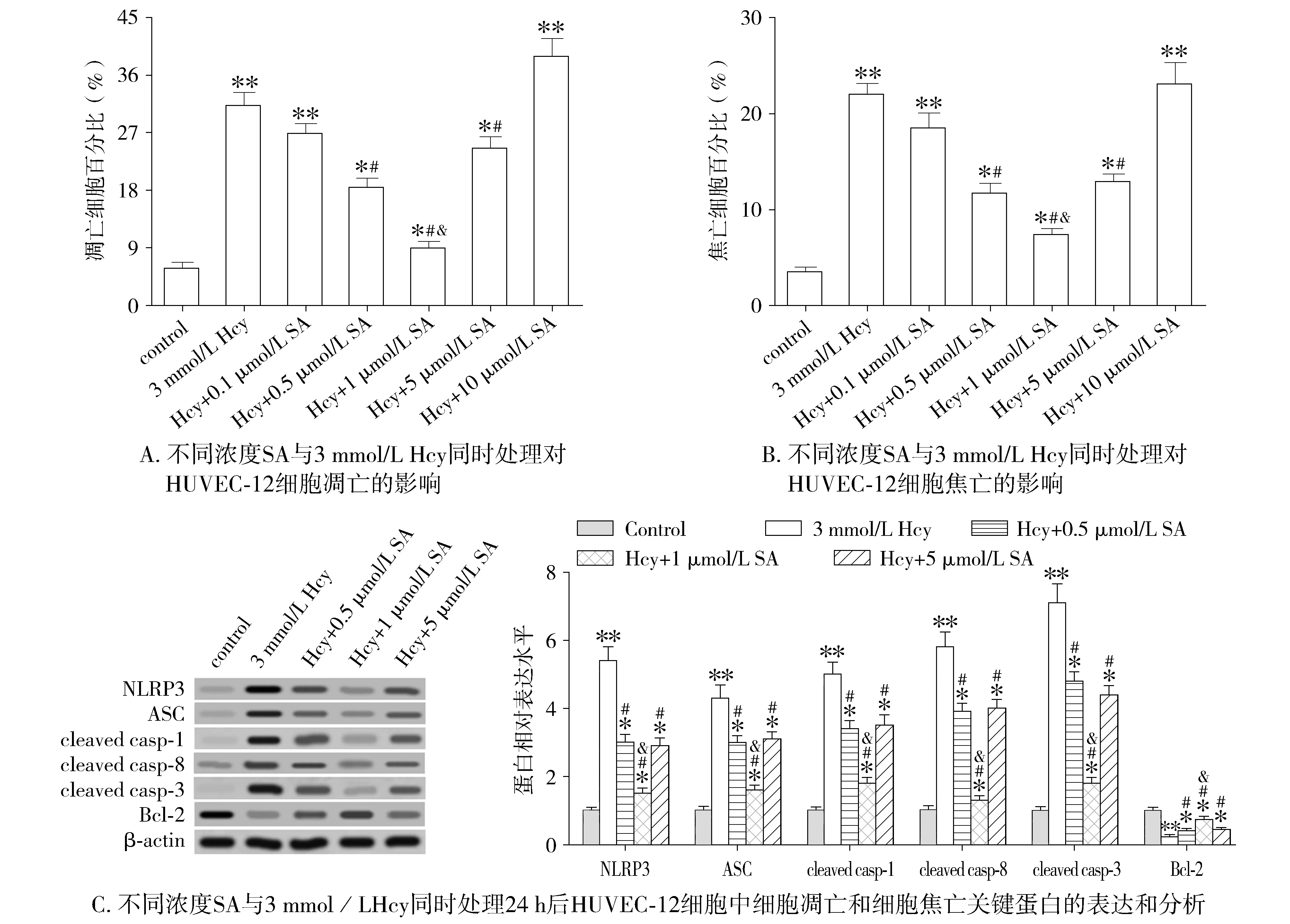
3 mmol/L Hcy与不同浓度SA同时处理HUVEC-12细胞,结果显示,随着SA浓度的增加,细胞凋亡和焦亡水平逐渐降低,浓度为1 μmol/L时降到最低;之后,随着SA浓度的增加,细胞凋亡和焦亡水平又逐渐增加(见图2A和2B)。NOD样受体蛋白3(NOD-like receptor 3,NLRP3)、凋亡相关点样蛋白(ASC)、半胱氨酸天冬蛋白酶-1(cysteine-aspartic acid protease,caspase-1)、caspase-8和caspase-3等蛋白的变化趋势与细胞凋亡和焦亡的变化趋势基本一致,抗凋亡蛋白Bcl-2表达变化趋势则相反(见图2C)。因此我们选择1 μmol/L SA浓度作为后续研究。

与对照比较,*P<0.05,**P<0.01;与Hcy+0.1 μmol/L SA比较,#P<0.05;与Hcy+0.5 μmol/L SA比较,&P<0.05图2 不同浓度SA对Hcy诱导的细胞凋亡和焦亡的影响Figure 2 Effects of different concentrations of SA on Hcy-induced apoptosis and pyroptosis of HUVEC-12
2.3 SA拮抗高Hcy诱导的内皮素和炎症因子分泌
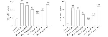
3 mmol/L Hcy与不同浓度SA同时处理HUVEC-12细胞时,炎症因子ET-1和IL-1β分泌水平的变化趋势与细胞凋亡和焦亡的变化趋势基本一致,随着SA浓度的增加,ET-1和IL-1β分泌逐渐减少,浓度为1 μmol/L时降到最低;之后,随着SA浓度的增加,ET-1和IL-1β分泌又逐渐增加(图3A和3B)。
2.4 SA与caspase抑制剂对细胞凋亡、焦亡影响的比较
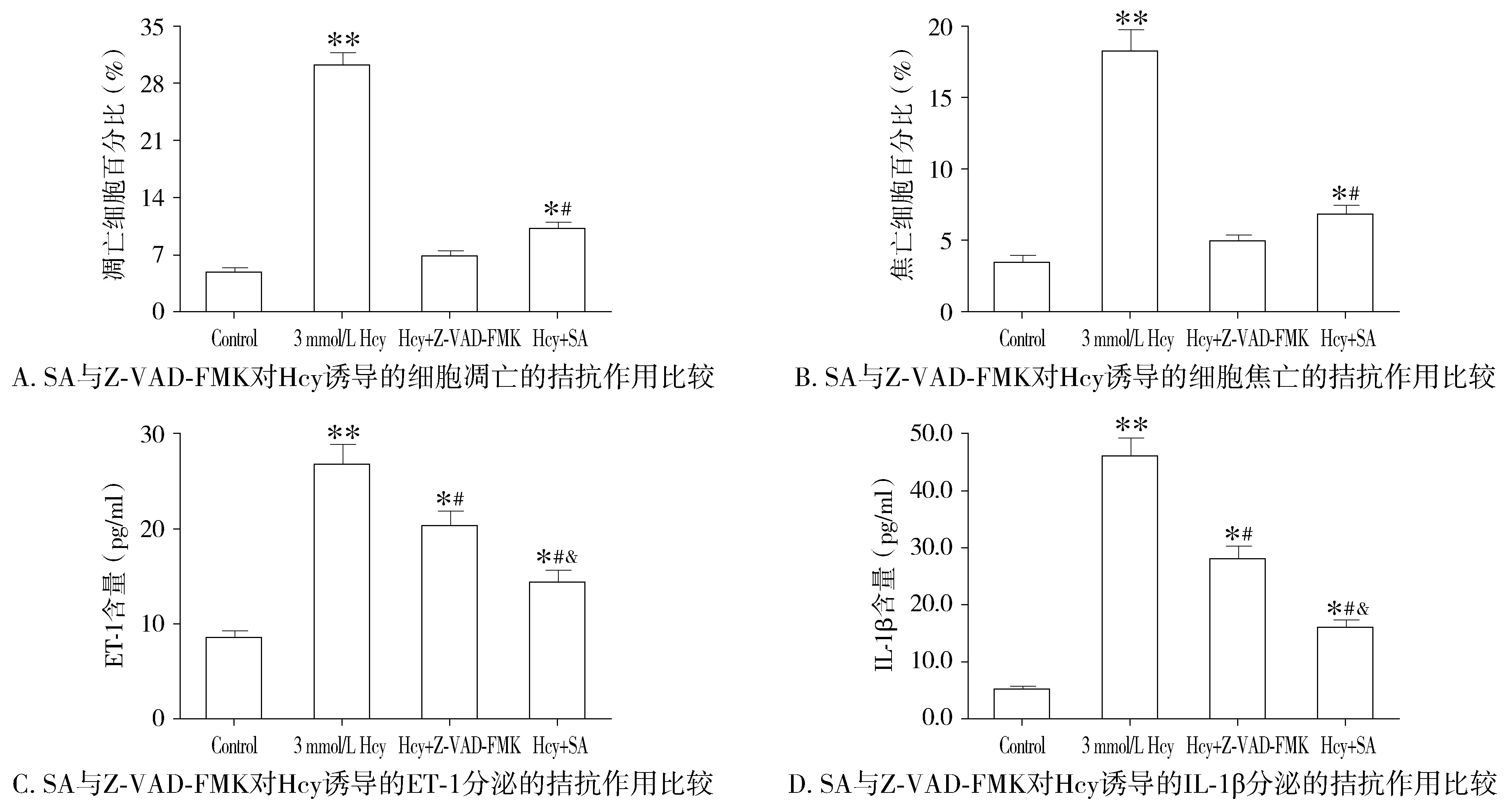
3 mmol/L的Hcy处理后,细胞凋亡及焦亡数量显著增加,1 μmol/L SA可拮抗Hcy诱导的细胞凋亡及焦亡,但这种作用稍弱于Z-VAD-FMK(见图4A,4B)。3 mmol/L的Hcy处理后,ET-1和IL-1β的分泌量也显著增高,1 μmol/L SA可拮抗Hcy诱导的ET-1和IL-1β的分泌,但该拮抗作用稍强于Z-VAD-FMK(见图4C,4D)。
3 讨论
细胞凋亡(apoptosis)和细胞焦亡(pyroptosis)都是细胞自主性死亡的过程。细胞凋亡时,胞内的显著特征为染色质DNA降解成大小为200 bp的整数倍的片段,主要由裂解激活的caspase-3调节[4]。细胞焦亡,又称细胞炎性坏死,表现为细胞不断胀大直至胞膜破裂,同时伴随强烈的炎症反应,该过程起始于炎性小体NLRP3等与ASC等结合后激活下游的caspase-1,从而催化炎症因子IL-1β等的成熟和释放[5]。现有研究表明,动脉粥样硬化等心血管疾病过程中炎症因子ET-1和IL-1β释放量增加,血管内皮细胞的凋亡和焦亡水平提高。本研究亦发现高水平Hcy诱导的内皮细胞功能障碍过程中细胞凋亡和焦亡水平显著升高,提示细胞凋亡和焦亡在内皮功能障碍过程中均发挥重要推动作用。

A. 不同浓度SA与3 mmol/LHcy同时处理对HUVEC-12细胞ET-1分泌的影响 B. 不同浓度SA与3 mmol/LHcy同时处理对HUVEC-12细胞IL-1β分泌的影响与对照比较,*P<0.05,**P<0.01;与Hcy+0.1 μmol/L SA比较,#P<0.05;与Hcy+0.5 μmol/L SA比较,&P<0.05图3 不同浓度SA对Hcy诱导的ET-1和IL-1β分泌的影响Figure 3 Effects of different concentrations of SA on Hcy-induced production of ET-1 and IL-1β in HUVEC-12 culture media

与对照比较,*P<0.05,**P<0.01;与3 mmol/LHcy比较,#P<0.05;与Hcy+Z-VAD-FMK比较,&P<0.05图4 SA与Z-VAD-FMK对Hcy诱导的细胞凋亡、焦亡和炎症因子分泌拮抗作用的比较Figure 4 Comparison of the antagonistic effects of SA versus Z-VAD-FMK on Hcy-induced apoptosis, pyroptosis, and production of inflammatory factors
高水平Hcy是动脉粥样硬化等心血管疾病的独立危险因素[6]。血浆Hcy浓度每升高3 μmol/L,机体发生卒中的概率将增加19%,而缺血性心脏的概率也增加11%[7]。高Hcy水平使得机体过多的产生活性氧物质,导致动脉血管内皮细胞损伤导致内皮细胞凋亡,并加重局部免疫反应。本研究表明,内皮细胞受到不同浓度Hcy处理时,细胞凋亡和焦亡水平随Hcy浓度升高而逐渐上升,当Hcy浓度≥0.5 mmol/L时细胞凋亡水平与对照组相比差异显著,Hcy浓度≥1 mmol/L时细胞焦亡水平与对照组相比差异显著,且3 mmol/L Hcy处理可引起细胞大量凋亡和焦亡。现有多项报道与本研究结果基本一致:人脐静脉内皮细胞在0.5 mmol/L Hcy浓度刺激下细胞骨架成分发生显著变化,细胞内氧化应激水平升高,当Hcy浓度达到3 mmol/L时,细胞发生大面积凋亡[8,9];最近亦有研究表明,0.5 mmol/L Hcy与10 μg/ml LPS共同处理内皮细胞后,细胞中活性caspase-3和活性caspase-1表达量显著增加,细胞凋亡和焦亡水平显著上升[10]。
芥子酸(又称3,5-二甲氧基-4-羟基肉桂酸)是一种天然酚酸类化合物,具有一定的刺激性。心肌梗死、肝损伤和庆大霉素诱导的急性肾损伤小鼠模型中,低浓度的SA表现出一定程度的抗炎、缓解脂质过氧化作用,起到保护心血管和肝肾的作用[11-13]。体内外研究也表明,低浓度的SA可减少ET-1的分泌,促进NO的合成,起到保护血管内皮、舒张血管、降低血压的作用[14]。本研究亦表明,SA以剂量依赖的方式影响内皮细胞的功能,低浓度的SA抑制caspase-1和caspase-3的表达,抑制Hcy诱导的细胞凋亡、焦亡和炎症反应,其效果与泛caspases抑制剂有类似之处。然而,高浓度的SA对细胞程序性死亡和炎症的缓解作用明显降低,甚至消失,这是因为酚酸类化合物本身就具有一定的刺激性和细胞毒性。可见,SA可作为一种天然的caspase-1/3抑制剂,在内皮细胞的病理性凋亡、焦亡、炎症和氧化应激的治疗过程中有巨大的潜在应用价值,但是一定要严格控制用量。
[1] Jeon SB, Kang DW, Kim JS,etal. Homocysteine, small-vessel disease, and atherosclerosis: an MRI study of 825 stroke patients[J]. Neurology, 2014, 83(8): 695-701.
[2] Ansari MA, Raish M, Ahmad A,etal. Sinapic acid mitigates gentamicin-induced nephrotoxicity and associated oxidative/nitrosative stress, apoptosis, and inflammation in rats[J]. Life Sci, 2016, 165 (11):1-8.
[3] Silambarasan T, Manivannan J, Priya MK,etal. Sinapic acid protects heart against ischemia/reperfusion injury and H9c2 cardiomyoblast cells against oxidative stress[J]. Biochem Bioph Res Co, 2015, 456(4): 853-859.
[4] Dunnick JK, Brix A, Sanders JM,etal. Apoptosis: a review of programmed cell death[J]. Toxicol Pathol,2007,35(4):495-516.
[5] Bergsbaken T, Fink SL, Cookson BT. Pyroptosis: host cell death and inflammation[J]. Nat Rev Microbiol,2009,7(2):99-109.
[6] Kumakura H, Fujita K, Kanai H,etal. High-sensitivity C-reactive protein, lipoprotein(a) and homocysteine are risk factors for coronary artery disease in Japanese patients with peripheral arterial disease[J]. J Atheroscler Thromb, 2015, 22(4): 344-354.
[7] Zhong C, Lv L, Liu C,etal. High homocysteine and blood pressure related to poor outcome of acute ischemia stroke in Chinese population[J]. PLoS One, 2014, 9(9): e107498.
[8] Kanazawa I, Tomita T, Miyazaki S,etal. Bazedoxifene ameliorates homocysteine-induced apoptosis and accumulation of advanced glycation end products by reducing oxidative stress in MC3T3-E1 cells[J]. Calcif Tissue Int, 2017, 100(3): 286-297.
[9] El-Hawli A, Qaradakhi T, Hayes A,etal. IRAP inhibition using HFI419 prevents moderate to severe acetylcholine mediated vasoconstriction in a rabbit model[J]. Biomed Pharmacother, 2017, 86(1): 23-26.
[10] Xi H, Zhang Y, Xu Y,etal. Caspase-1 inflammasome activation mediates homocysteine-induced pyrop-apoptosis in endothelial cells[J]. Circ Res, 2016, 118 (10):1525-1539.
[11] Shin DS, Kim KW, Chung HY,etal. Effect of sinapic acid against carbon tetrachloride-induced acute hepatic injury in rats[J]. Arch Pharm Res, 2013, 36(5): 626-633.
[12] Ansari MA, Raish M, Ahmad A,etal. Sinapic acid mitigates gentamicininduced nephrotoxicity and associated oxidative/nitrosative stress, apoptosis, and inflammation in rats[J]. Life Sci, 2016, 165(11):1-8.
[13] Zeng X, Zheng J, Fu C,etal. A newly synthesized sinapic acid derivative inhibits endothelial activation in vitro and in vivo[J]. Mol Pharmacol, 2013, 83(5): 1099-1108.
[14] Silambarasan T, Manivannan J, Krishna Priya M,etal. Sinapic acid prevents hypertension and cardiovascular remodeling in pharmacological model of nitric oxide inhibited rats[J]. PLoS One, 2014, 9(12): e115682.
Antagonisticeffectofsinapicacidonhomocysteine-inducedapoptosisandpyroptosisinendothelialcells
PEI Xing1#,HAN Yong1,QIU Hong1,FAN Yigang1,GENG Jie2,TIAN Hongyan2*
(1DepartmentofInternalMedicine,HonghuiHospitalAffiliatedtoXi’anJiaotongUniversity,Xi’an710054,China;2PeripheralVascularSection,FirstAffiliatedHospitaltoMedicalCollegeofXi’anJiaotongUniversity;*Correspondingauthor,E-mail:hytgxy@163.com;#Co-correspondingauthor,E-mail:gqgalbert@163.com)
ObjectiveTo investigate the effect of homocysteine(Hcy) on the promotion of caspases-mediated apoptosis and pyroptosis in endothelial cells and the antagonistic effect of sinapic acid(SA) during this process.MethodsHcy at the concentrations of 0, 0.1, 0.3, 0.5, 1, 3, 5 mmol/L were respectively used to incubate human umbilical vein endothelial HUVEC-12 cells for 24 h. Cell apoptosis and pyroptosis were detected to determine the optimal concentration of Hcy. Then the cells were incubated with the optimal concentration of Hcy together with SA at the concentrations 0.1, 0.5, 1, 5 and 10 μmol/L for 24 h,respectively. Early cell apoptosis was detected with the Annexin-Ⅴ/PI staining method. Cell pyroptosis was detected by the caspase-1-FLICA/PI method. Western blot was used to detect the expression of pyroptotic proteins NRLP3, ASC and caspase-1, the apoptotic proteins caspase-8 and caspase-3, and the anti-apoptotic protein Bcl-2. In the culture media, the production of ET-1 and IL-1β was determined by ELISA.ResultsCell apoptosis and pyroptosis were both gradually increased with the elevation of the Hcy concentration. Compared with 0 mmol/L Hcy, the cell apoptosis and pyroptosis were significantly increased when Hcy concentration≥0.5 mmol/L and ≥1 mmol/L, respectively(P<0.05), and 3 mmol/L and 5 mmol/L Hcy further promoted cell apoptosis, pyroptosis and expression of related proteins(P<0.05). So 3 mmol/L Hcy was selected as the optimal concentration for subsequent experiments. The 1 μmol/L SA antagonized the promoting effect of Hcy on cell apoptosis and pyroptosis and decreased the secretion of endothelin-1(ET-1) and IL-1β. Its antagonistic effect was consistent with Z-VAD-FMK, a pan-caspase inhibitor.ConclusionHcy can induce the apoptosis and pyroptosis of HUVEC-12 cells through the caspase pathways, which can be partially antagonized by low-dose of SA.
sinapic acid; homocysteine; vascular endothelial cells; apoptosis; pyroptosis
西安市红会医院科研基金资助项目(YJ2016003)
裴星,男,1964-06生,硕士,副主任医师,E-mail:gqgalbert@163.com
2017-06-05
R331.32;R364.5
A
1007-6611(2017)11-1096-06
10.13753/j.issn.1007-6611.2017.11.003
