微血管侵犯与孤立性大肝癌病理特征及根治术后疗效的关系研究*
2017-08-12高聪韩勇林大勇张宇陈云飞
高聪,韩勇,林大勇,张宇,陈云飞
(四川省人民医院 1.急诊科,2.药剂科,3.麻醉科,4.肝胆外科,四川 成都 610072)
微血管侵犯与孤立性大肝癌病理特征及根治术后疗效的关系研究*
高聪1,韩勇2,林大勇3,张宇4,陈云飞4
(四川省人民医院 1.急诊科,2.药剂科,3.麻醉科,4.肝胆外科,四川 成都 610072)
目的探讨微血管侵犯(MVI)与孤立性大肝癌的病理特征及其与根治术后疗效的相关性。方法回顾性分析2011年1月-2014年12月在该院普外科住院治疗的159例孤立性大肝癌的病理资料,根据患者有无发生MVI将其分为无MVI组(n=97)和有MVI组(n=62),分析两组患者的肿瘤大小、肿瘤分化、卫星结节与术后无进展期的关系。结果孤立性大肝癌根治术后的1、3和5年生存率分别为88.2%、56.7%和41.4%。单因素分析显示,年龄、性别、MVI、甲胎蛋白(AFP)、肿瘤分化程度、肿瘤直径和数量、卫星结节与孤立性大肝癌术后无进展期有关;多因素分析显示,AFP>400 ng/ml、MVI、卫星结节、肿瘤数量及肿瘤直径是孤立性大肝癌术后无进展期的危险因素。MVI发生率为43.7%。其中无MVI患者的中位无进展生存期为47个月,1、3和5年无进展生存率分别为91.7%、67.5%和56.0%;有MVI患者的中位无进展生存期为35个月,1和3年无进展生存率分别为84.7%和45.8%。结论AFP>400 ng/ml、MVI、卫星结节、肿瘤数量及肿瘤直径是影响孤立性大肝癌根治术后无进展生存期的重要因素。
孤立性大肝癌;微血管侵犯;风险因素
Abstract:ObjectiveTo investigate the relationship between microvessel invasion(MVI)and solitary large hepatocellular carcinoma (HCC)and its correlation with curative effect after radical operation.Methods159 cases of isolated large hepatocellular carcinoma were retrospectively analyzed.The patients were divided into MVI group (n=97)and MVI group (n=62)according to the presence or absence of MVI.Further analysis and comparison of two groups of patientsincluding tumor size,tumor differentiation,MVI,satellite nodules and postoperative non-progression(P<0.05).And the relationship between MVI and the above pathological indexes were analyzed.ResultsThe 1,3 and 5 year survival rates of solitary large hepatocarcinoma were 88.2%,56.7%and 41.4%,respectively.Univariate analysis showed that MVI,low tumor differentiation and tumor diameter>4 cm were the risk factors for the progression of solitary hepatocellular carcinoma without progression.Univariate analysis showed that MVI was a non-progression independent risk factors.The incidence of MVI was 43.7%.The median progression-free survival was 47 months in patients without MVI,and 91.7%,67.5%,56.0%in 1,3 and 5 years.The median progression-free survival in MVI patients was 35 month,1 and 3-year progression-free survival rates were 84.7%and 45.8%.Univariate analysis showed that the low degree of tumor differentiation is a risk factor for MVI.Multivariate analysis showed that low tumor differentiation was an independent risk factor for MVI.ConclusionsAFP>400 ng/ml,MVI,satellite nodules,tumor volume and tumor diameter are important factors that affect the progression-free survival of solitary large hepatocellular carcinoma after radical operation.Among them,MVI is the most significant factor.
Keywords:solitary large hepatocellular carcinoma;microvascular invasion;risk factor
作为最常见的消化系统恶性肿瘤之一[1],肝癌具有非常高的死亡率[2]。其中,孤立性大肝癌占所有肝癌的约50%[3]。目前,孤立性大肝癌的主要治疗手段为手术切除[4]。但是,手术后的复发和转移仍然是一个重要的问题[5]。其中,微血管侵犯(microvascular invasion,MVI),也称微血管癌栓,是指门静脉和肝静脉小分支在显微镜下发现癌细胞团。研究发现,其是肝癌术后复发和转移的高危因素,也是影响肝癌患者术后生存率的重要因素[6-7]。本研究旨在探讨MVI与孤立性大肝癌的病理特征及其与根治术后疗效的相关性。
1 资料与方法
1.1 一般资料
选取2011年1月-2014年12月在本院普外科治疗的159例孤立性大肝癌患者的临床资料。其中,男性138例,女性21例;年龄36~75岁,平均(45.5±11.7)岁;肿瘤直径(7.3±3.5)cm;肿瘤低分化65例,有MVI者62例,有卫星结节者20例。见表1。
1.2 随访
通过电话随访,了解患者出院后是否发生复发或者转移。以首次发现复发或转移的时间作为复发、转移的具体时间。有病历的患者以病历记录的时间为准;无病历的患者以患者本人或者家属叙述的时间为准。
1.3 统计学方法
数据分析采用SPSS 17.0统计软件,计量资料以均数±标准差(±s)表示,采用Studentt检验进行比较。分类变量以百分比或率表示,用χ2检验。采用Kaplan-Meier法计算无进展生存率,用Log-rank检验进行单因素分析及比较两组的生存率,Cox比例风险模型进行多因素分析,P<0.05为差异有统计学意义。
2 结果
2.1 一般临床特征
约1/4的年龄>60岁,以男性为主,90%左右的患者为HBsAg阳性,50%左右患者的甲胎蛋白(alpha fetoprotein,AFP)水平 >400 ng/ml。其他特征,如肿瘤数量、直径和分化程度分布。见表1。
2.2 两组患者的一般临床特征比较
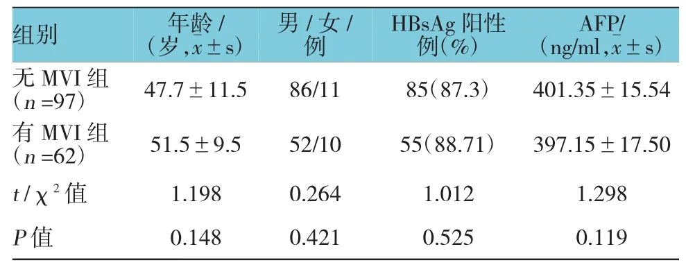
根据患者有无发生MVI将其分为无MVI组(n=97)和有MVI组(n=62)。两组患者在年龄、性别和HBsAg阳性的百分比以及血清AFP水平等方面差异无统计学意义(P>0.05),具有可比性。见表2。
2.3 无进展生存期的影响因素
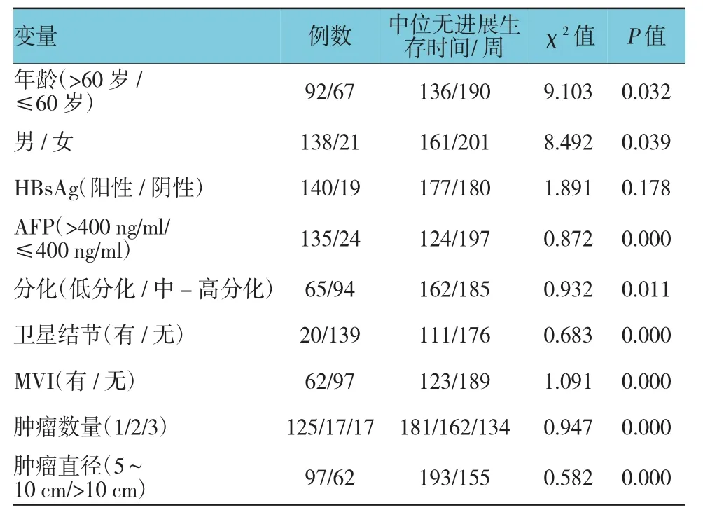
159例孤立性大肝癌根治术后的1、3和5年生存率分别为88.2%、56.7%和41.4%。单因素分析显示,年龄、性别、AFP水平、肿瘤分化程度、卫星结节、MVI、肿瘤数量和直径影响无进展生存期,差异有统计学意义(P<0.05)。见表3。

表1 患者的一般临床特征

表2 两组患者的一般临床特征比较
2.4 MVI与患者无进展生存期的关系
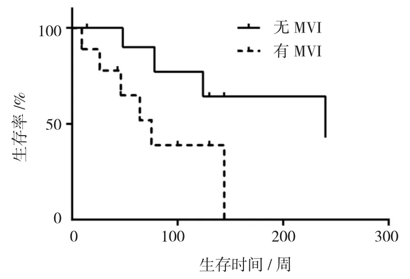
159例患者中,62例患者发生MVI,其发生率为39.0%。无MVI患者中位无进展生存期为47个月,1、3和5年无进展生存率分别为91.7%和67.5%和56.0%;有MVI患者中位无进展生存期为35个月,1和3年无进展生存率分别为84.7%和45.8%。Log-rank检验结果表明,有MVI患者根治术后的生存率低于无MVI患者,两者比较差异有统计学意义(χ2=7.98,P=0.003)。两组患者术后生存曲线。以单因素分析中有统计学意义的结果变量为自变量,以术后无进展生存期为因变量,采用多因素COX生存分析无进展生存期的影响因素。逐步法进行变量的筛选,引入水准为ɑ=0.05,剔除水准β=0.1。结果显示,AFP>400 ng/ml、有 MVI、卫星结节、肿瘤数量和肿瘤直径影响无进展生存期。见表4和附图。

表3 159例孤立性大肝癌患者术后影响无进展生存期的单因素分析

表4 孤立性大肝癌患者术后影响无进展生存期的多因素分析

附图 有MVI和无MVI患者根治术后的生存率比较
3 讨论
目前,手术切除仍然是未发生转移的孤立性大肝癌的主要治疗方式[1,4]。近年来,国内外的很多医院都主张对于米兰标准以外的孤立性大肝癌也进行手术切除[4-5]。因此,对于疗效和预后的预测就显得尤为重要,其对于临床上决定是否在术后需要进行化疗具有非常重要的意义。临床上,患者由于肿瘤分期、肝功能状态及一般健康状况等不同,其预后可能会存在较大的差异。临床实践发现即使是中晚期的肝癌患者也可能达到Child-Pugh A级[4-5,7]。
本研究评估的肝癌的特征包括:AFP水平、肿瘤分化程度、MVI、卫星结节、肿瘤数量以及肿瘤直径。根据以往的研究成果,本研究选择400 ng/ml作为阈值[8-10]。本研究结果显示,AFP>400 ng/ml(P=0.031),MVI(P=0.000),卫星结节(P=0.028)。根据以往的研究结果,本研究选取肿瘤数量1、2以及≥3作为判断的标准[11-12]。结果显示,肿瘤数量的P=0.000。另外,由于既往研究对于肿瘤直径的阈值的选取差异较大[13-14],因此本研究采用连续变量作为肿瘤直径的判断标准,结果显示,P=0.000。因此,AFP>400ng/ml、MVI、卫星结节、肿瘤数量及肿瘤直径是影响孤立性大肝癌根治术后无进展生存期的主要因素。这与国内外的研究成果是一致的[15-16]。
另外,本研究结果显示,MVI是影响孤立性大肝癌根治术后无进展生存期的最为显著的因素。以往的研究结果显示,MVI是影响肝癌切除术后复发的最重要的因素之一。TORZILLI等人[13]的研究结果显示,肝癌细胞侵犯门静脉和肝静脉小分支的肌肉层,肝癌细胞出现在距离肿瘤包膜>1 cm的门静脉和肝静脉小分支内,以及肝癌细胞出现在>5处的门静脉和肝静脉小分支均与肝癌切除术后复发正相关。另外,肝癌细胞侵犯门静脉和肝静脉小分支的肌肉层是影响肝癌切除术后复发的独立危险因素。AN等人[14]的研究结果显示,在门静脉和肝静脉小分支内出现>50个肝癌细胞是影响肝癌切除术后复发的独立危险因素。与<50个肝癌细胞和无MVI的患者相比较,>50个肝癌细胞的患者的术后生存率降低。BRUIX等人[15]的结果显示,无MVI的肝癌患者的生存率高于1~5处门静脉和肝静脉小分支发生MVI和>5处的患者。另外,>5处发生MVI的患者复发率和转移率高于无MVI和1~5处MVI的患者。MINAGAWA等人[16]的Meta分析结果表明,肝癌MVI患者的3、5年生存率低于无MVI的患者。另外,该类患者即使接受肝移植,其3年生存率也明显降低。这与本研究成果是一致的,提示术前评估MVI对于准确预测孤立性大肝癌根治术后患者预后的重要性。
本研究也存在着一些不足之处。首先,本研究仅在本院开展,因此样本的数量和种类都受到一定的限制。如果能够开展多中心研究的话,本研究结果和结论将更为可靠和准确。其次,本研究仅调查接受根治手术的孤立性肝癌患者的情况,需要进一步的研究使得本研究结果的适用范围更广。最后,本研究的样本数量有限(n=159),需要进一步的研究以扩大样本量以使本研究结果的适用范围更广。
综上所述,AFP>400 ng/ml、MVI、卫星结节、肿瘤数量及肿瘤直径是影响孤立性大肝癌根治术后无进展生存期的重要因素。
[1]WANG F S,FAN J G,ZHANG Z,et al.The global burden of liver disease:the major impact of China[J].Hepatology,2014,60(6):2099-2108.
[2]LIU P H,HSU C Y,HSIA C Y,et al.Surgical resection versus radiofrequency ablation for single hepatocellular carcinoma≤2 cm in a propensity score model[J].Annals of Surgery,2016,263(3):538-545.
[3]VITALE A,BURRA P,FRIGO A C,et al.And italian liver cancer g.survival benefit of liver resection for patients with hepatocellular carcinoma across different barcelona clinic liver cancer stages:a multicentre study[J].Journal of Hepatology,2015,62(3):617-624.
[4]ZHONG J H,KE Y,GONG W F,et al.Hepatic resection associated with good survival for selected patients with intermediate and advanced-stage hepatocellular carcinoma[J].Annals of Surgery,2014,260(2):329-340.
[5]LIM K C,CHOW P K,ALLEN J C,et al.Microvascular invasion is a better predictor of tumor recurrence and overall survival following surgical resection for hepatocellular carcinoma compared to the Milan criteria[J].Annals of Surgery,2011;254(1):108-113.
[6]HUNG H H,LEI H J,CHAU G Y,et al.Milan criteria,multi-nodularity and microvascular invasion predict the recurrence patterns of hepatocellular carcinoma after resection[J].Journal of Gastrointestinal Surgery,2013,17(4):702-711.
[7]KIM J M,KWON C H,JOH J W,et al.Differences between hepatocellular carcinoma and hepatitis B virus infection in patients with and without cirrhosis[J].Annals of Surgical Oncology,2014,21(2):458-465.
[8]GOH B K,TEO J Y,CHAN C Y,et al.Importance of tumor size as a prognostic factor after partial liver resection for solitary hepatocellular carcinoma:implications on the current AJCC staging system[J].Journal of Surgical Oncology,2016,113(1):89-93.
[9]YANG S L,LIU L P,YANG S,et al.Preoperative serum alpha-fetoprotein and prognosis after hepatectomy for hepatocellular carcinoma[J].British Journal of Surgery,2016,103(6):716-724.
[10]ELNEKAVE E,ERINJERI J P,BROWN K T,et al.Long-term outcomes comparing surgery to embolization-ablation for treatment of solitary HCC<7 cm[J].Annals of Surgical Oncology,2013,20(9):2881-2886.
[11]LI J,ZHOU J,YANG P H,et al.Nomograms for survival prediction in patients undergoing liver resection for hepatitis B virus related early stage hepatocellular carcinoma[J].European Journal of Cancer,2016,62:86-95.
[12]LI Y,XIA Y,LI J,et al.Prognostic nomograms for pre-and postoperative predictions of long-term survival for patients who underwent liver resection for huge hepatocellular carcinoma[J].Journal of the American College of Surgeons,2015,221(5):962-974.
[13]TORZILLI G,DONADON M,BELGHITI J,et al.Predicting individual survival after hepatectomy for hepatocellular carcinoma:a novel nomogram from the“HCC East&West Study Group”[J].Journal of Gastrointestinal Surgery,2016,18(6):1154-1162.
[14]AN C,KIM D W,PARK Y N,et al.Single hepatocellular carcinoma:preoperative MR imaging to predict early recurrence after curative resection[J].Radiology,2015,276:433-443.
[15]BRUIX J,HAN K H,GORES G,et al.Liver cancer:approaching a personalized care[J].Journal of Hepatology,2015,62(1):S144-156.
[16]MINAGAWA M,IKAI I,MATSUYAMA Y,et al.Staging of hepatocellular carcinoma:assessment of the Japanese TNM and AJCC/UICC TNM systems in a cohort of 13,772 patients in Japan[J].Annals of Surgery,2007;245(6):909-922.
Relationship between microvascular invasion and pathological features of solitary large hepatocellular carcinoma and curative effect after radical*
Cong Gao1,Yong Han2,Da-yong Lin3,Yu Zhang4,Yun-fei Chen4
(1.Department of Emergency;2.Department of Pharmacy;3.Department of Anesthesiology,4.Department of Hepatobiliary Surgery,Sichuan Provincial People's Hospital,Chengdu,Sichuan 610072,China)
R735.7
A
10.3969/j.issn.1005-8982.2017.15.017
1005-8982(2017)15-0082-05
2017-02-09
四川省卫生厅课题(No:30305030258)
