Wnt/β-catenin信号通路在鼻咽癌组织的异常激活
2017-07-03黄孝文崔伟伟王宝凤崔永华
黄孝文, 崔伟伟, 王宝凤, 崔永华
华中科技大学同济医学院附属同济医院耳鼻咽喉头颈外科,武汉 430030
实验研究
Wnt/β-catenin信号通路在鼻咽癌组织的异常激活
黄孝文, 崔伟伟, 王宝凤, 崔永华
华中科技大学同济医学院附属同济医院耳鼻咽喉头颈外科,武汉 430030
目的 探讨Wnt/β-catenin信号通路的异常激活在鼻咽癌发生发展机制中可能的作用。方法 以10例正常鼻咽组织为对照,运用免疫组织化学技术检测24例鼻咽癌组织中Wnt2和β-catenin的表达水平及其在癌细胞中的定位情况;运用实时定量PCR技术和蛋白免疫印迹技术分别检测鼻咽癌组织中Wnt2和β-catenin基因和蛋白的表达水平。结果 免疫组化检测结果显示,在鼻咽癌标本中,Wnt2的表达明显高于对照组织,且阳性染色主要位于细胞质内;β-catenin在细胞膜上的表达量显著低于对照组织,而在细胞质和细胞核中的表达呈明显上调。实时定量PCR检测结果显示,Wnt-2 mRNA在鼻咽癌组织中的表达量明显高于对照组织(P<0.01);β-catenin mRNA表达量则低于对照组织(P=0.016)。蛋白免疫印迹技术检测结果表明,Wnt2蛋白在鼻咽癌组织中的表达高于对照组织,而β-catenin蛋白在受检肿瘤标本中的含量亦呈明显上调。结论 Wnt2、β-catenin在鼻咽癌组织中存在明显的异常表达,提示Wnt/β-catenin信号通路的异常激活可能是鼻咽癌发生发展的重要分子机制之一。
鼻咽癌; Wnt/β-catenin信号通路; 异常激活
鼻咽癌(nasopharyngeal carcinoma,NPC)是我国南部地区常见的头颈部恶性肿瘤。目前,鼻咽癌发生发展的分子机制尚未完全阐明。有研究发现β-连环蛋白(β-catenin)在鼻咽癌组织存在异常表达,但Wnt(wingless-type MMTV integration site family member)/β-catenin信号通路的激活在其病理机制中的作用尚未见报道。本研究应用免疫组织化学技术、实时定量PCR技术和蛋白免疫印迹技术分别检测鼻咽癌组织中Wnt2和β-catenin的表达情况,探讨Wnt/β-catenin信号通路的异常激活在鼻咽癌发生发展中可能的作用,进而为其分子靶向治疗提供实验依据。
1 材料与方法
1.1 主要试剂与仪器
①免疫组织化学实验:羊抗兔单克隆抗体Wnt-2(Abcam,美国),羊抗兔多克隆抗体β-catenin(三鹰公司,武汉),过氧化物酶标记的亲和素-生物素-酶复合体二抗试剂盒(博士德公司,武汉)等。②实时定量PCR实验:RNA提取液Trizol(Fermentas公司,加拿大),RT-PCR mix(TaKaRa公司,大连),RT-PCR引物(生物工程技术服务有限公司,上海),RT-PCR SYBR green(Fermentas公司,加拿大),RNA逆转录试剂盒(TaKaRa公司,大连),RT-PCR仪(罗氏诊断应用科学公司,德国),PCR扩增仪(Roche公司,瑞士),荧光显微镜(Olympus公司,日本)等。③Western blot实验:RIPA裂解液(碧云天公司,济南),Cooktail(Roche公司,瑞士),SDS-PAGE凝胶试剂盒(博士德公司,武汉),蛋白上样缓冲液(博士德公司,武汉),SDS(Sigma公司,美国),Tris-base(Sigma公司,美国),PVDF膜(博士德公司,武汉)等。
1.2 研究对象
1.2.1 病例资料 从2012年11月到2014年12月在武汉同济医院耳鼻咽喉头颈外科门诊就诊的病例中,随机选取疑诊鼻咽肿瘤的患者30例。患者主诉包括颈部包块10例(33.3%),头痛6例(20.0%),耳鸣耳闭5例(16.7%),涕中带血5例(16.7%),鼻塞4例(13.3%);所有病例的电子鼻咽镜检查结果均提示鼻咽新生物可能。另随机选取非咽喉病患者10例作为正常对照组。
1.2.2 标本采集与处理 在患者知情并同意的情况下,对每一病例分别于表面麻醉下行内镜辅助鼻咽组织活检术。对所获取的病理标本,取部分组织以多聚甲醛固定后送临床病理学检查,剩余组织立即放入消毒的离心管,并迅速放入液氮中,随之保存于-80℃冰箱备用。
1.2.3 病理学检查结果及研究分组 对所有研究对象的鼻咽活检组织进行临床病理学检测,根据最终病理结果分组:30例疑癌患者中6例确诊为鼻咽慢性炎症,予以剔除;余24例确诊为不同分化程度和角化类型的鼻咽鳞状细胞癌,为实验组。非咽喉病患者10例,病理检查均排除鼻咽肿瘤和炎症,为对照组。实验组24例中男18例,女6例,平均年龄50.1岁;对照组10例中男6例,女4例,平均年龄44.8岁。
1.3 Wnt2、β-catenin的免疫组织化学染色
对全部标本行常规石蜡包埋切片。以0.01 mmol/L PBS缓冲液来代替一抗作为阴性对照,以Wnt2、β-catenin蛋白表达阳性的乳腺癌组织作为阳性对照,对全部鼻咽组织标本进行免疫组织化学检测。显微镜下观察并采图,记录染色部位和染色程度。
1.4 Wnt2、β-catenin mRNA的实时定量PCR检测
Trizol法提取鼻咽组织标本的总RNA,按反转录试剂盒说明书进行反转录,反应结束后,将产物cDNA放入-20℃冰箱保存。按试剂盒说明进行Wnt2、β-catenin的实时定量PCR反应。Wnt2、β-catenin及GAPDH基因片段的引物序列分别为:Wnt2上游引物5′-GCTCCCTCTGCTCTTGACCT-3′,下游引物5′-GCACATTATCGCACATCACC-3′;β-catenin上游引物5′-GGGTCCTCTGTGAACTTGCT-3′,下游引物5′-AATCTTGTGGCTTGTCCTCA-3′;GAPDH上游引物5′-ACCCAGAAGACTGTGGATGG-3′,下游引物5′-TTCTAGACGGCAGGTCAGGT-3′。采集RT-PCR数据,用2-ΔΔCt方法求得目的基因相对于内参基因的相对表达量。
1.5 Wnt2、β-catenin蛋白的Western blot检测
参照试剂盒说明书提取Wnt2和β-catenin的总蛋白,进行免疫印迹检测,将所得印迹放于曝光仪内曝光,并进行图像分析。
1.6 统计学分析
利用SPSS 17.0统计软件,采用Mann-Whitney U检验对实验组和对照组两独立样本的实验数据行非参数检验分析,以P<0.05为差异有统计学意义。
2 结果
2.1 Wnt2、β-catenin的免疫组织化学染色及定位
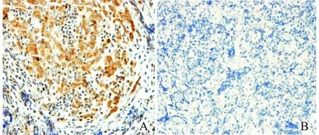
由图1可见,Wnt2在鼻咽癌组织中呈阳性表达,其染色定位于细胞质内,即胞质内出现均匀一致的棕黄色颗粒,其中强阳性62.5%(15/24),中等阳性37.5%(9/24)。Wnt2在正常鼻咽组织细胞质内基本无表达。
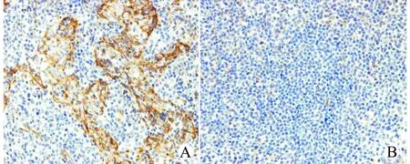
由图2可见,β-catenin在鼻咽癌组织中胞膜阳性表达减弱或消失,胞质或胞核呈阳性表达,其中强阳性54.2%(13/24),中等阳性33.3%(8/24)。在正常鼻咽组织中呈胞膜阳性表达,即细胞膜出现均匀的着色,胞质和胞核无染色(7/10)。

图1 鼻咽癌组织(A)及正常鼻咽组织(B)Wnt2免疫组织化学染色(×400)Fig.1 Immunohistochemical staining of Wnt2 in NPC(A)and control nasopharyngeal tissues(B)(×400)

图2 鼻咽癌组织(A)及正常鼻咽组织(B)β-catenin免疫组织化学染色(×400)Fig.2 Immunohistochemical staining of β-catenin in NPC(A)and control nasopharyngeal tissues(B)(×400)
2.2 Wnt2、β-catenin mRNA表达
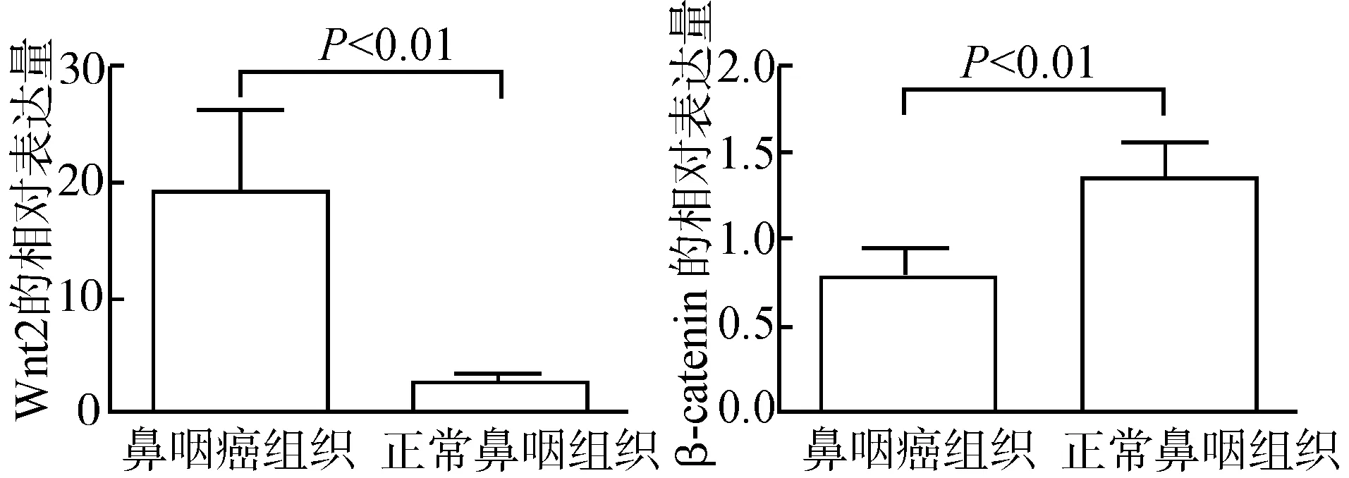
采用实时荧光定量PCR检测鼻咽组织Wnt2、β-catenin mRNA表达,结果如图3所示:与正常鼻咽组织比较,鼻咽癌组织中Wnt2 mRNA相对表达量增高(P<0.01),β-catenin mRNA相对表达量降低(P=0.016)。

图3 实时荧光定量PCR检测Wnt2、β-catenin mRNA表达Fig.3 Real time fluorescence-based quantitative PCR detection of Wnt2 and β-catenin mRNA in NPC and control tissues
2.3 Wnt2、β-catenin蛋白表达
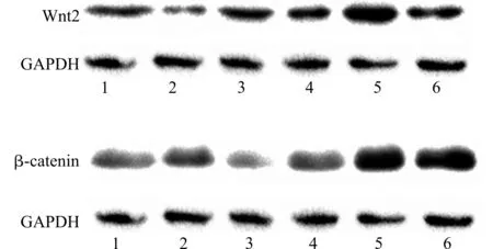
采用Western blot技术检测Wnt2、β-catenin蛋白表达,结果如图4所示:Wnt2、β-catenin蛋白在鼻咽癌组织中的表达量均高于正常鼻咽组织。

1~3:正常鼻咽组织;4~6:鼻咽癌组织图4 Western blot检测Wnt2、β-catenin蛋白表达Fig.4 Wnt2 and β-catenin protein expression in NPC and control tissues detected by Western blotting
3 讨论
肿瘤的发生发展与细胞内某些信号通路的异常激活有着密切关系,而Wnt/β-catenin信号通路是近年来学者们研究的热点。Wnt蛋白是一类富含丝氨酸且有19种家族成员的分泌型糖蛋白,以自分泌或旁分泌的方式产生后,Wnt蛋白与细胞膜上的特异性受体结合,激活经典的Wnt信号通路,作用于下游的靶基因,参与细胞生命活动的调控、细胞极化以及迁移等[1-2]。Wnt信号通路还可能通过抑制细胞凋亡来促进肿瘤细胞的增殖[3]。临床研究发现,该通路中的Wnt2蛋白参与了多种肿瘤的发生,如乳腺癌[4]、结直肠癌和胃癌[5-6]、食管癌[7]、胰腺癌[8]等。而β-catenin是一种多功能细胞骨架蛋白,在正常情况内,它与E-钙黏蛋白及α-catenin等形成复合体,介导细胞粘附,在维持细胞稳定、防止细胞迁移等方面发挥重要作用[9]。此外,作为Wnt通路十分重要的信号分子,β-catenin在基因转录过程中发挥重要作用。当Wnt信号被异常激活时,原本定位于细胞膜的β-catenin则在细胞质内大量积聚,同时其降解受到抑制,进而可能易位进入细胞核,激活下游的靶基因,导致后者转录增加,从而促进肿瘤的发生及发展。已有研究报道β-catenin在多种肿瘤中存在异常表达,而且与部分肿瘤的进展相关联[10-13]。
基于上述研究结果,我们推测Wnt/β-catenin信号通路在鼻咽癌中也极可能存在异常激活。为了证实这一假设,本研究运用免疫组织化学技术、实时定量PCR技术和Western blot法分别检测了鼻咽癌标本中Wnt2和β-catenin的表达情况。实验结果显示,一方面,在鼻咽癌组织中Wnt2呈高表达,在正常鼻咽组织标本中呈显著的低表达,而实时定量PCR检测结果也证实,Wnt2 mRNA在鼻咽癌标本中表达量明显高于正常鼻咽组织,差异有统计学意义(P<0.01),这与其他作者对结肠、直肠癌组织的Wnt2检测结果[5]是一致的。Western blot检测的结果也进一步证实了Wnt2的表达趋势,即在鼻咽癌中表达升高。另一方面,与正常鼻咽标本比较,β-catenin在鼻咽癌标本胞膜上的表达显著降低,而在胞质和胞核中表达升高,表明β-catenin在胞质出现了异常积聚且易位进入了胞核。实时定量PCR检测结果显示β-catenin mRNA在鼻咽癌组织中的表达量明显低于正常鼻咽组织(P=0.016)。这个结果似乎与免疫组化所见不尽一致。如上所述,正常情况下,β-catenin与E-钙黏蛋白在细胞表面形成复合物共同参与细胞间的粘附,这是β-catenin的主要存在形式。当发生癌变时,细胞表面的E-钙黏蛋白受到破坏,与其结合的β-catenin明显减少,后者大量进入胞质内,加之降解受阻,导致β-catenin在胞质内大量积聚,并可能易位进入细胞核。因此β-catenin在胞质内的积聚和增多是该蛋白从胞膜转移至胞质且降解受阻所导致的结果,并非由于基因表达量的增高所造成。β-catenin在胞质积聚且易位进入胞核是否反馈抑制其基因表达,有待进一步研究。本研究的Western blot检测发现β-catenin蛋白在鼻咽癌组织中的表达较正常鼻咽组织明显上调,这一现象可能是β-catenin蛋白在胞质和胞核异常分布且降解受阻的结果。这一结果与其他作者在其他类型肿瘤的研究结果[10-12]一致。
综上所述,本研究结果显示,在鼻咽癌组织中,Wnt2基因及蛋白的表达均上调,同时β-catenin蛋白含量升高,并且出现胞质和胞核的异常聚集,提示异常激活的Wnt/β-catenin信号通路极可能是鼻咽癌发生发展的重要机制之一。随着相关研究的不断深入和突破,Wnt/β-catenin分子通路有望成为鼻咽癌基因靶向治疗的新目标。
[1] Miller J R.The wnts[J].Genome Biol,2002,3(1):1-15.
[2] Cadigan K M,Nusse R.Wnt signaling:a common theme in animal development[J].Genes Dev,1997,11(24):3286-3305.
[3] Zeng Z Y,Zhou Y H,Zhang W L,et al.Gene expression profiling of NPC reveals the abnormally regulated Wnt signaling pathway[J].Hum Pathol,2007,38(1):120-133.
[4] 郭变琴,吴立翔,汪先桃,等.Wnt2在乳腺癌组织及患者血清中的表达及意义[J].细胞与分子免疫学杂志,2013,29(6):633-636.
[5] Katoh M.Frequent up-regulation of WNT2 in primary gastric cancer and colorectal cancer[J].Int J Oncol,2001,19(5):1003-1007.
[6] 刘龑航,刘映辉,董锡钓.Wnt2,APC及c-myc在胃癌中的表达[J].中国老年学杂志,2013,33(23):5838-5840.
[7] Fu L,Zhang C,Zhang L Y,et al.Wnt2 secreted by tumour fibroblasts promotes tumour progression in oesophageal cancer by activation of the Wnt/β-catenin signalling pathway[J].Gut,2011,60(12):1635-1643.
[8] Jiang H,Li Q,He C,et al.Activation of the Wnt pathway through Wnt2 promotes metastasis in pancreatic cancer[J].Am J Cancer Res,2014,4(5):537-544.
[9] Morin P J.β-catenin signaling and cancer[J].Bioessays,1999,21(12):1021-1030.
[10] Dong B,Lee J S,Park Y Y,et al.Activating CAR and β-catenin induces uncontrolled liver growth and tumorigenesis[J].Nat Commun,2015,6:5944.
[11] Xu X,Kim J E,Sun P L,et al.Immunohistochemical demonstration of alteration of β-catenin during tumor metastasis by different mechanisms according to histology in lung cancer[J].Exp Ther Med,2015,9(2):311-318.
[12] Nie E,Zhang X,Xie S,et al.β-catenin is involved in Bex2 down-regulation induced glioma cell invasion/migration inhibition[J].Biochem Biophys Res Commun,2015,456(1):494-499.
[13] Jamieson C,Sharma M,Henderson B R.Targeting the β-catenin nuclear transport pathway in cancer[J].Semin Cancer Biol,2014,27:20-29.
(2017-02-23 收稿)
Aberrant Activation of Wnt/β-catenin Signaling Pathway in Nasopharyngeal Carcinoma Tissues
Huang Xiaowen,Cui Weiwei,Wang Baofengetal
DepartmentofOtolaryngologyHeadandNeckSurgery,TongjiHospital,TongjiMedicalCollege,HuazhongUniversityofScienceandTechnology,Wuhan430030,China
Objective To investigate expression of Wnt2 and β-catenin in nasopharyngeal carcinoma (NPC),so as to explore the possible role of aberrant activation of Wnt/β-catenin signaling pathway in the mechanism of tumorigenesis and development of NPC.Methods With 10 normal nasopharyngeal tissue samples as control,Wnt2 and β-catenin expression levels and localization in the NPC cells of 24 cases were detected by using immunohistochemistry.The mRNA and protein expression levels of Wnt2 and β-catenin in the NPC tissues were detected respectively by using real-time quantitative PCR and Western blotting.Results Immunohistochemistry illustrated that the expression of Wnt2 was significantly higher in the NPC than in the control nasopharyngeal tissues,and the positive staining was mainly located in the cytoplasm;the expression of β-catenin on the cell membrane in the NPC was significantly lower than that of the control tissues,however,β-catenin in the cytoplasm and the nucleus of NPC was significantly up-regulated.Real-time quantitative PCR showed that the expression of Wnt2 mRNA in the NPC tissue was significantly higher than that in the control tissues(P<0.01),and the expression of β-catenin mRNA was significanlty lower than that of the control tissues(P=0.016).Western blotting revealed that the expression of Wnt2 protein in the NPC tissue was higher than that in the control tissues,and the content of β-catenin protein was significantly increased in the tested NPC samples.Conclusion There are significantly abnormal expression levels of Wnt2 and β-catenin in NPC,suggesting that aberrant activation of Wnt/β-catenin signaling pathway may be one of important molecular mechanisms of tumorigenesis and development of NPC.
nasopharyngeal carcinoma; Wnt/β-catenin signaling pathway; aberrant activation
R739.63
10.3870/j.issn.1672-0741.2017.03.011
黄孝文,男,1962年生,主任医师,E-mail:xwhuang@tjh.tjmu.edu.cn
