干细胞转染CMV-Luciferase-PGK-Puro慢病毒后化学发光成像
2016-12-29阮光萍刘菊芬李自安王金祥吕燕波庞荣清潘兴华
阮光萍,刘菊芬,李自安,王金祥,吕燕波,庞荣清,潘兴华
干细胞转染CMV-Luciferase-PGK-Puro慢病毒后化学发光成像
阮光萍,刘菊芬,李自安,王金祥,吕燕波,庞荣清,潘兴华
目的研究用化学发光成像的方法观察脐带间充质干细胞转染CMV-Luciferase-PGK-Puro慢病毒后的情况,代替活体成像仪的可行性。方法用CMV-Luciferase-PGK-Puro慢病毒转染树鼩脐带间充质干细胞,用最适浓度的嘌呤霉素筛选,存活的细胞用6孔板的3个孔培养,贴壁后,3个孔依次加入底物D-荧光素钾盐,用化学发光成像仪拍照,再用软件进行活体成像转换。将转染成功的细胞注入麻醉后的树鼩皮下,树鼩静脉注射底物。结果细胞加入底物后有生物发光,发光强度随底物作用时间延长而减弱。树鼩皮下也观察到发光细胞。结论CMV-Luciferase-PGK-Puro慢病毒能成功转染树鼩脐带间充质干细胞,转染成功的细胞用于动物模型治疗后,可进行活体成像,观察细胞在动物体内的分布。
树鼩;脐带;间充质干细胞;CMV-Luciferase-PGK-Puro慢病毒;转染;化学发光成像
近年来,小动物活体成像技术得到了较大推广,广泛应用于动物模型、肿瘤机制等研究[1-4]。各种细胞在小动物体内的定位示踪进一步推广了这一技术的应用。细胞通常用CMV-Luciferase-PGK-Puro慢病毒转染,细胞回输后在动物体内定位、扩增后,可通过给动物注射底物D-荧光素钾盐后,带有CMV-Luciferase-PGK-Puro基因的细胞就会与底物作用而发光,通过活体成像仪可检测到发光现象。但活体成像仪价格昂贵,只有少数单位能够购买。本实验室有化学发光成像仪,可以观察到生物发光。于是本研究用化学发光成像仪观察CMV-Luciferase-PGK-Puro慢病毒转染的树鼩脐带间充质干细胞的情况,探讨其代替活体成像仪的可行性,为下一步的树鼩疾病模型研究奠定基础。
1 材料与方法
1.1 试剂 CMV-Luciferase-PGK-Puro慢病毒和底物D-荧光素钾盐购自上海吉满生物技术有限公司,化学发光成像仪为上海天能科学仪器厂生产 (型号:Tanon 5200);树鼩购自中科院昆明动物所,实验动物生产许可证号为SCXK(滇)K2013-0005,实验动物使用许可证号为SYXK(滇)K2013-0012。
1.2 树鼩脐带间充质干细胞的分离培养 收集进行剖腹产的树鼩的脐带,用盐水冲洗,用青、链霉素双抗浸泡,用小剪刀尽量将脐带剪碎,放入培养瓶中贴壁培养。第2 d可见单个细胞从组织块爬出贴壁,第7 d左右细胞长满可传代;传代后为第1代细胞,第7 d左右细胞再次长满,可传代为第2代细胞;第3代细胞用于CMV-Luciferase-PGK-Puro慢病毒转染。
1.3 CMV-Luciferase-PGK-Puro慢病毒转染树鼩脐带间充质干细胞 200 μl CMV-Luciferase-PGK-Puro慢病毒,滴度2×10⁷TU/ml,每瓶4×106个树鼩脐带间充质干细胞,加入50 μl(1×10⁶TU)CMV-Luciferase-PGK-Puro慢病毒,转染 3 d后,加入嘌呤霉素2~3 μg/m l。加入嘌呤霉素后,只有转染成功的细胞能够存活。待细胞筛选成功后,取5×10⁵个细胞接种6孔板,共接种3个孔。
1.4 化学发光仪成像观察慢病毒转染干细胞结果
接种后第2 d、细胞贴壁后,6孔板每孔有2 ml培养基,3个孔依次加20 μl D-荧光素钾盐(15 mg/ml),先在A1孔加入20 μl底物D-荧光素钾盐,化学发光仪检测生物发光度;再在A2孔加入20 μl底物D-荧光素钾盐,化学发光仪检测生物发光度;最后在A3孔加入20 μl底物D-荧光素钾盐,化学发光仪检测生物发光度。3个孔依次加入底物,可以证明生物发光的特异性。用仪器自带软件进行活体成像合成,最后可观察到6孔板中的发光现象。
1.5 慢病毒转染成功的树鼩脐带间充质干细胞的体内实验 将一只10 w的树鼩背部用脱毛膏脱毛,将树鼩麻醉后,背部皮下注入3.7×10⁶个慢病毒转染成功的树鼩脐带间充质干细胞,同时尾静脉注射1.3 ml底物D-荧光素钾盐,化学发光仪成像拍照检测树鼩背部皮下的生物发光,用仪器自带软件进行活体成像合成。树鼩的背部用脱毛膏脱毛,方便观察结果。
2 结果
2.1 树鼩脐带间充质干细胞的生长与形态 原代培养的树鼩脐带间充质干细胞从剪碎的组织块爬出,贴壁生长(图1A)。随着培养时间的延长,贴壁生长的细胞越来越多,呈长梭形,长满后可传代(图1B)。
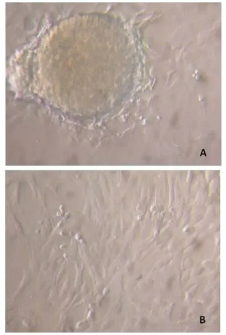
图1 树鼩脐带间充质干细胞的生长与形态
2.2 细胞生物发光检测结果 A1孔加入20 μl底物后,化学发光成像仪拍照观察到生物发光(图2)。
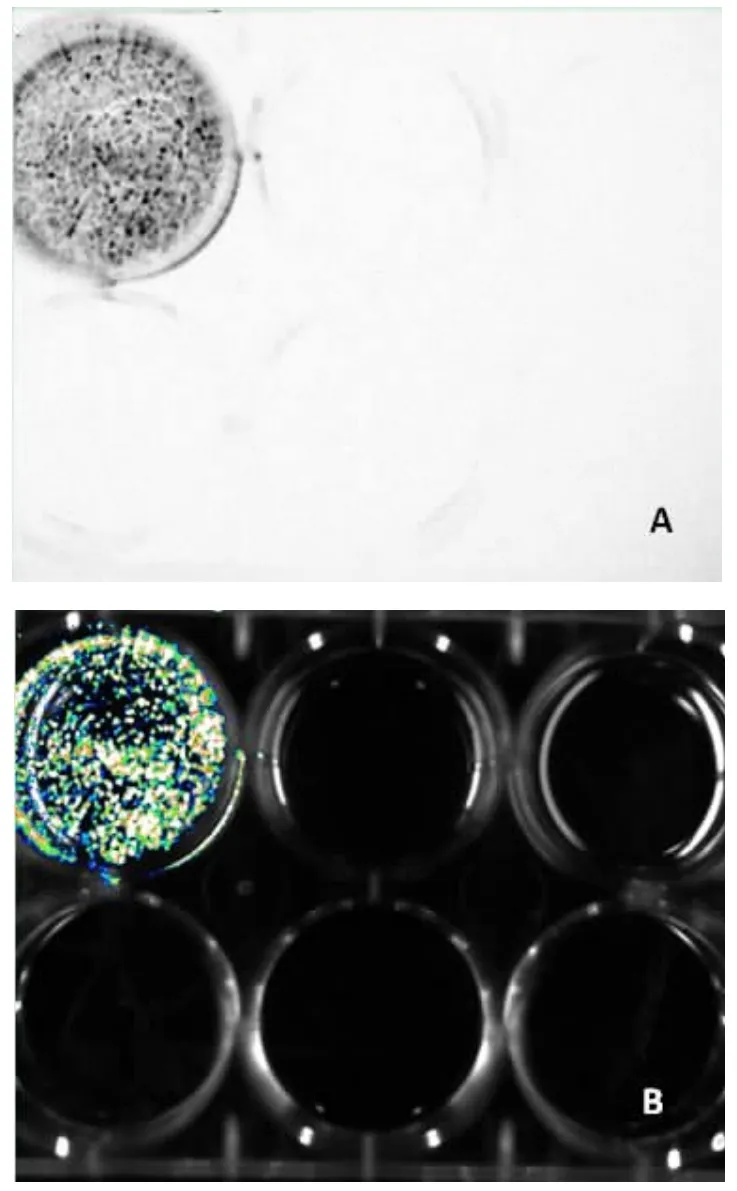
图2 细胞生物发光检测结果
A2孔加入20 μl底物后,化学发光成像仪观察到A1和A2孔均发光,新加入底物的A2孔发光更强(图3)。
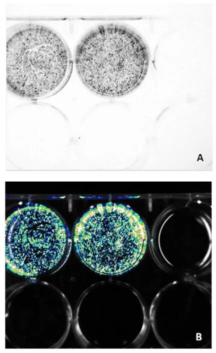
图3 化学发光成像仪观察结果
A3孔加入20 μl底物后,化学发光成像仪观察到A1、A2和A3孔均发光,新加入底物的A3孔发光更强,而A2孔发光强于A1孔(图4)。
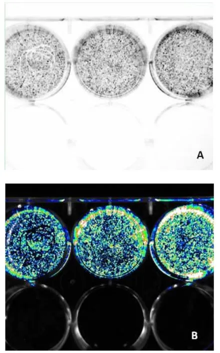
图4 化学发光成像仪观察结果
通过平行3个孔的研究,证明细胞生物发光是特异的,并且细胞发光强弱与D-荧光素钾盐底物的加入时间有关,随加入时间延长,细胞生物发光逐渐减弱。
2.3 树鼩皮下化学发光成像 在树鼩皮下注射慢病毒转染成功的树鼩脐带间充质干细胞、静脉注射底物后,化学发光成像仪观察到生物发光,用软件合成后类似活体成像(图5)。
3 讨论
本实验通过用普通的化学发光成像仪观察到细胞转染CMV-Luciferase-PGK-Puro慢病毒是成功的,通过平行3个孔的研究,证明细胞生物发光是特异的,并且细胞发光强弱与D-荧光素钾盐底物的加入时间有关,随加入时间延长,细胞生物发光逐渐减弱。通过本实验研究,证明CMV-Luciferase-PGK-Puro慢病毒可以成功转染树鼩脐带间充质干细胞,转染成功的细胞可用于治疗各种动物疾病模型。近年来,越来越多的文献报道了脐带间充质干细胞可用于多种人类疾病的治疗,包括系统性红斑狼疮、糖尿病、代谢综合征等[5-8]。由于脐带间充质干细胞来源于生产后废弃的脐带,没有伦理学争论,培养容易,又具有干细胞多向分化与修复损伤的作用,已越来越受到研究者的重视[9-12]。
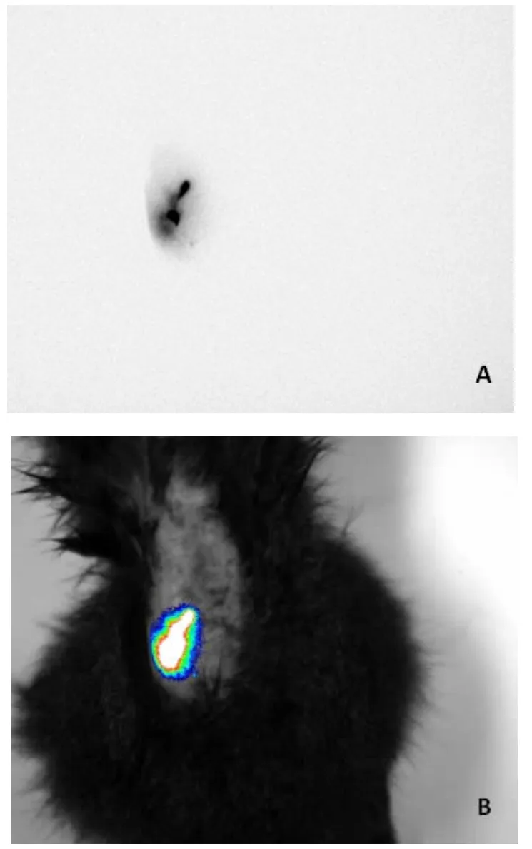
图5 树鼩皮下活体成像结果
转染CMV-Luciferase-PGK-Puro慢病毒后的树鼩脐带间充质干细胞,通过嘌呤霉素的筛选,转染成功的细胞存活下来,并可以继续传代扩增。有了种子细胞,今后用于各种树鼩动物模型治疗,可以更好地通过活体成像仪追踪细胞在体内的分布与定位,为脐带间充质干细胞治疗各种动物模型的疗效与机制研究打下了良好的基础。
通过软件转换,生物发光效果图可转化为活体成像效果图,转化后的发光图片与小动物活体成像仪观察到的效果一致。所以,用化学发光成像仪可以对细胞的转染效果进行观察,可在本单位完成实验,而不需要联系到外单位的小动物活体成像仪,避免细胞运输过程培养基逸出,细胞活性受到影响,使化学发光成像仪发挥更大的作用与价值。
[1] Vicidomini C,Panico M,Greco A,et al.In vivo imaging and characterization of[(18)F]DPA-714,a potential new TSPO ligand, in mouse brain and peripheral tissues using small-animal PET[J]. Nucl Med Biol,2015,42(3):309-316.
[2] Lee O,Lee G,Kim M,et al.Multimodal evaluation of xenograft tumors in mice with an in-vivo stereo imaging system and small-animal PET/CT[J].Melanoma Res,2013.
[3] Hag AM,Ripa RS,Pedersen SF,et al.Small animal positron emission tomography imaging and in vivo studies of atherosclerosis[J].Clin Physiol Funct Imaging,2013,33(3):173-185.
[4] Fuchs K,Kohlhofer U,Quintanilla-Martinez L,et al.In vivo imaging of cell proliferation enables the detection of the extent of experimental rheumatoid arthritis by 3'-deoxy-3'-18ffluorothymidine and small-animal PET[J].J Nucl Med,2013,54 (1):151-158.
[5] Zhuang Y,Li D,Fu J,et al.Comparison of biological properties of umbilical cord-derived mesenchymal stem cells from early and late passages:immunomodulatory ability is enhanced in aged cells[J].Mol Med Rep,2015,11(1):166-174.
[6] Zhu L,Ruan Z,Yin Y,et al.Expression and significance of DLL4--Notch signaling pathway in the differentiation of human umbilical cord derived mesenchymal stem cells into cardiomyocytes induced by 5-azacytidine [J].Cell Biochem Biophys,2015,71(1):249-253.
[7] Zhu JQ,Lu HK,Cui ZQ,et al.Therapeutic potential of human umbilical cord blood mesenchymal stem cells on erectile function in rats with cavernous nerve injury[J].Biotechnol Lett,2015,37 (7):1-11.
[8] Zhou X,Gu J,Gu Y,et al.Human umbilical cord-derived mesenchymal stem cells improve learning and memory function in hypoxic-ischemic brain-damaged rats via an IL-8-mediated secretion mechanism rather than differentiation pattern induction[J].Cell Physiol Biochem,2015,35(6):2383-2401.
[9] Todeschi MR,Backly R El,Capelli C,et al.Transplanted umbilical cord mesenchymal stem cells modify the in vivo microenvironment enhancing angiogenesis and leading to bone regeneration[J].Stem Cells Dev,2015,24(13):1570-1581.
[10] Shuai H,Shi C,Lan J,et al.Double labelling of human umbilical cord mesenchymal stem cells with Gd-DTPA and PKH26 and the influence on biological characteristics of hUCMSCs[J].Int J Exp Pathol,2015,96(1):63-72.
[11] Xie L,Lin L,Tang Q,et al.Sertoli cell-mediated differentiation of male germ cell-like cells from human umbilical cord Wharton's jelly-derived mesenchymal stem cells in an in vitro co-culture system[J].Eur J Med Res,2015,20(1):9.
[12] Ding DC,Chang YH,Shyu WC,et al.Human umbilical cord mesenchymal stem cells:a new era for stem cell therapy[J].Cell Transplant,2015,24(3):339-347.
Chemiluminescence imaging of conditions after tree shrews UC-MSCs are transfected with CMV-Luciferase-PGK-Puro lentivirus
Ruan Guangping,Liu Jufen,Li Zi'an,Wang Jinxiang,Lv Yanbo,Pang Rongqing,Pan Xinghua Cell Biological Therapy Center, Kunming General Hospital of Chengdu Military Command,Kunming,Yunnan,650032,China;National Joint Engineering Laboratory of Stem Cells and Immune Cells and Biological Medicine Technology,Kunming,Yunnan,650032,China;Key Laboratory of Cell Therapy Technology and Translational Medicine of Yunnan Province,Kunming,Yunnan,650032,China;Yunnan Stem Cell Engineering Laboratory,Kunming,Yunnan,650032,China;Key Laboratory of Stem Cell and Regenerative Medicine of Kunming, Kunming,Yunnan,650032,China
Objective To study the feasibility of chemiluminescence imaging instead of living imaging for observing the conditions after tree shrews umbilical cord mesenchymal stem cells(UC-MSCs)are transfected with CMV-Luciferase-PGK-Puro lentivirus.MethodsThe tree shrew UC-MSCs transfected with CMV-Luciferase-PGK-Puro lentivirus were screened with puromycin of optimum concentration.The survival cells were cultured with three holes of six-hole plate.Upon adherence,the substrate d-luciferin potassium salt was successively added to the three holes.A picture was taken by chemiluminescence imager,after which living imaging conversion was conducted.The cells successfully transfected were injected under the skin of postanesthetic tree shrew,which were injected with substrate by intravenous injection.ResultsUpon the addition of substrate,the cells had bioluminescence;the luminousintensity weakened with the extension of substrate action time.Subcutaneous luminous cells were also observed under the skin of tree shrew.ConclusionCMV-Luciferase-PGK-Puro lentivirus may successfully transfect tree shrews UC-MSCs.Successfully transfected cells used for the treatment of animal models can undergo living imaging to observe the distribution of cells in the animal body.
tree shrew;UC;MSC;CMV-Luciferase-PGK-Puro lentivirus;transfection;chemiluminescence imaging
R 318.1
A
1004-0188(2016)12-1378-04
10.3969/j.issn.1004-0188.2016.12.008
2016-06-28)
国家科技支撑计划项目(2014BI01B0);国家973计划项目(2012CB5181060);云南省科技计划重点项目(2013CA005)
650032昆明,成都军区昆明总医院细胞生物治疗中心,干细胞与免疫细胞生物医药技术国家地方联合工程实验室,云南省细胞治疗技术转化医学重点实验室,云南省干细胞工程实验室,昆明市干细胞与再生医学研究重点实验室
潘兴华,E-mail:xinghuapan@aliyun.com;庞荣清,E-mail:pangrq2000@aliyun.com
