良恶性乳腺肿瘤外周血基因差异表达研究①
2016-11-11何浪魏娜郭政王丹
何 浪 魏 娜 郭 政 王 丹
(电子科技大学生命科学与技术学院,成都610054)
良恶性乳腺肿瘤外周血基因差异表达研究①
何浪魏娜②郭政③王丹③
(电子科技大学生命科学与技术学院,成都610054)
目的:利用基因表达谱数据,探讨良恶性乳腺肿瘤患者外周血基因表达变化。方法:从GEO 数据库中获取良性和恶性乳腺肿瘤患者外周血单个核细胞(PBMCs)表达谱。GEO2R 在线工具筛选差异表达基因, DAVID 工具富集基因功能和通路。STRING数据库构建差异表达基因蛋白产物相互作用的网络,筛选核心基因。结果:良恶性乳腺肿瘤分别筛选到563和237个差异基因,乳腺癌差异基因涉及白细胞激活、血管生成等生物学过程以及白细胞跨内皮迁移信号通路。IL8、RHOB、ITGB1等为关键基因。结论:良恶性乳腺肿瘤患者外周血基因表达模式存在明显差异,为将外周血作为替代材料应用于乳腺肿瘤的诊断及监测研究开辟了新思路。
乳腺肿瘤;外周血单个核细胞;差异表达基因;通路富集分析
乳腺癌是常见的女性恶性肿瘤,已排在肺癌之后,成为女性第二位常见恶性肿瘤[1]。外周血被认为是无创性分子诊断的理想材料。外周血单个核细胞(Peripheral blood mononuclear cells,PBMCs)由众多免疫细胞组成,是机体免疫防御体系的基本要素,监控机体生理状态,其基因表达模式的改变是机体对病理刺激做出的及时反应[2],相比于手术中一次取样的肿瘤组织,外周血更具有动态性。基因芯片的广泛应用,为疾病诊断、疾病发展机制研究提供了大量高通量的基因表达数据,对这些数据的再分析可以为新的研究提供有价值的线索[3,4]。本研究比较了健康人群,良性、恶性乳腺肿瘤的外周血单个核细胞(PBMCs)mRNA基因表达谱,筛选分别与良恶性乳腺肿瘤表型相关的差异表达基因,并分析其涉及的关键通路,为寻找更为准确、简便、实用的生物标记,鉴别诊断和指导个体化治疗提供新思路。
1 材料与方法
1.1材料基因表达谱数据来源于基因表达综合数据库(GEO,Gene Expression Omnibus, http://www.ncbi.nlm.nih.gov/geo/)[5],材料为外周血,分离单个核细胞。采用Affymetrix公司出品的Affymet-rix Human Genome U133 Plus 2.0 Array生物芯片。GEO登陆号为GSE27562[6],由LaBreche HG等提交,包括57例乳腺癌患者,37例良性乳腺肿瘤和31例乳腺X线摄影诊断为阴性的健康人外周血标本。
1.2方法
1.2.1差异表达基因分析采用GEO 数据库的GEO2R在线分析工具进行差异表达基因分析(http://www.ncbi.nlm.nih.gov/geo/geo2r/)。该工具基于R程序语言,利用R语言中GEOquery和limma程序包,应用t检验筛选差异表达基因[7]。基因表达谱数据具有高维度(检测的基因数非常多,通常几千甚至上万)和小样本量的特点,需要多重假设检验(Multiple test)控制,本研究中多重假设检验控制采用Benjamini 和 Hochberg提出的假阳性率控制法[8]。差异基因筛选标准:P<0.05,校正P值(adjustedP-value) <0.05,基因表达值倍数变化(Fold change,FC)≥1.5。
1.2.2差异表达基因功能注释和通路分析采用注释及可视化整合分析数据库( The Database for Annotation,Visualization and Integrated Discovery,DAVID)平台,对差异表达的基因在基因本体(GO,Gene Ontology)中注释其参与的生物学过程[9],并进行京都基因与基因组百科全书 (Kyoto Encyclopedia of Genes and Genomes,KEGG)通路分析[10]。采用改良Fisher精确检验的统计学方法,分析差异表达基因是否在某个功能节点上出现过,得出有显著关联的基因功能类或通路,按其P值排序后输出功能类表格,对这些基因进行生物学解释。P<0.05 ,多重检验校正P值<0.05,富集到的基因数count>2为具有统计学意义。
1.2.3差异表达基因核心蛋白筛选利用互作基因检索工具(the Search Tool for the Retrieval of Interacting Genes,STRING,http://string-db.org/)蛋白质相互作用数据库[11],分析乳腺癌外周血差异表达基因所编码蛋白的相互作用关系,构建蛋白质互作网络(Protein protein interaction,PPI),互作评分combination score>0.4为存在互作的阈值条件。将STRING中所得蛋白互作网络数据导入Cytoscape软件[12],运用其网络分析(Network Analyzer) 插件计算节点的边(Degree,即互作连线的数量)筛选网络中心节点(Hub)。中心节点对应的蛋白质为具有重要生理调节功能的核心蛋白质 (基因)。
2 结果
2.1差异表达基因良性和恶性乳腺肿瘤差异表达基因中上调、下调及其共同差异的基因的数目见图1。二者共同的差异基因仅17个,均为表达上调基因。
2.2差异表达基因生物学功能注释及信号通路分析GO功能注释显示,恶性乳腺肿瘤PBMCs差异表达上调的基因共富集到生物学过程(Biological process,BP)39条,其中免疫相关的功能22条,按P值排序,见表1。主要涉及细胞信号转导、白细胞激活、细胞迁移、创伤应答、血管发育、细胞因子刺激反应、造血细胞分化等免疫相关的生物学过程。良性乳腺肿瘤PBMCs差异表达上调的基因共富集到4条,涉及淋巴造血器官发育。两种表型的乳腺肿瘤PBMCs差异表达下调的基因均未富集到符合阈值条件的BP功能类。
表1良性和恶性乳腺肿瘤分别相对于健康对照的差异表达基因功能注释
Tab.1Gene Ontology categories enriched by differentially expressed genes among benign,malignant breast tumor and healthy control (HC)

AnnotationIDBiologicalprocessestermBreastcancerGO:0006928cellmotionGO:0007242intracellularsignalingcascadeGO:0045321leukocyteactivationGO:0001944vasculaturedevelopmentGO:0042127regulationofcellproliferationGO:0016477cellmigrationGO:0009611responsetowoundingGO:0001568bloodvesseldevelopmentGO:0048514bloodvesselmorphogenesisGO:0051674localizationofcellGO:0048870cellmotilityGO:0009991responsetoextracellularstimulusGO:0008285negativeregulationofcellproliferationGO:0032496responsetolipopolysaccharideGO:0034097responsetocytokinestimulusGO:0001525angiogenesisGO:0007243proteinkinasecascadeGO:0002237responsetomoleculeofbacterialoriginGO:0007167enzymelinkedreceptorproteinsignalingpathwayGO:0006916anti-apoptosisGO:0042981regulationofapoptosisGO:0045637regulationofmyeloidcelldifferentiationBenignbreasttumorGO:0020027hemoglobinmetabolicprocessGO:0030097hemopoiesisGO:0048534hemopoieticorlymphoidorgandevelopment
表2良性和恶性乳腺肿瘤分别相对于健康对照的差异表达基因KEGG通路分布(P<0.05)
Tab.2KEGG pathways enriched by differentially expressed genes among among benign,malignant breast tumor and healthy control (HC) (P<0.05)

PhenotypeKEGGIDTermGenesymbolBreastcancerhsa05120EpithelialcellsignalinginHelicobacterpyloriinfectionADAM10,IL8,MAPK14,JUN,HBEGF,PTPN11hsa05130PathogenicEscherichiacoliinfectionACTB,YWHAZ,FYN,TLR4,ITGB1hsa04722NeurotrophinsignalingpathwayYWHAZ,MAP3K1,MAPK14,JUN,BAX,YWHAE,PTPN11hsa04310WntsignalingpathwayCSNK1A1,TBL1XR1,CTBP1,JUN,PPP3R1,PPP2R5E,TCF7L2hsa04670LeukocytetransendothelialmigrationACTB,CYBB,CXCR4,MAPK14,ITGB1,PTPN11Benignbreasttumorhsa04722NeurotrophinsignalingpathwayRPS6KA6,BCL2,YWHAB,YWHAQ,ARHGDIA,ARHGDIB,PTPN1
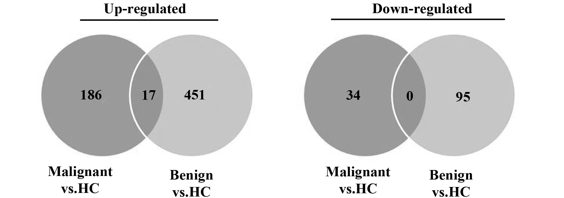
图1 良性和恶性乳腺肿瘤患者PBMCs较健康人群的差异表达基因Fig.1 Number of differentially expressed genes among benign,malignant breast tumor and healthy control (HC)Note: Significant differential expression is represented by an absolute FC ≥1.5,P<0.05,adjusted P<0.05.
表3乳腺癌外周血差异表达上调核心蛋白
Tab.3Hub genes selected based on up-regulated DEGs

GenesymbolDegreeJUN44MAPK1439FOS36EGR127IL822RHOB19ACTB18YWHAZ17ITGB113
Note:Node degree is topological parameter used for gene prioritization in the network.
良恶性乳腺肿瘤PBMCs差异表达下调的基因均没有富集到符合显著性阈值的通路。乳腺癌PBMCs差异表达上调的基因扰动的通路集中在致病菌感染、神经营养因子信号、幽门螺杆菌感染上皮细胞信号、Wnt信号及白细胞跨内皮迁移。良性乳腺肿瘤PBMCs差异表达上调的基因仅富集到1条KEGG通路,神经营养因子信号,涉及的基因与上述乳腺癌PBMCs涉及该通路的差异基因无交叠。通路按P值排序,见表2。
2.3乳腺癌外周血差异表达关键基因筛选以乳腺癌外周血PBMCs差异表达上调的203个基因构建蛋白质相互作用网络,除去孤立无互作的蛋白节点,筛选出157个上调基因编码的蛋白质具有相互作用关系,构成了包含有552个互作边关系的复杂网络。以节点的边Degree>10为标准筛选中心节点,获得Hub genes 9个,见表3。
3 讨论
肿瘤被认为是系统性疾病[13],肿瘤组织与宿主之间的相互作用,不仅局限于肿瘤微环境中,还涉及到远处组织器官和系统免疫反应。脉管系统作为运输工具,因其贯穿机体所有器官而成为肿瘤系统效应的中枢,其中最主要的是外周血,包含了大量免疫细胞,是机体免疫系统的重要组成。应用生物信息学的方法,本研究分别比较了良性和恶性乳腺肿瘤患者外周血单个核细胞与健康人群的差异,评估两种表型在外周血细胞是否存在表达模式的差异,并进一步了解乳腺癌在机体引发的系统效应,为肿瘤诊断、治疗及预后监测提供新策略[14-17]。
研究中发现,无论是良性还是恶性乳腺肿瘤,患者外周血单个核细胞中基因表达改变主要为表达上调,其中良性乳腺肿瘤PBMCs基因差异表达范围更为广泛,表明乳腺肿瘤组织在体内的存在,刺激了机体产生应答,在外周血中表现明显。差异表达基因的功能及通路分析表明,尽管良性乳腺肿瘤PBMCs差异表达基因数量较多,但其差异表达过于宽泛,缺乏针对性,涉及的功能较少,仅富集到血红蛋白代谢相关的3条功能类,而乳腺癌PBMCs差异表达基因从功能上看更多涉及免疫反应。肿瘤细胞恶性转化过程中产生肿瘤特异性抗原,能够诱导机体产生抗肿瘤免疫反应,良恶性乳腺肿瘤在免疫原性上的差异,可能是外周血产生应答差异的主要原因。进一步地,本研究通过构建蛋白互作网络,得到乳腺癌PBMCs差异表达上调的关键基因9个。
关键基因c-jun和c-fos均属于早期反应基因(Immediate early genes,IEGs),针对各种刺激迅速做出反应[18]。前炎症转录调节子在乳腺癌PBMCs差异表达基因中显著上调,并处于核心位置,表明宿主免疫系统状态倾向于固有免疫激活。活化的免疫细胞由外周血招募至肿瘤局部,穿越血管内皮进入肿瘤间质,成为肿瘤浸润性免疫细胞,发挥抗肿瘤效应的同时也为肿瘤细胞的生长和转移提供了生长因子、基质重塑因子和新生血管。免疫细胞招募及其趋化作用与乳腺癌进展和预后之间的相关性以及在治疗方面的应用也日益成为研究的热点[19,20]。
关键基因中IL-8、RHOB参与了血管生成(Angiogenesis)生物学过程。异常的血管增生是实体肿瘤的固有特征之一,促血管因子可来自于肿瘤细胞,也可来自于激活的免疫细胞。例如,参与固有免疫的细胞,尤其是巨噬细胞、中性粒细胞、肥大细胞以及骨髓造血祖细胞均在血管生成转换(Angiogenic switch)中扮演重要的角色[21]。IL-8,可直接促进内皮细胞的增殖、管腔形成及迁移,不依赖于血管内皮生长因子途径[22],降低IL-8表达可削弱肿瘤性血管生长[23]。RHOB属于GTP酶Ras超家族,可激活血管内皮细胞Akt信号通路,促进内皮细胞存活和生长,介导肿瘤血管生成[24]。IL-8,RHOB在乳腺癌外周血细胞差异表达基因中处于关键位置,可能介导了免疫细胞促血管生成作用,进而影响乳腺癌的预后。
关键基因中ACTB、MAPK14、ITGB1参与了白细胞跨内皮迁移通路(hsa04670:Leukocyte transendothelial migration)。外周血管中的白细胞穿越血管内皮细胞,向组织趋化的过程为跨内皮迁移(Transendothelial migration,TEM),是免疫监视和炎症反应中的第一步。TEM全过程依赖于白细胞与血管内皮细胞之间的识别、黏附和细胞运动,涉及大量的分子相互作用[25,26]。ITGB1基因编码整合素β1,是整合素家族的跨膜受体。整合素在多数白细胞表达,包括T淋巴细胞、单核细胞、粒细胞等,通过与纤连接蛋白(Fibronectin)相互作用,介导白细胞与血管内皮细胞识别、黏附及白细胞溢出循环系统。Integrin α4 和β1结合形成异二聚体VLA-4(Very Late Antigen-4),介导免疫细胞向炎症部位的趋化。最近的研究表明,骨髓来源的造血祖细胞通过VLA-4,经外周血循环向Fibronectin较丰富的部位趋化,协助形成适于肿瘤细胞生长的前转移微环境[27]。ITGB1在上调基因中的中心位置,表明其可能是协助乳腺癌外周血免疫细胞进入肿瘤局部微环境的主要分子,对其深入研究将有可能明确乳腺癌外周血与肿瘤组织局部免疫微环境之间的相互联系。
综上,本研究利用生物信息学工具,分析了良性和恶性乳腺肿瘤外周血单个核细胞的基因表达变化。结果显示,良恶性乳腺肿瘤患者的外周血基因表达变化模式存在明显的差异,乳腺癌患者外周血表现出免疫炎症反应,外周血可能作为替代材料,用于评估乳腺癌患者的免疫状态。筛选得到的关键基因,如IL-8、RHOB、ITGB1等,调节白细胞活化,血管生成及白细胞跨内膜迁移,在乳腺癌引发的系统免疫效应中可能起了重要作用,其在乳腺癌外周血细胞向肿瘤局部趋化过程中的作用值得深入研究,为乳腺癌的诊断、患者个体化治疗方案制定、监测等提供新的思路。
[1]Kohler BA,Sherman RL,Howlader N,etal.Annual report to the nation on the status of cancer,1975-2011,featuring incidence of breast cancer subtypes by race/ethnicity,poverty,and state[J].J Natl Cancer Inst,2015,107(6):v48.
[2]Balacescu O,Balacescu L,Virtic O,etal.Blood genome-wide transcriptional profiles of HER2 negative breast cancers patients[J].Mediators Inflamm,2016,2016:3239167.
[3]Amid A,Wan CW,Jamal P,etal.Microarray and quantitative PCR analysis of gene expression profiles in response to treatment with tomato leaf extract in mcf-7 breast cancer cells[J].Asian Pac J Cancer Prev,2012,13(12):6319-6325.
[4]Yang L,Tan J,O′Brien EJ,etal.Systems biology definition of the core proteome of metabolism and expression is consistent with high-throughput data[J].Proc Natl Acad Sci USA,2015,112(34):10810-10815.
[5]Barrett T,Wilhite SE,Ledoux P,etal.NCBI GEO:archive for functional genomics data sets--update[J].Nucleic Acids Res,2013,41(Database issue):D991-D995.
[6]Labreche HG,Nevins JR,Huang E.Integrating factor analysis and a transgenic mouse model to reveal a peripheral blood predictor of breast tumors[J].BMC Med Genomics,2011,4(1):61.
[7]Huber W,Carey VJ,Gentleman R,etal.Orchestrating high-throughput genomic analysis with Bioconductor[J].Nat Methods,2015,12(2):115-121.
[8]刘晋,张涛,李康.多重假设检验中FDR的控制与估计方法[J].中国卫生统计,2012,29(2):305-308.
[9]The Gene Ontology Consortium.Gene Ontology Consortium:going forward[J].Nucleic Acids Res,2015,43(D1):D1049-D1056.[10]Kanehisa M,Goto S,Furumichi M,etal.KEGG for representation and analysis of molecular networks involving diseases and drugs[J].Nucleic Acids Res,2010,38(Database issue):D355-D360.
[11]Franceschini A,Szklarczyk D,Frankild S,etal.STRING v9.1:protein-protein interaction networks,with increased coverage and integration[J].Nucleic Acids Res,2013,41(Database issue):D808-D815.
[12]Shannon P,Markiel A,Ozier O,etal.Cytoscape:a software environment for integrated models of biomolecular interaction networks[J].Genome Res,2003,13(11):2498-2504.
[13]Mcallister SS,Weinberg RA.The tumour-induced systemic environment as a critical regulator of cancer progression and metastasis[J].Nat Cell Biol,2014,16(8):717-727.
[14]Shaked Y,Mcallister S,Fainaru O,etal.Tumor dormancy and the angiogenic switch:possible implications of bone marrow-derived cells[J].Curr Pharm Des,2014,20(30):4920-4933.
[15]Hanahan D,Coussens LM.Accessories to the crime:functions of cells recruited to the tumor microenvironment[J].Cancer Cell,2012,21(3):309-322.
[16]Christopher MJ,Rao M,Liu F,etal.Expression of the G-CSF receptor in monocytic cells is sufficient to mediate hematopoietic progenitor mobilization by G-CSF in mice[J].J Exp Med,2011,208(2):251-260.
[17]Joyce JA,Fearon DT.T cell exclusion,immune privilege,and the tumor microenvironment[J].Science,2015,348(6230):74-80.
[18]Cattane N,Minelli A,Milanesi E,etal.Altered gene expression in schizophrenia:findings from transcriptional signatures in fibroblasts and blood[J].PLoS One,2015,10(2):e116686.
[19]Frankenberger C,Rabe D,Bainer R,etal.Metastasis suppressors regulate the tumor microenvironment by blocking recruitment of prometastatic tumor-associated macrophages[J].Cancer Res,2015,75(19):4063-4073.
[20]Palucka AK,Coussens LM.The Basis of Oncoimmunology[J].Cell,2016,164(6):1233-1247.
[21]Hanahan D,Weinberg RA.Hallmarks of cancer:the next generation[J].Cell,2011,144(5):646-674.
[22]Choi I,Lee YS,Chung HK,etal.Interleukin-8 reduces post-surgical lymphedema formation by promoting lymphatic vessel regeneration[J].Angiogenesis,2013,16(1):29-44.
[23]Passaro C,Borriello F,Vastolo V,etal.The oncolytic virus dl922-947 reduces IL-8/CXCL8 and MCP-1/CCL2 expression and impairs angiogenesis and macrophage infiltration in anaplastic thyroid carcinoma[J].Oncotarget,2016,7(2):1500-1515.
[24]Howe GA,Addison CL.RhoB controls endothelial cell morphogenesis in part via negative regulation of RhoA[J].Vasc Cell,2012,4:1.
[25]Heemskerk N,van Rijssel J,van Buul JD.Rho-GTPase signaling in leukocyte extravasation:an endothelial point of view[J].Cell Adh Migr,2014,8(2):67-75.
[26]Timmerman I,Daniel AE,Kroon J,etal.Leukocytes crossing the endothelium:a matter of communication[J].Int Rev Cell Mol Biol,2016,322:281-329.
[27]Shokeen M,Zheleznyak A,Wilson JM,etal.Molecular imaging of very late antigen-4 (alpha4beta1 integrin) in the premetastatic niche[J].J Nucl Med,2012,53(5):779-786.
[收稿2016-03-02修回2016-04-13]
(编辑许四平)
Gene expression profiles analysis identifies key genes of PBMCs in patients with benign and malignant breast tumor
HE Lang,WEI Na,GUO Zheng,WANG Dan.
School of Life Science and Technology,University of Electronic Science and Technology of China,Chengdu 610054,China
Objective:To observe the changes of gene expression in peripheral blood mononuclear cells(PBMCs) of benign and malignant breast tumor based on gene expression profiling. Methods: Datasets of gene expression profiling were downloaded from the GEO database,including PBMCs profilings of benign breast tumor,breast cancer and healthy controls.GEO2R tool was used to analyze the data to identify the differentially expressed genes (DEGs).Function of DEGs were annotated by DAVID.Protein interaction analysis and hub gene select were then performed using STRING database. Results: 563 and 237 DEGs respectively were identified.DEGs in breast cancer involved in biological process of leukocyte activation,angiogenesis and leukocyte transendothelial migration.The hub genes are IL8,RHOB,ITGB1. Conclusion: The data suggests that gene expression patterns of these two profilings are different at a certain degree.PBMCs maybe a better noninvasive material for biomarker detection of benign and malignant breast tumor.
Breast tumor;PBMCs;Differentially expressed gene;Pathway enrichment analysis
10.3969/j.issn.1000-484X.2016.10.004
①本文为国家自然科学基金(81201702)和四川省卫计委科研项目(No.120491;No.130295)。
,同时就职于福建医科大学基础医学院,福州350108,E-mail:gz0163@yeah.net;E-mail:helle@cmc.edu.cn。
何浪(1979年-), 女,博士,副教授,主要从事肿瘤免疫,抗肿瘤药物筛选,同时就职于成都医学院生物医学系,E-mail:helang79@sohu.com。
R730.3
A
1000-484X(2016)10-1424-05
②四川省肿瘤医院,成都 610041。