M′型LuTaO4∶Eu3+透明闪烁厚膜的制备及性能研究
2016-06-15邱志澈刘小林黄世明
邱志澈,顾 牡,刘小林,刘 波,黄世明,倪 晨
同济大学物理科学与工程学院,上海市特殊人工微结构材料与技术重点实验室,上海 200092
M′型LuTaO4∶Eu3+透明闪烁厚膜的制备及性能研究
邱志澈,顾 牡*,刘小林,刘 波,黄世明,倪 晨
同济大学物理科学与工程学院,上海市特殊人工微结构材料与技术重点实验室,上海 200092
X射线成像在生命科学和物质微结构分析等许多方面有着非常重要的应用,X射线成像仪器核心部件之一为X射线-可见光转换屏。透明闪烁薄膜是实现高空间分辨率X射线成像的一条有效途径。铕掺杂M′型LuTaO4是一种性能优越的闪烁材料,其密度高达9.75 g·cm-3,化学性质稳定,辐照硬度大,有望制备成透明薄膜型高空间分辨率X射线转换屏。以2-甲氧基乙醇为溶剂、PVP为胶粘剂,采用溶胶-凝胶法成功制备出M′型LuTaO4∶Eu3+透明闪烁厚膜,并对透射率、光致发光、X射线激发发射光谱和空间分辨率等一系列的薄膜性能进行表征。经过8次旋涂之后,膜层均匀、无裂纹,厚度为2.1 μm,发光波段的透射率为70%以上,成像空间分辨率达到1.5 μm。将厚膜作为X射线-可见光转换屏,成功对果蝇进行了X射线成像,其复眼结构清晰可见。此外,紫外和X射线激发下闪烁膜的发光特性研究表明,该厚膜具有优良的发光性能,已基本满足高分辨率X射线成像的要求,有望在显微X射线成像方面获得很好应用。
LuTaO4∶Eu3+厚膜; 溶胶-凝胶法; 发光性能; X射线成像
引 言
X射线成像在许多方面有着非常重要的应用,如: 生命科学和物质微结构分析等[1]。随着X射线成像技术的发展,人们发明了数字X射线成像(如: X射线电荷耦合器件(X射线CCD)[2-5]),器件的核心部件之一就是X射线-可见光转换屏。通常X射线-可见光转换屏由微米尺寸的荧光粉颗粒构成,但因为受颗粒光散射的影响,X射线CCD的空间分辨率一般在几十微米。为了克服散射问题,人们研制了一些特殊的闪烁转换屏,如透明薄膜[5-6]、柱状结构[8]和像素结构转换屏[8]。透明闪烁薄膜是实现高空间分辨率X射线成像的一条有效途径。为了兼顾薄膜对X射线的吸收及空间分辨率的要求,薄膜厚度应控制在1~3 μm之间[9]。铕掺杂M′型LuTaO4是一种性能优越的闪烁材料[10-13],其空间点群为P2/a,密度高达9.75 g·cm-3,化学性质稳定,辐照硬度大,有望制备成透明薄膜型高空间分辨率X射线转换屏。目前对稀土掺杂LuTaO4的研究主要都局限于粉末形态[10-13],尽管前期我们已制备出了厚度约为340 nm的M′型LuTaO4∶Eu3+薄膜[14],但从X射线成像应用的要求来说,该薄膜厚度有待进一步提高。
采用溶胶-凝胶法制备出厚度达2.1 μm的M′型LuTaO4∶Eu厚膜,通过对透射率、光致发光、X射线激发发射光谱和空间分辨率等薄膜性能的一系列表征,以及利用该厚膜进行的X射线成像实验等,证明了所制备的M′型LuTaO4∶Eu厚膜具有很好的闪烁和X射线成像性能,有望在高分辨率X射线成像领域获得应用。
1 实验部分
采用溶胶-凝胶法,结合旋涂技术,制备出M′型Lu0.9Eu0.1TaO4透明闪烁厚膜。将TaCl5(99.99%)溶于2-甲氧基乙醇(99%)和浓盐酸(36 Wt%)的混合液中,搅拌回流。将Lu(NO3)3·6H2O(99.99%)和Eu2O3(99.99%)完全溶于2-甲氧基乙醇溶液中。在上述两种混合溶液中加入PVP(polyvinyl pyrrolidone,平均分子量为1.3×10⁶),其中原料摩尔配比为Lu∶Ta∶Eu∶PVP=0.9∶1∶0.1∶0.4,室温下陈化2~3天,即得到透明溶胶。在洁净的石英基片上旋涂得到湿膜,将其置于马弗炉中,升温至600 ℃保温1 h。待其冷却后,再以同样的方式进行旋涂和热处理。多次旋涂后即得所需厚度膜层,膜层的最终热处理为温度1 200 ℃保温2 h,自然冷却后获得M′型Lu0.9Eu0.1TaO4透明闪烁厚膜。
采用Hitachi F7000荧光光谱仪测量样品的光致发光光谱。利用X射线衍射仪(铜靶Kα线,波长0.15 405 nm,电压40 kV,电流30 mA)对样品进行物相分析。通过Philips XL-30 扫描电镜(SEM)观察样品的表面和断面形貌。样品的透射谱采用日本分光(JASCO)公司的V-570型紫外/可见/近红外分光光度计测得。X射线激发发射谱由自行研制的X射线激发发射谱仪获得,其中X射线源为F30-Ⅲ X光机(工作电压和电流分别为80 kV和6 mA),光电探测器件为Hamamatsu CR131光电倍增管,单色仪为Zolix SBP300单色仪。膜层空间分辨率测量在上海光源BL13W1光束线站上进行,测量中采用了JIMA RT RC-02标准分辨率板和由PCO2000型CCD相机和显微系统构成的法国Optique Peter探测器,X射线的能量为12 keV,曝光时间为30 s。所有测试都在室温下进行。
2 结果与讨论
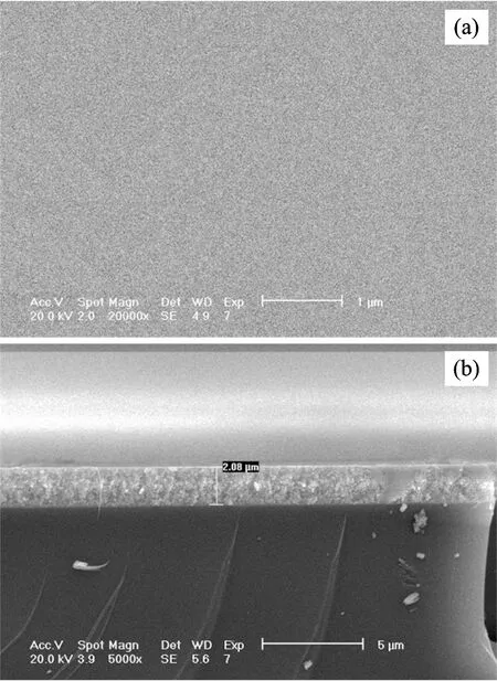
膜层厚度是反映闪烁薄膜性能的关键参数。图1为8次旋涂制备的M′型LuTaO4∶Eu3+厚膜表面和断面的SEM图。从图中可以看到,膜层表面整洁、无裂纹,没有形成大的颗粒,其厚度约2.1 μm,能很好满足微米量级空间分辨率X射线成像的要求。与之前的工作相比[14],由于引入了胶粘剂PVP,不仅有效提高了溶胶的粘度,而且还抑制了膜层形成过程中产生的应力,膜层厚度得到了很大的提高。

Fig.1 (a) surface morphology and (b) cross-sectional view of the M′-type LuTaO4∶Eu3+ thickfilm
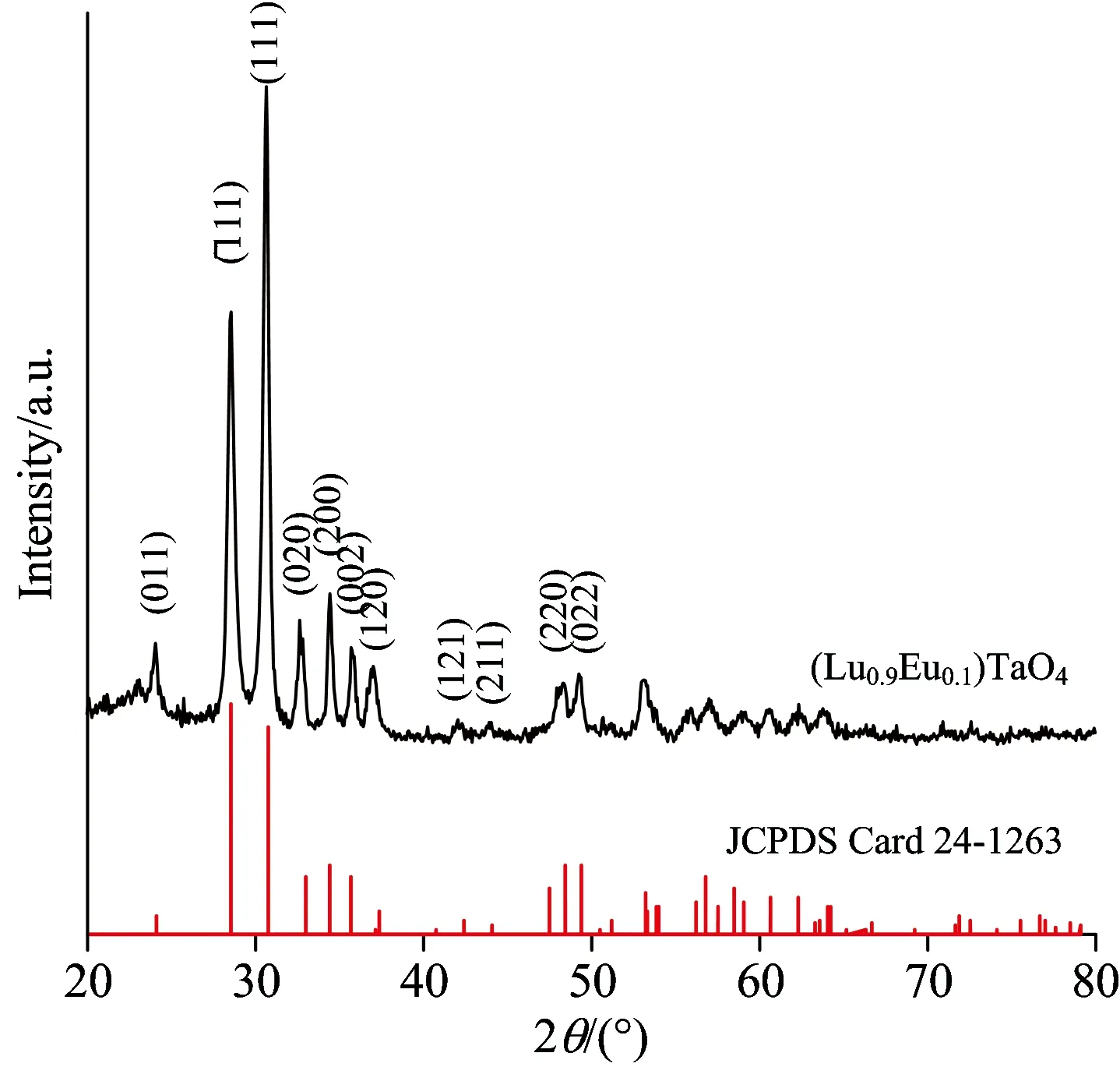
图2是样品的X射线衍射谱(XRD), 从中可以看出衍射峰与M′型LuTaO4的PDF标准卡片24-1263匹配,没有出现别的杂峰,说明所制备样品的结构为M′型LuTaO4。采用Scherrer公式:Dλ/(βcosθ),对膜层晶粒大小进行估算,式中λ为X射线波长(0.154 05nm),β为(111)主衍射峰的半高宽,θ为布拉格衍射角,为0.89。计算得厚膜晶粒的平均尺寸约为21 nm。运用Vegard定律:ax=(1-x)a1+xa2[15],可以较为精确地确定薄膜中Eu3+的实际掺杂浓度,式中ax为Lu1-xEuxTaO4的晶格常数,a1和a2分别表示LuTaO4和EuTaO4的晶格常数,其取值分别为5.237和5.426,x是Eu3+的实际掺杂浓度。用JADE软件对样品的XRD数据进行拟合,得到样品晶格常数ax=5.253 ,样品中Eu3+的实际掺杂浓度为0.085。可见,该浓度值比其化学配比略低,其原因可能是部分Eu3+并未进入LuTaO4晶格。

Fig.2 X-ray diffraction pattern of the M′-type LuTaO4∶Eu3+ thick film
膜层的透明性是影响闪烁薄膜光输出的关键因素之一。图3为所制备的M′型LuTaO4∶Eu3+厚膜的透射谱。可以看出,膜层在波长为500~800 nm范围内的透射率超过70%。利用透射谱进一步确定了样品的光学吸收边。吸收系数α=-lnT/d,式中T和d分别表示膜层的透过率和厚度。而α2∝(hν-Eopt),式中hν和Eopt分别为光子能量和光学吸收边。以(lnT)2和hν作图,如图3中的插图所示。从图中可得该厚膜的吸收边为5.5eV,与文献[14]的结果一致。

Fig.3 Transmission spectrum of the M′-type LuTaO4∶Eu3+thickfilm. The inset presentsits optical band gap


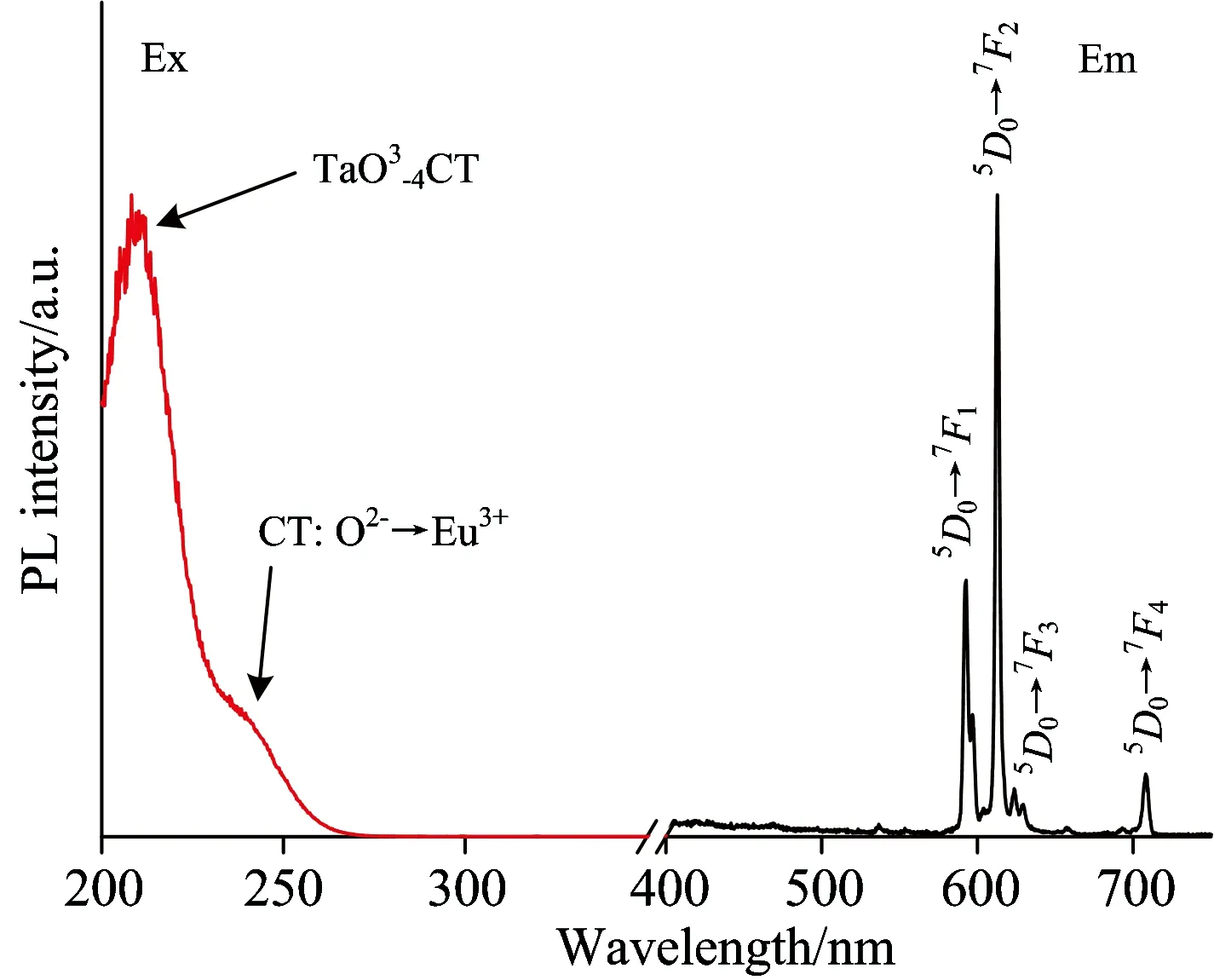
Fig.4 PL spectra of the M′-type LuTaO4∶Eu3+thick film(λex=210 nm,λem=613 nm)

Fig.5 XEL spectrum of the M′-type LuTaO4∶Eu3+thick film
X射线激发发射谱是闪烁材料的重要性能指标。图5为样品的X射线激发发射谱。可见,其发射谱与光致发光情况类似,为典型的Eu3+发射。发射谱具有良好的信噪比,表明该厚膜具有较高的发光效率和对X射线的截止本领。
图6为样品在12keV的X射线照射下,JIMA RT RC-02空间分辨率板的成像结果。从图像中可判断,膜层的分辨率为1.5 μm。利用该厚膜作为X射线转换屏,对一身长约2 mm的果蝇进行了成像,其中X射线能量为12 keV,曝光时间为30 s,果蝇离转换屏的距离为25 cm,其结果如图7所示。从图7(b)中可以较为清楚地看到果蝇的复眼结构。以上结果表明,所制备M′型LuTaO4∶Eu3+厚膜已具有良好的综合性能,基本达到实用化的要求。

Fig.6 Image of the X-ray spatial resolution of the M′-type LuTaO4∶Eu3+ thick film

Fig.7 (a) X-ray image of fruit fly by the M′-type LuTaO4∶Eu3+thick film, (b) the detail with enlarged scale of the compound eye
3 结 论
采用溶胶-凝胶法,以2-甲氧基乙醇为溶剂,PVP为胶粘剂,成功制备出厚度约2.1 μm的M′型LuTaO4∶Eu3+透明闪烁厚膜。膜层均匀、无裂纹,其发光波段的透射率在70%以上。X射线空间分辨率板测量结果表明,闪烁膜成像的空间分辨率达到1.5 μm,具备了优良的高分辨率X射线成像本领。同时,所制备M′型LuTaO4∶Eu3+透明闪烁厚膜发光性能良好,因此,它有望在显微X射线成像领域获得很好应用。
[1] Antonuk L E, Jee K W, El-Mohri Y, et al. Med. Phys., 2000, 27(2): 289.
[2] Momose A, Koyama I, Hirano K, et al. Proc. SPIE, Developments in X-Ray Tomography Ⅲ, 2002, 4503: 71.
[3] Butler L G, Ham K, Kurtz R L. Proc. of SPIE, Developments in X-Ray Tomography, 2002, 4503: 62.
[4] Rao D V, Seltzer S M, Hubbell J H, et al. Proceeding of SPIE. Vol. 4503(08) (Conf. on Developments in X-Ray Tomography Ⅲ, 2002, 4503: 62. July-3 August,2001, Journal of San Diego California, USA).
[5] Martin T, Koch A . Synchrotron Radiation, 2006, 13, 180.
[6] Koch A, Raven C, Spanne P, et al. Opt. Soc. Am. 1998, A 15(7), 1940.
[7] Nagarkar V, Gupta T, Miller S, et al. IEEE Trans. Nucl. Sci., 1998, 45(3): 492.
[8] Jung P G, Lee C H, Bae K M, et al. Opt. Express,2010, 18(14): 14850.
[9] Dujardin C, Luyer C Le, Martinet C, et al. Nuclear Instruments and Methords in Physics Research A, 2005, 537: 237.
[10] Issler S L, Torardi C C. Alloys Compd., 1995, 229: 54.
[11] Zhang H J, Wang Y H, Xie L C. Lumin.,2010, 130: 2089.
[12] Chen S W, Liu X L, Gu M, et al. Lumin.,2013, 140: 1.
[13] Blasse G, Dirksen G, Brixner L H, et al. Alloy. Comp.,1994, 209(1-2): 1.
[14] Liu X L, Chen S W, Gu M, et al. Optial Materials Express, 2014, 4(1): 172.
[15] Lambregts M J,Frank S. Talanta, 2004, 62(3): 627.
[16] Liu W R, Lin C C, Chiu Y C, et al. Opt. Express, 2009, 17(20): 18103.
[17] Blasse G,Gtabmaier B C. Luminescent Materials. Springer-Verlag, 1994, chap. 3.
Preparation and Performances of the M′-Type LuTaO4∶Eu3+Transparent Scintillator Films
QIU Zhi-che, GU Mu*, LIU Xiao-lin, LIU Bo, HUANG Shi-ming, NI Chen
Shanghai Key Laboratory of Special Artificial Microstructure Materials & Technology, School of Physics Science and Engineering, Tongji University, Shanghai 200092, China
X-ray imaging has a very important role in life sciences and material microstructure analysis and other applications. One of the core components of X-ray imaging equipment is the X-rays-visible light conversion screen. Flashing transparent film is an effective way to achieve high spatial resolution X-ray imaging. M′-type LuTaO4∶Eu3+is an excellent scintillation material. It has high light yield, high density, good radiation hardness and good chemical stability. Therefore, to research and develop the transparent conversion screen with M′-type LuTaO4∶Eu3+is very important for the application of X-ray detector in high spatial resolution X-ray imaging. In this paper, the M′-type LuTaO4∶Eu3+transparent scintillator films were successfully prepared from the inorganic salt and 2-methoxyethanol solution containing polyvinylpyrrolidone(PVP) via sol-gel technique, and transmittance, photoluminescence, X-ray excitation emission spectral and spatial resolution, and a series of film properties were characterized. A film thickness of about 2.1 μm was achieved after 8 coatings. The thick film was homogeneous and crack free, and the transmittance was approximately 70% in its emission region. The spatial resolution of the thick film was 1.5 μm, which measured by the standard spatial resolution panels. An X-ray imageof fruit fly was obtained by using this thick film. Additionally, thesol-gel derived M′-type LuTaO4∶Eu3+thick film revealed excellent photoluminescence and X-ray excited luminescence performances. All results indicated that the M′-type LuTaO4∶Eu3+thick films have satisfied the essential requirements for applications in high-spatial-resolution X-ray imaging.
LuTaO4∶Eu3+thick film; Sol-gel method; Luminescence property; X-ray imaging
Sep. 18, 2014; accepted Dec. 20, 2014)
2014-09-18,
2014-12-20
国家自然科学基金项目(11475128, 11375129,11475127)和科学技术部国家重大科学仪器设备开发专项项目(2011YQ13001902)资助
邱志澈,1989年生,同济大学物理科学与工程学院研究生 e-mail: giuzhiche@aliyun.com *通讯联系人 e-mail: mgu@tongji.edu.cn
O482.3
A
10.3964/j.issn.1000-0593(2016)02-0336-04
*Corresponding author
