腰骶椎融合术后继发骶骨骨折的手术治疗一例
2016-05-12胡永凯刘宪义李淳德王宇于峥嵘
胡永凯 刘宪义 李淳德 王宇 于峥嵘
作者单位:100034 北京大学第一医院骨科
Surgical treatment of sacral fractures following lumbosacral vertebral fusion:1 case report
HU Yong-kai,LIU Xian-yi,LI Chun-de,WANG Yu,YU Zheng-rong.Department of Orthopedics,Peking University First Hospital,Beijing,100034,PRC
腰骶椎融合术后继发骶骨骨折的手术治疗一例
胡永凯 刘宪义 李淳德 王宇 于峥嵘
作者单位:100034 北京大学第一医院骨科
Surgical treatment of sacral fractures following lumbosacral vertebral fusion:1 case report
HU Yong-kai,LIU Xian-yi,LI Chun-de,WANG Yu,YU Zheng-rong.Department of Orthopedics,Peking University First Hospital,Beijing,100034,PRC
【关键词】脊柱融合术;腰骶部;脊柱骨折;骶骨;骨钉
临床上诊断为继发于腰骶椎体后路融合术的骶骨骨折不常见,相关文献报道为数不多。尽管该类骶骨骨折报道较少,但其发生率可能很高。该类骨折在 X 线片中很难被发现,确诊一般依赖于 CT 或 MRI,因此易致漏诊。漏诊的另一个原因是大多数该类骨折未予手术干预也能在数月内痊愈,一般很难引起骨科医生的重视。根据文献报道的经验,大多数该类骨折发生在腰骶椎融合术后 3 个月内,绝大多数患者经保守治疗效果较好。当存在神经功能损害、椎弓根钉明显错置、严重的疼痛或骨折不愈合时,手术干预很有必要。
本病例为 1 例应用糖皮质激素长达 15 年的 65 岁女性患者,在我科行 L4~S1后路融合术后继发骶骨完全性骨折,采用切开复位和髂骨螺钉固定手术治疗,报告如下。
临床资料
患者,女,65 岁,体重超重 (BMI 指数为 25.63),主诉为左下肢疼痛进行性加重伴行走困难 5 年,经保守治疗后反复发作。查体左下肢肌力 4 级,Hoffmann征、Babinski 征均为阴性。患者因患哮喘应用糖皮质激素 15 年。髋关节骨密度检测 T 值为 -3.7,为重度骨质疏松。
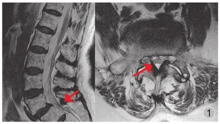
腰椎 X 线片示 L5椎体向前 2 度滑脱。腰椎 MRI 显示L5~S1平面中央椎管狭窄,L4~S1椎间孔狭窄 (图 1)。予以手术治疗,即在 L4~S1椎体双侧置入多轴万向螺钉和两根 XIA II 连接棒 (Stryker,美国) 进行后方融合,并将术中咬除的自体腰椎椎板及棘突咬成小骨粒进行椎体后外侧植骨。此外,切除 L5~S1椎间盘并置入一个 cage。
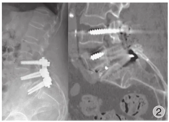
患者对手术耐受良好,术后第 1 天即下床活动。但术后第 5 天,患者诉双侧臀部突然疼痛加重,并伴有左下肢放射痛,二便不受控制。查体左下肢肌力 3 级。CT 扫描结果示 S1~2平面有一水平裂缝骨折,S2完全性移位 (图 2)。
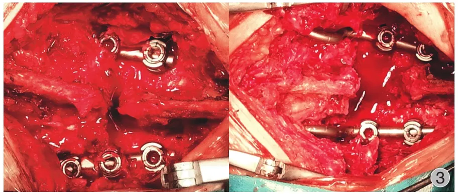
笔者团队尝试通过牵引进行骨折复位但未成功。考虑存在明显的神经功能损害及 S2严重移位,在牵引复位后1 周施行后路神经解压及切开复位内固定术,应用髂骨螺钉将固定融合范围延至髂骨翼 (图 3、4)。

图 1 腰椎 MRI 示 L5~S1平面中央椎管狭窄,L4~S1椎间孔狭窄 (所示)Fig.1 The MRI of the spine found central lumbar spinal stenosis at L5-S1level and L4-S1foraminal narrowing

图 2 CT 扫描结果示 S1~2平面有一水平裂缝骨折,S2完全性移位Fig.2 A CT scanning revealed a horizontal fracture at the S1-2level with S2being totally displaced

图 3 显露后发现骶骨棘突间完全移位,即骶骨棘突向后移位、骶骨椎板位于内固定背面。为了更好地复位,用撑开器将 S1和 L4撑开,结果证明很有效。撑开后,S1和 L4的距离明显增大Fig.3 After the exposure,we found that the interspinous process of the sacrum was totally displaced.It moved backward and cranially onto the backside of the instrumentation.To reduce the fracture,we used a spreader to distract the spinal processes of the S1and L4,which turned out to be effective.After the distraction,the distance between the spinal processes of the S1and L4increased significantly
切开复位内固定融合术麻醉苏醒后,患者诉臀部及左下肢疼痛较术前明显减轻。返病房后经后续治疗,查体左下肢肌力从 3 级逐渐恢复至 5 级。切开复位内固定术后13 天出院。
矢状位影像学参数见表 1,术前患者的骨盆入射角(pelvic incidence,PI) 和骶骨倾斜角 (sacral slope,SS) 都很大,分别为 64.1°、37.1°。

表 1 侧位 X 线片矢状位参数 (°)Tab.1 The Sagittal Parameters in the lateral radiographs (°)
讨 论
腰骶后路融合术后骶骨骨折很少见,已发表的文献中一直缺乏该类资料的报道。经检索发现在 2013 年之前只有 34 个病例被报道,目前没有一篇文章病例数超过5 例[1-6]。许多脊柱外科医生可能从未遇到或注意到该类骨折。
一、发生率及诊断
腰椎术后继发骶骨骨折的发生率一直被低估,因为绝大多数该类骨折在 X 线片上分辨不出,诊断主要依赖于CT、MRI 或骨扫描。最近有 2 篇病例数较多的文章发表。Meredith 等[7]回顾性研究其中心 2002 年至 2011 年所有行腰椎后路内固定融合术的病例,共 392 例,其中 24 例有影像学依据 (CT、MRI 或骨扫描) 证实在内固定以下存在骶骨骨折,发生率为 6.1%。这 24 例中只有 1 例在常规X 线片中可明确辨认;通过 CT 确诊骶骨骨折的有 22 例,2 例无移位性骶骨骨折是通过骨扫描确诊。Wilde 等[8]通过研究其中心近 5 年的 CT、核医学及 MRI 检查报告,共有 23 例腰骶椎融合术后继发骶骨骨折,同样发现在 X 线片上可见骶骨骨折的只有 1 例,在 CT 上所有骶骨骨折均可确诊。腰骶椎融合术后骶骨骨折的漏诊率很高,CT 能发现大部分该类骨折,因此,当患者在腰骶椎融合术后3 个月内臀部再发新的疼痛时,CT 应作为常规检查。
二、风险因素和预防
在现有文献报道中,大部分学者认为高龄、女性、骨质疏松、肥胖、长期应用糖皮质激素和多节段腰骶椎融合是腰骶椎融合术后继发骶骨骨折的风险因素[1,3,5,8-10]。生物力学研究表明,多节段腰骶椎融合改变了脊柱的应力分布,显著增加固定节段的末节椎体及相邻平面的应力[11-12]。当合并有其它因素如骨质疏松时,骨的抗骨折能力降低,合并肥胖时传导的重力载荷增大,这些应力的分布异常可能会导致骨折发生[3,7,13]。本病例中的患者为 65 岁老年女性,BMI 25.63,有哮喘病史,应用糖皮质激素 15 年,髋关节骨密度测量 T 值为 -3.7,提示重度骨质疏松。这些均为腰骶椎融合术后继发骶骨骨折的风险因素。
此外,脊柱骨盆力线异常也是骶骨骨折形成的一个风险因素。Odate 等[14]研究 2010 年至 2011 年所有从 L2或更高位椎体至骶骨的腰骶内固定融合术的病例,将女性患者分为 2 组,骨折组 5 例,非骨折组 64 例。比较发现骨折组的 PI [ 平均 (72±8) ° ]大于骨折组 [ 平均 (51±12) ° ],差异有统计学意义 (P<0.01)。作者建议脊柱外科医生应尽量恢复患者腰椎曲线,这不仅可以使脊柱骨盆相协调,还可以避免术后发生骶骨骨折。对于需长节段内固定的患者,单纯腰骶椎内固定融合很难提供足够的力量以维持腰椎正常曲线,因此应该进行骶髂关节固定以保护骶骨。本病例骶骨骨折后 PI (64.1°) 相对较大,骶骨倾斜较大,SS值为 37.1°。腰骶交界处的剪切力、肥胖、骨质疏松和多节段固定导致的应力集中共同作用于骶骨,引起腰骶椎融合术后骶骨发生骨折。对于长节段融合或骨质较差的患者合并有骶骨骨折危险因素时,为了预防术后骶骨骨折,可以考虑将融合的范围预防性延至髂骨翼。
三、手术治疗
文献表明,腰骶椎融合术后骶骨骨折采用保守治疗效果较好,包括减少活动、外固定及抗骨质疏松治疗[2-3,5,15]。当出现骶骨不全性骨折明显移位、矢状位失衡、神经损伤症状及骨折不愈合时,施行切开复位内固定手术则非常必要。最常用的手术方式是用髂骨螺钉将融合范围延至髂骨翼。在现有文献中所有采用手术治疗的病例,治疗效果显示骨折愈合良好、疼痛明显缓解[2-3,5,7-8,11,14-15]。
四、结论
腰骶椎融合术后骶骨骨折的发生率可能被明显低估,因为大多数该类骨折在 X 线片中很难被发现。CT 能发现大部分该类骨折,因此,当患者在腰骶椎融合术后 3 个月内臀部再发新的疼痛时,CT 应作为常规检查。绝大多数患者经保守治疗可痊愈,但并发骨折移位、矢状位失衡、出现神经损害症状及疼痛剧烈的骨折不愈合时,应采用髂骨螺钉将融合范围延至髂骨翼的手术治疗。
参考文献
[1]Vavken P,Krepler P.Sacral fractures after multi-segmental lumbosacral fusion:a series of four cases and systematic review of literature.Eur Spine J,2008,17(Suppl 2):S285-290.
[2]Fourney DR,Prabhu SS,Cohen ZR,et al.Early sacral stress fracture after reduction of spondylolisthesis and lumbosacral fixation:case report.Neurosurgery,2002,51(6):1507-1511.
[3]Khan MH,Smith PN,Kang JD.Sacral insufficiency fractures following multilevel instrumented spinal fusion:case report.Spine,2005,30(16):E484-488.
[4]Koh YD,Kim JO,Lee JJ.Stress fracture of the pelvic wingsacrum after long-level lumbosacral fusion:a case report.Spine,2005,30(6):E161-163.
[5]Mathews V,McCance SE,O’Leary PF.Early fracture of the sacrum or pelvis:an unusual complication after multilevel instrumented lumbosacral fusion.Spine,2001,26(24):E571-575.
[6]Pennekamp PH,Kraft CN,Stutz A,et al.Sacral fracture as a rare early complication of lumbosacral spondylodesis.Z Orthop Ihre Grenzgeb,2005,143(5):591-593.
[7]Meredith DS,Taher F,Cammisa FJ,et al.Incidence,diagnosis,and management of sacral fractures following multilevel spinal arthrodesis.Spine J,2013,13(11):1464-1469.
[8]Wilde GE,Miller TT,Schneider R,et al.Sacral fractures after lumbosacral fusion:a characteristic fracture pattern.AJR Am J Roentgenol,2011,197(1):184-188.
[9]Shono Y,Kaneda K,Abumi K,et al.Stability of posterior spinal instrumentation and its effects on adjacent motion segments in the lumbosacral spine.Spine,1998,23(14):1550-1558.
[10]Wood KB,Geissele AE,Ogilvie JW.Pelvic fractures after long lumbosacral spine fusions.Spine,1996,21(11):1357-1362.
[11]Wood KB,Schendel MJ,Ogilvie JW,et al.Effect of sacral and iliac instrumentation on strains in the pelvis.A biomechanical study.Spine,1996,21(10):1185-1191.
[12]Erickson MA,Oliver T,Baldini T,et al.Biomechanical assessment of conventional unit rod fixation versus a unit rod pedicle screw construct:a human cadaver study.Spine,2004,29(12):1314-1319.
[13]Gotis-Graham I,McGuigan L,Diamond T,et al.Sacral insufficiency fractures in the elderly.J Bone Joint Surg Br,1994,76(6):882-886.
[14]Odate S,Shikata J,Kimura H,et al.Sacral fracture after instrumented lumbosacral fusion:analysis of risk factors from spinopelvic parameters.Spine,2013,38(4):E223-229.
[15]Elias WJ,Shaffrey ME,Whitehill R.Sacral stress fracture following lumbosacral arthrodesis.Case illustration.J Neurosurg,2002,96(Suppl 1):135.
(本文编辑:王萌)
【Abstract】Objective To present a case of totally displaced sacral fracture following posterior L4-S1fusion.Methods This article presented a case of totally displaced sacral fracture following posterior L4-S1fusion in a 65-year-old female patient with a 15-year history use of corticosteroid.She underwent open reduction and internal fixation by iliac screws.Results Immediately after the revision surgery,the patient’s pain in buttock and left leg was relieved significantly.The weakness of the left lower extremity was also improved from 3/5 to 5/5.Conclusions The incidence of postoperative sacral fractures has been rather underestimated,because most of these fractures are not visible on plain radiographs.CT has been proved to be able to detect most of such fractures and should be performed routinely when patients complain of renewed buttock pain within 3 months after the lumbosacral fusion.The majority of the patients respond well to the conservative treatment.The fusion construction to the iliac wings by iliac screws will be needed when concurrent fracture displacement,sagittal imbalance,neurologic symptoms,or painful nonunion exist.
【Key words】Spinal fusion; Lumbosacral region; Spinal fractures; Sacrum; Bone nails
(收稿日期:2015-05-15)
Corresponding author:LI Chun-de,Email:lichunde@medmail.com.cn
通信作者:李淳德,Email:lichunde@medmail.com.cn
DOI:10.3969/j.issn.2095-252X.2016.01.017
中图分类号:R683.6
