Intermittent blood flow restriction does not reduce atrophy following anterior cruciate ligament reconstruction
2016-04-25ErikIversenViekestdArneLrmoNorweginOlympicndPrlympicCommitteendConfedertionofSportsOslo0840NorwyCurtontgenSndvikOslo0050NorwyReceived29July2014revised20Octoer2014ccepted15Decemer2014Avilleonline18April2015
Erik Iversen*,Vieke RøstdArne LrmoNorwegin Olympic nd Prlympic Committee nd Confedertion of Sports,Oslo 0840,NorwyCurto Røntgen,SndvikOslo 0050,NorwyReceived 29 July 2014; revised 20 Octoer 2014; ccepted 15 Decemer 2014 Aville online 18 April 2015
Intermittent blood flow restriction does not reduce atrophy following anterior cruciate ligament reconstruction
Erik Iversena,*,Vibeke Røstada,Arne LarmobaNorwegian Olympic and Paralympic Committee and Confederation of Sports,Oslo 0840,Norway
bCurato Røntgen,Sandvika,Oslo 0050,Norway
Received 29 July 2014; revised 20 October 2014; accepted 15 December 2014 Available online 18 April 2015
Peer review under responsibility of Shanghai University of Sport.
* Corresponding author.
E-mail address: Erik.Iversen@Olympiatoppen.no (E.Iversen).
http://dx.doi.org/10.1016/j.jshs.2014.12.005
2095-2546/©2016 Production and hosting by Elsevier B.V.on behalf of Shanghai University of Sport.
Abstract
Background: A previous study has reported a 50% reduction in disuse atrophy of the quadriceps during the first 14 days after anterior cruciate ligament (ACL) reconstruction.A follow-up trial is needed to confirm these promising results.The present study aims to investigate the effect of an occlusion stimulus on quadriceps atrophy after ACL reconstruction.
Methods: A total of 24 subjects participated in the study.They were randomized into two groups.Starting the 2nd day after surgery,the occlusion group received an occlusion stimulus for 5 min,followed by removal of the occlusive pressure for 3 min.This was repeated five times in one training session,twice daily.During the period of occlusive stimulus,the subjects performed 20 low load exercises for the quadriceps.The control group followed the same exercise protocol,but without the occlusion stimulus.Changes in quadriceps anatomical cross section area (ACSA) were measured using axial magnetic resonance (MR) images at 40% and 50% of the length of the femur.
Results: Both groups had a significant reduction of quadriceps ACSA from 2 days before surgery to 16 days after surgery.During the intervention period,the occlusion group lost 13.8%±1.1% (mean±SEM) and the control group lost 13.1%±1.0% of their quadriceps ACSA,respectively.There was no significant difference between the occlusion and control groups with regards to atrophy of the quadriceps muscles.
Conclusion: In conflict with other studies using a similar protocol,application of blood flow restriction the first 14 days after ACL reconstruction did not reduce quadriceps ACSA muscle atrophy measured by MR in a population of athletes.
©2016 Production and hosting by Elsevier B.V.on behalf of Shanghai University of Sport.
Keywords:ACL reconstruction; Ascular occlusion; Hypoxia ischemia; Quadriceps atrophy
1.Introduction
Recovery from lower leg trauma or surgery often requires a period of unloading,resulting in loss of muscle mass.In the thigh,the quadriceps is most affected,while the muscle mass of the hamstrings and adductors are mainly maintained.1Even short periods of disuse have been shown to cause substantial loss of muscle mass in the anti-gravity muscles.2-5For example,in healthy subjects,a reduction in quadriceps muscle volume of 8.4% after 14 days of lower limb unloading has been reported.3Rate of atrophy is higher after arthroscopic knee surgery.Gerber et al.6found a reduction in quadriceps muscle volume of 20%-33% from time of injury to 3 weeks after surgery.Muscle mass lost due to short term unloading can be restored relatively quickly with active recovery.The quadriceps muscle mass lost due to 3 weeks of unloading can be restored in the same amount of time in uninjured subjects using traditional resistance training.5However,the quadriceps atrophy after anterior cruciate ligament (ACL) reconstruction is harder to regain.A difference of 8.6% in quadriceps anatomical cross section area (ACSA) of the injured limb compared to the un-injured limb 49 months after ACL reconstruction has been reported.7
Despite conflicting evidence,a majority of studies suggest that specific atrophy among the individual muscle bellies of the quadriceps does not occur.1,5However,disuse atrophy is notuniformly distributed along the length of the muscle.Loss of muscle mass is greater around the region of the peak ACSA,rather than towards the proximal and distal ends of the muscle.8,9Persistent atrophy of the quadriceps muscles following ACL reconstruction can be a major challenge.Preventive measures,such as neuromuscular electrical stimulation (NMES),are often used in an attempt to reduce muscle atrophy afterACL reconstruction.A recent study investigating the effect of NMES on prevention of quadriceps atrophy after 5 days of lower limb unloading,found no significant change in quadriceps ACSA in the group using NMES for 40 min twice daily.In the control group,ACSA of the quadriceps was reduced by 3.5%±0.5%.2A number of studies reporting hypertrophic changes in skeletal muscle after low load resistance training with restricted blood flow have been published in the past decade.10Blood flow restriction has also been used to reduce muscle atrophy and loss of muscle strength after lower limb surgery and disuse.Studies have shown impressive results using a protocol consisting of five repetitions of vascular occlusion for 5 min,followed by release of occlusion for 3 min,twice daily.They reported a reduction in quadriceps atrophy after ACL surgery by 50%,11and maintenance of thigh circumference and strength after unloading.12
To our knowledge,the effect of this intervention has not been evaluated on a population of athletes before.The aim of this study was to repeat these promising results in a population of athletes during the first 16 days after ACL reconstruction.Our hypothesis was that the occlusion group would have less relative reduction in quadriceps ACSA compared to the control group.
2.Methods
2.1.Subjects
A total of 24 patients planned for ACL reconstruction surgery were included in the study.All subjects were physically active,and had sustained their injury while participating in sports.Subjects between 18 and 40 years of age,with no prior knee injuries,ACL injury not more than 6 months before surgery,and planned for reconstruction using hamstring tendon graft were eligible to participate in the study.Prior to inclusion,all participants were given information about the purpose of the study,and gave their written informed consent.The research project was conducted according to the Declaration of Helsinki and was approved by the Norwegian Ethics Committee.
2.2.Experimental procedure
A total of 24 patients participated in the study.Twelve patients (7 men,5 women) were randomized to intermittent blood flow restriction and exercises (occlusion group),and 12 patients (7 men,5 women) were randomized to exercises only (control group).There was no statistical difference between the groups at baseline (Table 1).During the pre-operation visit,all subjects were instructed in how to perform the exercise protocol.In addition,the occlusion group were trained in operating the occlusion cuff.Magnetic resonance (MR) imaging was performed on the injured leg 2 days before,and 16 days after surgery.The intervention started at Day 2 after surgery.In the experimental group,a 14-cm wide contoured pneumatic occlusion cuff (Delphi low pressure cuff 9-7450-003) was applied to the most proximal part of the thigh.Inflation of the cuff was administered with a portable blood pressure hand pump (Trigger Aneroid DS66; Welch Allyn,Skaneateles Falls,NY,USA)fitted to the cuff.The cuff was inflated to give an occlusion stimulus for 5 min,followed by removal of the occlusive pressure for 3 min.This was repeated five times in one training session.Subjects were sitting with their upper body inclined to about 45°during the session.Starting pressure was set at 130 mmHg with an increase in pressure of 10 mmHg every second day,up to a maximum pressure of 180 mmHg.If the occlusive stimulus caused unbearable pain,the subjects were instructed to use the highest tolerable pressure.The experimental group performed the occlusion protocol twice daily.During the occlusion intervals the patients performed quadriceps exercises consisting of isometric quadriceps contractions,progressing to leg extension over a knee-roll,and straight leg-raises.The patient performed 20 repetitions during each 5-min occlusion period.Adding up to 100 repetitions per training session,and 200 repetitions per day.The occlusion stimulus was terminated 14 days after surgery,to avoid muscle swelling to interfere with the MR measurements.The control group followed the same exercise regime,but without the occlusion stimulus.Patients in the occlusion group received training in the use of the occlusion cuff prior to surgery.After surgery they performed the occlusion protocol as a home exercise.All patients had two consultations during the experimental period,5 and 10 days after surgery.During these consultations degree of swelling and knee mobility was subjectively evaluated.In addition,performance of the quadriceps exercises,and use of the occlusion cuff was controlled.All participants recorded every training session,and the occlusion group also recorded cuff pressure.
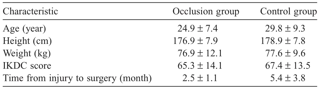
Table 1Baseline characteristics of subjects (mean±SD).
2.3.MR
To obtain anatomical cross sectional images,MR was performed using a Toshiba Excelart Vantage Atlas (1.5 T,Toshiba Medical Systems Corp.,Tochigi,Japan).During the first MR,a coronal-plane T1-weighted echo-sequence of the entire femur was performed.When necessary,markers attached tothe skin were used as a reference for the measurement of the length of the femur.The MR investigations were performed 2 days before surgery,and 16 days after surgery.During the MR investigation,a sagittal-plane T1-weighted spin echo-sequence was performed.This sequence covered the distal 2/3 of the femur,and proximal 1/3 of the calf of the injured leg.In addition,an axial-plane T1-weighted spin echo-sequence with 10-mm sectional thickness was performed.This sequence was started at the lateral joint line of the knee,and covered 2/3 of the distal thigh.The following variables were used: TR 775 ms,TE 15 ms,FOV 25×30.The patient was placed in a supine position.Fixation and cushions were used to avoid movement during the examination.The length of the femur was determined using the lateral joint line as reference point.Axial images at 40% and 50% of the femur length,measured distal to proximal from the lateral joint line of the knee,were obtained.ACSA of the quadriceps were measured using software included in Sectra PACS.An experienced radiologist (A.L.) blinded to group allocation performed the ACSA calculations.
2.4.Statistical analysis
Descriptive data are presented as means±SD.All analyses were performed using statistical software (Prism 6; GraphPad Software,Inc.,La Jolla,CA,USA).Comparison between changes in variables obtained form the occlusion and control groups were made with a parametric unpaired t test.Our null hypothesis was that there would be no difference between the occlusion group and the control group with regards quadriceps ACSA 16 days after surgery.
3.Results
At baseline,2 days before surgery,there was no statistically significant difference between the groups in quadriceps ACSA.From 2 days before surgery to 16 days after surgery there was a statistically significant reduction in quadriceps ACSA for both the occlusion (p<0.0001) and control groups (p<0.0001).
MR taken at 40% the femur length,with reference to the lateral joint line of the knee,showed a reduction in quadriceps ACSA of−9.7±1.0 cm2(mean±SEM) and−9.2±0.8 cm2for the occlusion and control groups,respectively.MR taken at the midpoint of the femur showed a reduction in quadriceps ACSA of−13.7±0.9 cm2and−11.5±0.7 cm2for the occlusion and control groups,respectively.Mean change in quadriceps ACSA (40% and 50% of the length of the femur) was−13.8%±1.1% and−13.1%±1.0% for the occlusion and control groups,respectively.Thus,there was no significant difference in loss of quadriceps ACSA between the groups after the intervention period (p = 0.6265) (Fig.1).
When analyzing men and women separately,in the occlusion group,men lost 12.8%±1.1%,and women lost 15.3%±2.1% of their quadriceps ACSA,with no statistical difference (p = 0.28).In the control group,men lost−12.2%±0.8%,and women lost−14.3%±2.2%,with no statistically significant difference (p = 0.31).Changes in quadriceps ACSA are summarized in Table 2.
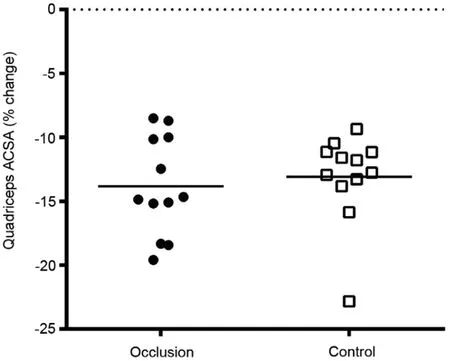
Fig.1.Mean % change in quadriceps anatomical cross sectional area (ACSA).
4.Discussion
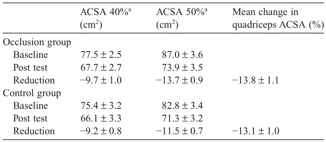
Table 2Change in anatomical cross sectional area (ACSA) of quadriceps from baseline to 16 days after surgery for the occlusion and control groups (mean±SD).
Our main finding was that intermittent restriction of blood flow does not reduce quadriceps atrophy during the first 16 days after ACL reconstruction in an athletic population.This is in contrast with previous studies using the same inflation/deflation protocol.11,12For example,Takarada et al.11reported a reduction in quadriceps ACSA of 9.4%±1.6% and 20.9%±2.2% in the occlusion and control groups,respectively.In our study,we did not find a significant difference between groups.From 2 days before surgery to 16 days after surgery,the occlusion group had a reduction of 13.8%±1.1%,and the control group a reduction of 13.1%±1.0% in quadriceps ACSA.The rate of quadriceps atrophy in our study was slightly more than reported in studies using unloading of the lower limb to study the effects of disuse.This is expected as the surgical procedure traumatizes the knee far more than pure unloading.However,theamount of quadriceps atrophy in our study was markedly less than what was previously reported after ACL surgery.6Previous studies have shown a slight difference between male and female with regards to quadriceps atrophy after ACL surgery,with female losing more muscle mass.11,13There was a tendency towards women losing more muscle mass in our study,however this was not statistically significant.
We used a wider cuff than the previous study investigating the effect of vascular occlusion on muscle atrophy after ACL reconstructive surgery.Takarada et al.11used a 90-mm wide cuff with a maximal pressure of 238±8 mmHg.They assumed that the occlusive pressure effectively inhibits both the arterial inflow and venous outflow through the occluded area.Wide occlusion cuffs restrict arterial blood flow at a lower pressure than narrow cuffs.14-16Laurentino et al.17using a 140-mm wide cuff reported cessation of the tibial artery pulse at pressures ∼130 mmHg.However,the same cuff may not restrict the same amount of blood flow on all individuals.Thigh circumference has been shown to be the largest determinant of arterial occlusion pressure.16We did not use individual adjustment of the restrictive cuff pressure in our study,which must be taken in to account when interpreting our data.
A recent study18reported that the intensity of the exercises during blood flow restricted training is important.They suggest that approximately 10% of maximal strength is the minimum intensity for muscle hypertrophy to occur.This is linked to both muscle activation and muscle cell swelling.Thus,it is possible that the training intensity in our study is below or at the lower border necessary to obtain a reduction in quadriceps atrophy.Based on the number of studies showing effect on muscle hypertrophy,a research model combining NMES with sufficient muscle activation and blood flow restriction may be indicated.
We used two ACSA measurements to quantify change in quadricepsmusclesize,at40%and50%ofthelengthofthefemur withreferencetothelateraljointlineoftheknee.Thus,weshould be able to detect area specific atrophy.However,muscle volume hasbeensuggestedasamoreaccuratemethodtoestimatemuscle size.1FuturestudiesshouldusemusclevolumeratherthanACSA asoutcomemeasureforquadricepsatrophy.Anotherlimitationis the low number of subjects in the study,particularly as there was a substantial variation in atrophy between subjects (−8.5% to −22.8%).A larger RCT is needed to establish the effect of intermittent blood flow restriction on reduction in quadriceps atrophy the first 2 weeks after ACL reconstruction.
5.Conclusion
In conflict with other studies using a similar protocol,application of blood flow restriction the first 14 days after ACL reconstruction did not reduce quadricepsACSA muscle atrophy measured by MR in a population of athletes.
Authors’contributions
EI conceived of the study,participated in the design of the study,contributed to the patient intervention,performed the statistical analyses,and drafted the manuscript.VR participated in the design of the study,contributed to the patient intervention,and helped to draft the manuscript.AL designed the MR protocol and performed the ACSA measurements.All authors have read and approved the final version of the manuscript,and agree with the order of presentation of the authors.
Competing interests
None of the authors declare any competing interests.
References
1.Akima H,Furukawa T.Atrophy of thigh muscles after meniscal lesions and arthroscopic partial menisectomy.Knee Surg Sports Traumatol Arthrosc 2005;13:632-7.
2.Dirks ML,Wall BT,Snijders T,Ottenbros CL,Verdijk LB,van Loon LJ.Neuromuscular electrical stimulation prevents muscle disuse atrophy during leg immobilization in humans.Acta Physiol 2014;210:628-41.
3.Wall BT,Dirks ML,Snijders T,Senden JM,Dolmans J,van Loon LJ.Substantial skeletal muscle loss occurs during only 5 days of disuse.Acta Physiol 2014;210:600-11.
4.Wall BT,van Loon LJ.Nutritional strategies to attenuate muscle disuse atrophy.Nutr Rev 2013;71:195-208.
5.Campbell EL,Seynnes OR,Bottinelli R,McPhee JS,Atherton PJ,Jones DA,et al.Skeletal muscle adaptations to physical inactivity and subsequent retraining in young men.Biogerontology 2013;14:247-59.
6.Gerber JP,Marcus RL,Leland ED,Lastayo PC.The use of eccentrically biased resistance exercise to mitigate muscle impairments following anterior cruciate ligament reconstruction: a short review.Sports Health 2009;1:31-8.
7.Arangio GA,Chen C,Kalady M,Reed 3rd JF.Thigh muscle size and strength after anterior cruciate ligament reconstruction and rehabilitation.J Orthop Sports Phys Ther 1997;26:238-43.
8.Akima H,Kubo K,Imai M,Kanehisa H,Suzuki Y,Gunji A,et al.Inactivity and muscle: effect of resistance training during bed rest on muscle size in the lower limb.Acta Physiol Scand 2001;172:269-78.
9.Miokovic T,Armbrecht G,Felsenberg D,Belavy DL.Heterogeneous atrophy occurs within individual lower limb muscles during 60 days of bed rest.J Appl Physiol (1985) 2012;113:1545-59.
10.Wernbom M,Augustsson J,Raastad T.Ischemic strength training: a low-load alternative to heavy resistance exercise? Scand J Med Sci Sports 2008;18:401-16.
11.Takarada Y,Takazawa H,Ishii N.Applications of vascular occlusion diminish disuse atrophy of knee extensor muscles.Med Sci Sports Exerc 2000;32:2035-9.
12.Kubota A,Sakuraba K,Sawaki K,Sumide T,Tamura Y.Prevention of disuse muscular weakness by restriction of blood flow.Med Sci Sports Exerc 2008;40:529-34.
13.Arvidsson I,Arvidsson H,Eriksson E,Jansson E.Prevention of quadriceps wasting after immobilization: an evaluation of the effect of electrical stimulation.Orthopedics 1986;9:1519-28.
14.Reilly CW,McEwen JA,Leveille L,Perdios A,Mulpuri K.Minimizing tourniquet pressure in pediatric anterior cruciate ligament reconstructive surgery: a blinded,prospective randomized controlled trial.J Pediatr Orthop 2009;29:275-80.
15.Younger AS,McEwen JA,Inkpen K.Wide contoured thigh cuffs and automated limb occlusion measurement allow lower tourniquet pressures.Clin Orthop Relat Res 2004;428:286-93.
16.Loenneke JP,Fahs CA,Rossow LM,Sherk VD,Thiebaud RS,Abe T,et al.Effects of cuff width on arterial occlusion: implications for blood flow restricted exercise.Eur J Appl Physiol 2012;112:2903-12.
17.Laurentino G,Ugrinowitsch C,Aihara AY,Fernandes AR,Parcell AC,Ricard M,et al.Effects of strength training and vascular occlusion.Int J Sports Med 2008;29:664-7.
18.Abe T,Loenneke JP,Fahs CA,Rossow LM,Thiebaud RS,Bemben MG.Exercise intensity and muscle hypertrophy in blood flow-restricted limbs and non-restricted muscles: a brief review.Clin Physiol Funct Imaging 2012;32:247-52.
杂志排行
Journal of Sport and Health Science的其它文章
- Assessing proprioception: What do you really want to know? —Response to Krewer et al.
- Commentary on:“Assessing proprioception: A critical review of methods”by Han et al.
- Author biographies
- Performance on the Functional Movement Screen in older active adults
- Single dose of intra-muscular platelet rich plasma reverses the increase in plasma iron levels in exercise-induced muscle damage: A pilot study
- Time-dependent gene expression analysis after mouse skeletal muscle contusion