Effect of maxillary expansion on orthodontics
2015-10-31ZhenTangLiPingJiangJianYongWu
Zhen Tang, Li-Ping Jiang, Jian-Yong Wu
Department of Orthodontics, Affiliated Stomatological Hospital of Nanchang University, Nanchang 330006, China
Effect of maxillary expansion on orthodontics
Zhen Tang, Li-Ping Jiang, Jian-Yong Wu*
Department of Orthodontics, Affiliated Stomatological Hospital of Nanchang University, Nanchang 330006, China
ARTICLE INFO
Article history:
in revised form 20 September 2015
Accepted 15 October 2015
Available online 20 November 2015
Maxillary expansion
Objective: To explore the effect of maxillary expansion on orthodontics. Methods: Eight beagle dogs were randomly divided into two groups, with 4 dogs in each group. Dogs in group 1 were executed immediately and received the direct physical measurement. The magnetic expansion appliance was used in group 2 for the maxillary expansion. After the expansion,the model was taken again and they were executed after cone beam CT (CBCT) scanning. The model measurement method was adopted in group 1 to measure the dental measurement indicators and width of base bone arch. The CBCT measurement method was employed to measure the above dental indicators and bone indicators. The difference in the indicators measured by different methods was compared and analyzed. Results: Before the expansion,there was no significant difference in the bone measurement indicators between the CBCT measurement method and direct physical measurement method. After the expansion, there was no significant difference in indicators between the CBCT measurement method and direct physical measurement. But there was significant difference among the model measurement method, CBCT measurement method and direct physical measurement method. There was the significant difference in the dental indicators between the CBCT measurement method and model measurement, as well as the bone indicators of posterior marginal spacing of greater palatine foramen, posterior marginal spacing of incisive foramen, width of base bone arch and spacing of implant anchorage. Conclusions: There is no significant difference between the effect of CBCT measurement method and direct physical measurement method, but CBCT is significantly better than the model measurement.
Document heading doi:10.1016/j.apjtm.2015.10.005
1. Introduction
The maxillary expansion is a common treatment for oral orthodontics, mainly for the treatment of maxillary arch constriction, anterior and posterior crossbite, maxillary protrusion and dental crowding[1]. In 1860, Angle found that the maxillary could be laterally expanded through the separation of median suture, which thus proved the clinical effect of rapid maxillary expansion[2]. Because of the exact effect and easy preparation, themaxillary expansion has become a common orthopedic method in the oral orthodontics. However, some obvious drawbacks have limited its application in the clinical practice. For instance, patients should apply the force in the mouth themselves and the operation is complex and risky, which is especially difficult for child patients. Meanwhile, the initial force for the expansion is relatively strong and its value is indefinite, which may easily cause the side effects of root resorption and bone fenestration[3,4]. Therefore, the optimization of expansion methods has always been the research focus in the field of oral orthodontics. In recent years, many scholars tried to use other methods such as the magnetic force to perform the expansion and hoped to study and control the optimal value of force to expansion in a more accurate way.
The relatively uniform evaluation standard of therapeutic effect is the precondition to objectively and correctly evaluate the different expansion methods. But there has been no uniform standard forthe evaluation of therapeutic effect. Presently, according to the measurement methods, the common evaluation means of maxillary expansion include the model method, two-dimensional imaging method, three-dimensional imaging method and direct physical method. According to the measurement indicators, they can be divided into the bone indicator difference and dental indicator difference. To reflect the effect of expansion and the relationship between the dental effect and bone effect, the molar angulation plays a unique role in the effect evaluation of maxillary expansion[5-7].
The model measurement method is to measure the mark points of perfused model, with the characteristics of low cost and independence of equipments. However, its disadvantages of low accuracy, vulnerability of material shrinkage and wearing have limited its development[8,9]. The two-dimensional imaging method is characterized by the convenient operation and easy acquisition of image information, which can qualitatively observe the change in the width of median palatine suture[10]. To improve the twodimensional imaging method that has the disadvantages of image overlay, artifact and distorted deformation, the helical CT appears as the three-dimensional imaging method. The helical CT is capable to evaluate the expansion from the three-dimensional angle and then perform the quantitative evaluation[11,12]. Besides, the cone beam CT (CBCT) greatly increases the scope of application for the three-dimensional imaging method. The scanning time for CBCT is only 10-70 s, which can scan the whole maxillofacial region and especially the specific regions; meanwhile, the radiation dose is controlled within 36.9-50.3 μSv. However, there are some defects for CBCT, such as the unsatisfactory examination and evaluation of soft tissue lesion[13-15]. The direct physical measurement is to perform the measurement on the living or dead animal body directly. Because of the relatively accurate measurement data, it is usually regarded as the gold standard for the measurement of animal experiment. But such method also has the disadvantages. For instance, it's difficult to repeatedly measure the living animal body and there would be errors in the process of treating the dead body or dry skull[16].
There have been limited researches on the exact comparison of animal experiment using the different measurement methods. In this study, the beagle dog was selected as the laboratory animal and the magnetic expansion appliance was used for the maxillary expansion. The direct physical measurement method was chosen as the gold standard and the comparative study was performed on the accuracy between the model measurement method and CBCT measurement method. Meanwhile, it observed the measurement indicators, including the difference of bone and dental measurement indicators before and after the expansion and the molar angulation,in order to evaluate the difference in the accuracy and precision between different measurement methods, which has the certain value of application.
2. Materials and methods
2.1. Laboratory animals
Eight healthy male beagle dogs with the age of 6 months and weight of about 8 kg (in the initial stage of permanent dentition,without obvious oral and maxillofacial defects) were purchased from Shanghai Xingang Laboratory Animal Farm. The laboratory dogs were fed with the standard diet in the standard animal cage, allowing the free drinking and eating. The feeding room had the good ventilation and natural lighting day and night. The room temperature was maintained at 18-25 ℃. The experiment was performed after one week of adaptive feeding.
2.2. Equipments, reagents and instruments
The dental self-threading pin was purchased from Hangzhou Westlake Biomaterial Co., Ltd. (311107); the sintered N52 Nd-Fe-B magnets from Beijing Zhongke Sanhuan Hi-tech Co., Ltd.;the fine silicone rubber impression material from 3M (7312); the glass ionomer orthodontic band cement from Fuji; and pentobarbital sodium from Merck (TF110).
The clean bench was purchased from Sujing Protective Atmosphere Co., Ltd.; the electronic universal testing machine from Instron(2343); VBCT machine from Danaher, KaVo 3D eXam; the precision balance from Sartorius, 124S; the imaging system from Olympus, CX71; and the vernier caliper Guilin Guanglu Measuring Instrument Co., Ltd.
2.3. Mechanical performance testing of magnetic expansion appliance
To test the magnetic expansion appliance made of high energy product Nd-Fe-B magnets by this research group (dimension of magnet: 11.50×mm×2.73 mm×2.30 mm), Instron electronic universal testing machine was employed to test the torque curve after two times of thrust boosting by magnetic expansion appliance.
2.4. Animal grouping and modeling
After being numbered, 8 beagle dogs aged 6-month were intraperitoneally injected with 2% pentobarbital sodium (1 mL/1 kg) for the anaesthesia respectively. At the maxillary first and fourth premolar buccal line and both sides of median palatine suture, the 3mm bone surface was incised and the self-threading dental pin was screwed in perpendicular to the bone surface for the location marker. The prefabricated maxillary individual tray was used to prepare the silicone rubber impression and then the CBCT scan was performed. The laboratory dogs were randomly divided into two groups,with 4 dogs in each group. Four dogs in group one were executed immediately and then the direct physical measurement was taken. The diamond burs were employed for 4 dogs in group two to perform the tooth preparation at the maxillary fourth premolar and first molar on both sides. The silicone rubber impression was prepared and then the die stone was perfused to make the plaster model. An adjustable integrated magnetic expansion device was made on the model. Afterwards, the beagle dogs were anaesthetized again. The wetinsulated dental surface was cleaned and the expansion device was worn and adhered. Two weeks after the expansion, the laboratory dogs were anaesthetized again. The magnetic expansion device wasremoved. After taking the model, CBCT scan was performed on the whole skull. Animals were executed and then the direct physical measurement was carried out (Figure 1).
2.5. Measurement methods and indicators
The model measurement method was adopted for animals in group one to measure the dental measurement indicators, namely the spacing between teeth (C-C), spacing of the fourth premolar(P-PM4), spacing of the first molar (M-M1) and width of base bone arch. The CBCT measurement method was employed to measure the above dental indicators and bone indicators (spacing of implant anchorage, posterior marginal spacing of greater palatine foramen,posterior marginal spacing of incisive foramen, and width of base bone arch); the direct physical measurement was performed on animals for the above dental indicators and bone indicators. Before the expansion in group two, the model measurement method was adopted to measure the dental measurement indicators and width of base bone arch; the CBCT measurement method was employed to measure the above dental indicators, bone indicators and molar angulation; after the expansion, the model measurement method was adopted to measure the dental measurement indicators and width of base bone arch; and the CBCT measurement method to measure the above dental indicators, bone indicators and molar angulation;the direct physical measurement was performed on animals for the above dental indicators and bone indicators. Indicator difference = postoperative indicator measurement value-preoperative indicator measurement value. The specific measurement methods and indicators are shown as follows.
Model measurement method: The prefabricated maxillary individual tray was used to prepare the silicone rubber impression and then the die stone was perfused to make the plaster model. The plaster model was placed on the horizontal worktable surface to keep the occlusal plane parallel to the horizontal plane and following indicators were measured. (1) spacing between teeth (C-C): the divider was used to locate the peak point of bilateral cuspid and the width was transferred to the blank paper, which was measured and read by the electronic digital caliper (accurate to 0.01 mm);(2) spacing of the fourth premolar (P-PM4): the divider was used to locate the peak point of bilateral fourth premolar mesial cusp and the width was transferred to the blank paper, which was measured and read by the electronic digital caliper. (3) spacing of the first molar(M-M1): the divider was used to locate the peak point of bilateral first molar mesial cusp and the width was transferred to the blank paper, which was measured and read by the electronic digital caliper.(4) width of base bone arch: the divider was used to locate the width of base bone arch and the width was transferred to the blank paper,which was measured and read by the electronic digital caliper.
CBCT measurement method: the self-made skull location device was used to fix the skull of animals. A senior technician performed the whole skull CBCT scan and recording for the laboratory dogs,with the same scanning conditions every time: exposure parameters 120 kVp, 20.27 mAs, 14.7 s, resolution of 0.25 voxel, and reconstruction volume of 16 mm×16 mm×10 mm. All Dicom data of scanning were input in InVivo5 (Anatomage, San Jose, Calif). The indicators were measured after the reconstruction.
Direct physical measurement method: animals were executed and the soft tissue attachment in the maxillofacial region was stripped. The skull sample was placed on the horizontal worktable surface to keep the occlusal plane parallel to the horizontal plane and following indicators were measured. (1) spacing between teeth(C-C): the divider was used to locate the peak point of bilateral cuspid and the width was transferred to the blank paper, which was measured and read by the electronic digital caliper; (2) spacing of the fourth premolar (P-PM4): the divider was used to locate the peak point of bilateral fourth premolar mesial cusp and the width was transferred to the blank paper, which was measured and read by the electronic digital caliper; (3) spacing of the first molar (M-M1): the divider was used to locate the peak point of bilateral first molar mesial cusp and the width was transferred to the blank paper, which was measured and read by the electronic digital caliper. (4) spacing of implant anchorage: it included the spacing of location pin in PM1region and spacing of location pin in PM1region. For the measurement of spacing of implant anchorage, the divider was used to locate the central neck of location pin in the bilateral first premolar region (the junction with the bone surface) and the width was transferred to the blank paper, which was measured and read by the electronic digital caliper. For the measurement of spacing of location pin in PM4region, the divider was used to locate the central neck of location pin in the bilateral fourth premolar region (the junction with the bone surface) and the width was transferred to the blank paper, which was measured and read by the electronic digital caliper; (5) posterior marginal spacing of incisive foramen: the divider was used to locate the bilateral posterior marginal spacing of incisive foramen and the width was transferred to the blank paper, which was measured and read by the electronic digital caliper; (6) posterior marginal spacing of greater palatine foramen: the divider was used to locate the bilateral posterior marginal spacing of greater palatine foramen and the width was transferred to the blank paper, which was measured and read by the electronic digital caliper; (7) width of base bone arch: the divider was used to locate the concave part of the alveolar bone at the transitional folds of the buccal side of bilateral first molarmesial cusp and the width was transferred to the blank paper, which was measured and read by the electronic digital caliper.
2.6. Statistical analysis
The experimental data was treated by SPSS19.0. Results were expressed by mean±SD. The analysis of variance was performed for the comparison of multiple groups, while t-test was used for the comparison between two groups. P<0.05 indicated the significant difference. The multi-factor repeated measures analysis of variance was performed for the repeated comparison of measurement methods, with the significant level of α=0.05.
3. Results
3.1. Mechanical performance of magnetic expansion appliance
Instron electronic universal testing machine was employed to test the torque curve after two times of thrust boosting by magnetic expansion appliance. After two times of thrust boosting by magnetic expansion appliance, it had the similar torque curves, with the force range of 10.38 N-1.63 N and magnet spacing of 0 mm-3 mm (as shown in Figure 2).
3.2. Comparison of measurement indicators using different measurement methods before expansion
The analysis of variance with compatibility groups was performed on the comparison of three measurement methods that were used to measure the dental measurement indicators before expansion, including the model measurement, CBCT measurement and direct physical measurement, with results shown in Table 1. Results indicated that there was significant difference in P-PM4between model group and CBCT group (P=0.010), but the difference was not significant between model group and physical group (P=0.160),or between CBCT group and physical group (P=0.076). There was significant difference in M-M1between model group and CBCT group (P=0.002), and between model group and physical (P=0.014);but the difference was not significant between CBCT group and physical group (P=0.153). CBCT result was shown in Figure 3.

Table 1Comparison of measurement indicators among three measurement methods before expansion.
As the bone measurement indicators could not be measured in the model, the comparison was only performed between the CBCT measurement and direct physical measurement. The t-test was adopted for the analysis. Results showed that there was no significantdifference in five bone measurement indicators (spacing of location pin in PM1region, spacing of location pin in PM4region, width of base bone arch, posterior marginal spacing of incisive foramen, and posterior marginal spacing of greater palatine foramen) between CBCT measurement and direct physical measurement (P>0.05)(Table 2). group (P>0.05).

Table 2 Comparison of bone measurement indicators among different measurement methods before expansion.

Table 3 Comparison of dental measurement indicators among three measurement methods after expansion.
3.3. Comparison of measurement indicators using different measurement methods after expansion
The analysis of variance with compatibility groups was performed on the comparison of three measurement methods that were used to measure the dental measurement indicators after expansion,including the model measurement, CBCT measurement and direct physical measurement, with results shown in Table 3. Results indicated that there was the difference in three indicators of C-C,P-PM1 and P-PM4 between model group and CBCT group, and between model group and physician group (P<0.05); but there was no significant difference between CBCT group and direct physical
As the bone measurement indicators could not be measured in the model, the comparison was only performed between the CBCT measurement and direct physical measurement. The t-test was adopted for the analysis. Results showed that there was no significant difference in five bone measurement indicators after expansion between CBCT measurement and direct physical measurement(P>0.05) and also no significant difference in the accuracy of bone measurement indicators between two methods (P>0.05) (Table 4).
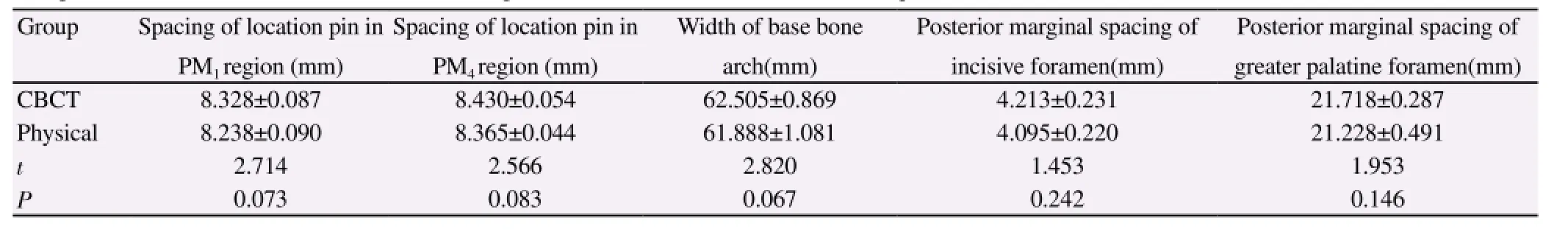
Table 4 Comparison of bone measurement indicators among different measurement methods after expansion.
3.3. Comparison of difference in change of measurement indicators before and after expansion
Results indicated that there was the significant difference in change of spacing between teeth, spacing of the fourth premolar and spacing of the first molar (P<0.05), which meant that there was the significant difference between model measurement method and CBCT measurement method (Table 5).

Table 5 Comparison of change of measurement indicators between model measurement and CBCT measurement before and after expansion.
The CBCT method was employed to measure the difference of five bone measurement indicators (difference in spacing of location pin in PM1region, difference in spacing of location pin in PM4region, difference in width of base bone arch, difference in posterior marginal spacing of incisive foramen, and difference in posterior marginal spacing of greater palatine foramen) before and after the expansion. The t-test was performed on the comparison of these five bone measurement indicators each other. It indicated that there was the significant difference in the spacing of location pin in PM1region and posterior marginal spacing of incisive foramen, the spacing of location pin in PM4and posterior marginal spacing of greater palatine foramen, and spacing of location pin in PM4and width of base bone arch (P<0.05), which meant that the implant location markers could not be replaced by above bone indicators.
4. Discussion
Because of the exact effect and easy preparation, the maxillary expansion has become a common orthopedic method in the oral orthodontics. Relying on the separation of median palatine suture using the mechanical orthopedic force, it has been mainly employed for the treatment of maxillary arch constriction, anterior and posterior crossbite, maxillary protrusion and dental crowding[1]. The optimization of expansion methods has always been the research focus in the field of oral orthodontics. In recent years, many scholars tried to use other methods such as the magnetic force to perform the expansion and hoped to study and control the optimal value of force to expansion in a more accurate way. However, because of no uniform standard for the evaluation of therapeutic effect, it is quite difficult to evaluate the effect of expansion. Presently, according to the measurement methods, the common evaluation means of maxillary expansion include the model method, two-dimensional imaging method, three-dimensional imaging method and direct physical method. According to the measurement indicators, they can be divided into the bone indicator difference and dental indicatordifference. To reflect the effect of expansion and the relationship between the dental effect and bone effect, the molar angulation plays a unique role in the effect evaluation of maxillary expansion[5-7, 17]. To confirm an evaluation standard, it started from the angles of evaluation method and measurement indicator to study the effect of different measurement evaluation methods and indicators on the expansion. The model measurement is a traditional method, the model was prepared from the animals or human before and after the expansion and the expansion effect was evaluated at mark points in the model. It is the method that is still widely applied at present. Magnusson et al measured the width of maxillary cuspid and dental arch in molar region in the model before and after the experiment directly to evaluate the long-term stability after the surgically-aided maxillary[18]. Such measurement has the easy operation, but does not consider the dental inclination, with the big error. Namara et al[19] located points on four surfaces along the mesial-distal and buccolingual directions with the assistance of advanced threedimensional model measurement system. These points were used to confirm the geometric center and center of mass of each tooth. Such method of point location would not consider the change in the tooth rotation and thus promote the measurement accuracy of dental arch width. In this experiment, there was the significant difference in the measurement of spacing between teeth and width of base bone in the model between CBCT method and direct physical method. The reason might be that the experiment adopted the contact measurement and the shrinkage of molding materials could cause the error.
The direct physical measurement is to perform the measurement on the living or dead animal body directly. Because of the accurate measurement data, it is usually regarded as the gold standard for the measurement of animal experiment[16]. In this experiment, 4 animals were executed before the expansion and then the measurement was taken for related indicators as the comparison standard of accuracy research.
CBCT is a new three-dimensional medical imaging technique appeared in the end of 20th century, which has been widely applied in the field of oral medicine. More and more scholars have adopted CBCT to evaluate the effect of maxillary expansion. Kanomi et al used CBCT to evaluate the effect of rapid expansion on 89 patients. Results showed that there was good effect for 6-15 year-old patients using two different expansion appliances (bone supporting and dental supporting). Lagravère et al randomly adopted the dental supporting and bone supporting expansion appliances to perform the maxillary expansion for 62 patients. The change in the dental inclination before and after the expansion was observed through CBCT. Results showed that there was the change in molar angulation using two expansion methods, but no change in the premolar using the bone supporting appliance[20,21]. In this study, CBCT was employed to evaluate the change in the bone and dental measurement indicators and molar angulation of beagle dogs before and after the expansion. Results showed that there was no significant difference in the measurement of spacing between teeth, width of base bone arch and spacing of bone marker points between CBCT method and direct physical one, but the significant difference between CBCT and model measurement. According to results, CBCT measurement was accurate and had the better effect in the evaluation of expansion than the model measurement.
Previous researches usually adopted the cuspid, premolar and spacing of the first molar as the dental measurement indicators of maxillary expansion. Magnusson et al measured the width of maxillary cuspid and dental arch in molar region in the model before and after the experiment directly to evaluate the long-term stability after the surgically-aided maxillary. Canut et al measured the peak point in the bilateral cuspid region, fovea in the premolar region and mesial buccal spacing in the molar region to evaluate the effect of expansion. In this study, the adjustable integrated magnetic expansion appliance was fixed in the cuspid, fourth premolar and first molar of beagle dogs. Accordingly, the spacing of cuspid, fourth premolar and first molar were regarded as the dental measurement indicators to evaluate the effect before and after the expansion[22].
Presently, the common bone measurement indicators to evaluate the effect of maxillary expansion include the spacing of anterior nasal spine, spacing of incisive foramen and spacing of palatal glazing cementum that reflect the effect of anterior expansion; and the spacing of greater palatine foramen, spacing of posterior nasal spine, spacing of palatal alveolar ridge in the maxillary first molar region, and width of base bone arch that reflect the effect of posterior expansion[16,18,22,23]. In this study, the difference in the posterior marginal spacing of incisive foramen that had the good repeatability in CBCT imaging of beagle dogs was chosen as the bone indicator to evaluate the effect of anterior expansion; while the difference in posterior marginal spacing of greater palatine foramen and difference in width of base bone arch that had the good repeatability in CBCT imaging of beagle dogs to evaluate the effect of posterior expansion. Because of the large volume of dental implant, it's quite difficult to set points in CBCT correctly and it also cost much. The amalgam pin would cause the great trauma and it was not suitable for the clinical research. But as the marker, the self-threading dental pin was characterized by the easy operation, small diameter, easy location and low cost. Therefore, at the maxillary first and fourth premolar buccal line and both sides of median palatine suture, the 3 mm bone surface was incised and the location pin was screwed in. The difference in the spacing of location pin before and after the expansion was chosen as the gold standard to study the accuracy of difference in the posterior marginal spacing of incisive foramen;meanwhile at the maxillary fourth premolar buccal line and both sides of median palatine suture, the 3 mm bone surface was incised and the location pin was screwed in. The difference in the spacing of location pin before and after the expansion was chosen as the gold standard to study the accuracy of difference in the posterior marginal spacing of greater palatine foramen and width of base bone arch.
In this study, three methods of model measurement, CBCT measurement and direct physical measurement were performed to measure the spacing between teeth, spacing of the fourth premolar and spacing of the first molar before and after the expansion of beagle dogs. They were compared each other. The direct physical measurement method was chosen as the gold standard and the comparative study was performed on the accuracy between the model measurement method and CBCT measurement method. Results showed that there was no significant difference in the width of cuspid before the expansion between CBCT and model and direct physical measurement. There was the difference in the width of molar between the model measurement and CBCT and direct physical measurement, but no difference between CBCT measurement and direct physical measurement, which was in line with findings of Sun et al[16]. In consequence, in the measurement of dental indicators, CBCT method was better than the modelmeasurement method, which could replace the direct physical measurement.
The bone indicators include the spacing of location pin in PM1region and posterior marginal spacing of incisive foramen that reflect the effect of anterior expansion; and the spacing of location pin in PM4region, posterior marginal spacing of greater palatine foramen and width of base bone arch that reflect the effect of posterior expansion. The CBCT measurement and direct physical measurement were performed; meanwhile, the physical measurement was chosen as the gold standard to study the accuracy of CBCT measurement method. Results showed that there was no significant difference in above 5 bone indicators between CBCT measurement method and direct physical measurement method and it indicated that, for the measurement of these indicators, the accuracy of CBCT method was close to direct physical measurement method.
As the direct physical measurement did not allow the matching study on the difference in dental indicators, the comparison study was only performed on the difference in dental indicators by the model measurement and CBCT measurement. According to the comparison between the dental indicators by the model measurement and CBCT measurement and the one by direct physical measurement before and after the expansion showed that there was no significant difference between CBCT measurement method and direct physical measurement method, which were better than the model measurement method. Accordingly, it can be regarded that the CBCT measurement method has the better effect than the model measurement method in the measurement of difference in dental indicators.
Conflict of interest statement
We declare that we have no conflict of interest.
[1] Liu S, Xu T, Zou W. Effects of rapid maxillary expansion on the midpalatal suture: a systematic review. Eur J Orthod 2015; 10.1093/ejo/ cju100.
[2] Timms DJ. The dawn of rapid maxillary expansion. Angle Orthod 1999;69(3): 247-250.
[3] Leung CC, Palomo L, Griffith R, Hans MG. Accuracy and reliability of cone-beam computed tomography for measuring alveolar bone height and detecting bony dehiscences and fenestrations. Am J Orthod Dentofacial Orthop 2010; 137(4 Suppl): S109-S119.
[4] Baysal A, Uysal T, Veli I, Ozer T, Karadede I, Hekimoglu S. Evaluation of alveolar bone loss following rapid maxillary expansion using conebeam computed tomography.Korean J Orthod 2013; 43(2): 83-95.
[5] Davis WM, Kronman JH. Anatomical changes induced by splitting of the midpalatal suture. Angle Orthod 1969; 39(2):126-132.
[6] Wagemans PA, van de Velde JP, Kuijpers-Jagtman AM. Sutures and forces: a review. Am J Orthod Dentofacial Orthop 1988; 94(2): 129-141.
[7] Bazargani F, Feldmann I, Bondemark L. Three-dimensional analysis of effects of rapid maxillary expansion on facial sutures and bones. Angle Orthod 2013 ; 83(6): 1074-1082.
[8] Magnusson A, Bjerklin K, Nilsson P, Marcusson A. Surgically assisted rapid maxillary expansion: long-term stability. Eur J Orthod 2009; 31(2):142-149.
[9] Anttila A, Finne K, Keski-Nisula K, Somppi M, Panula K, Peltomäki T. Feasibility and long-term stability of surgically assisted rapid maxillary expansion with lateral osteotomy. Eur J Orthod 2004; 26(4): 391-395.
[10] Gurgel Jde A, Malmström MF, Pinzan-Vercelino CR. Ossification of the midpalatal suture after surgically assisted rapid maxillary expansion. Eur J Orthod 2012; 34(1): 39-43.
[11] Lee KJ, Park YC, Park JY, Hwang WS. Miniscrew-assisted nonsurgical palatal expansion before orthognathic surgery for a patient with severe mandibular prognathism. Am J Orthod Dentofacial Orthop 2010; 137(6):830-839.
[12] Akyalcin S, Schaefer JS, English JD, Stephens CR, Winkelmann S. A cone-beam computed tomography evaluation of buccal bone thickness following maxillary expansion. Imaging Sci Dent 2013; 43(2): 85-90.
[13] Adibi S, Zhang W, Servos T, O'Neill PN. Cone beam computed tomography in dentistry: what dental educators and learners should know. J Dent Educ 2012; 76(11): 1437-1442.
[14] Li M, Ballhausen H, Hegemann NS, Ganswindt U, Manapov F, Tritschler S, et al. A comparative assessment of prostate positioning guided by three-dimensional ultrasound and cone beam CT. Radiat Oncol 2015;10(1): 82.
[15] Celikten B, Uzuntas CF, Kursun S, Orhan AI, Tufenkci P, Orhan K, et al. Comparative evaluation of shaping ability of two nickel-titanium rotary systems using cone beam computed tomography. BMC Oral Health 2015;15(1): 32.
[16] Sun Z, Smith T, Kortam S, Kim DG, Tee BC, Fields H. Effect of bone thickness on alveolar bone-height measurements from cone-beam computed tomography images. Am J Orthod Dentofacial Orthop 2011;139(2): e117-e127.
[17] Laudemann K, Santo G, Revilla C, Harth M, Kopp S, Sader RA, et al. Assessment of surgically assisted rapid maxillary expansion regarding pterygomaxillary disjunction using thin volume-rendering technique:in variance analysis and in reliability, accuracy, and validity. J Oral Maxillofac Surg 2011; 69(10): 2631-2643.
[18] da Silva Filho OG, Lara TS, de Almeida AM, da Silav HC. Evaluation of the midpalatal suture during rapid palatal expansion in children: a CT study. J Clin Pediatr Dent 2005; 29(3): 231-223.
[19] Pinheiro FH, Garib DG, Janson G, Bombonatti R, de Freitas MR. Longitudinal stability of rapid and slow maxillary expansion. Dental Press J Orthod 2014; 19(6): 70-77.
[20] Kanomi R, Deguchi T, Kakuno E, Takano-Yamamoto T, Roberts WE. CBCT of skeletal changes following rapid maxillary expansion to increase arch-length with a development-dependent bonded or banded appliance. Angle Orthod 2013; 83(5): 851-857.
[21] Lagravère MO, Gamble J, Major PW, Heo G. Transverse dental changes after tooth-borne and bone-borne maxillary expansion. Int Orthod 2013;11(1): 21-34.
[22] Carrillo R, Buschang PH, Opperman LA, Franco PF, Rossouw PE. Segmental intrusion with mini-screw implant anchorage: a radiographic evaluation. Am J Orthod Dentofacial Orthop 2007; 132(5): 576.e1-576.e 6.
[23] Khan A, Fareed WM, Tandon P, Zafar MS. Distraction osteogenesis for correction of post ankylosis mandibular deformities. J Biomed Res 2015;4: 332-336.
15 August 2015
Jian-Yong Wu, PhD., Attending Physician, Professor,Department of Orthodontics, Affiliated Stomatological Hospital of Nanchang University, Nanchang, China.
Tel: 13970078639
E-mail: wjyyrx@163.com
Foundation project: It was supported by National Natural Science Foundation of China (81160138).
Magnetic expansion
Orthodontics
杂志排行

Asian Pacific Journal of Tropical Medicine的其它文章
- Demographic, socioeconomic and environmental changes affecting circulation of neglected tropical diseases in Egypt
- Phenolic profile and biological potential of Endopleura uchi extracts
- Roots extracts of Adenophora triphylla var. japonica improve obesity in 3T3-L1 adipocytes and high-fat diet-induced obese mice
- Anti TB drug resistance in Tanga, Tanzania: a cross sectional facility base prevalence among pulmonary TB patients
- In vitro inhibitory effects of plumbagin, the promising antimalarial candidate, on human cytochrome P450 enzymes
- Vibrio spp. from Macrobrachium amazonicum prawn farming are inhibited by Moringa oleifera extracts