高频彩色多普勒超声检查在甲状腺良恶性结节诊断中的临床应用价值
2015-09-14董应梅等
董应梅等
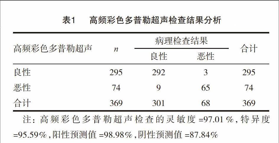
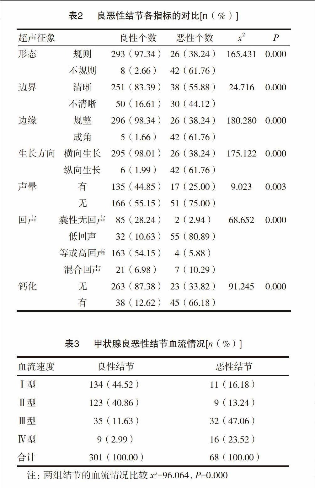
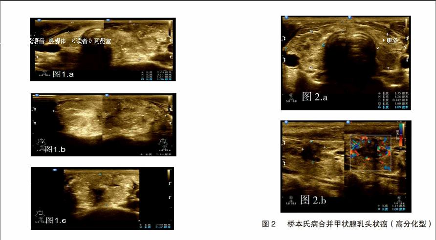
[摘要] 目的 探讨在甲状腺良恶性结节诊断中采用高频彩色多普勒超声检查的临床价值。 方法 对185例甲状腺结节患者(共369个结节)采用高频彩色多普勒超声检查,分析其影像学特点及对比良恶性结节征象。 结果 采用高频彩色多普勒超声检查甲状腺结节,灵敏度为97.01%,特异度为95.59%,阳性预测值为98.98%,阴性预测值为87.84%;超声检查发现,良性结节形态多规则,边界清晰,伴有或不伴有声晕,横向生长,内部回声多为中等回声、高回声、囊性无回声,多数不伴有钙化,同恶性结节相比较,差异具有统计学意义(P<0.05);对血流进行分析,301个良性结节中Ⅰ型血流134例,Ⅱ型血流123例,Ⅲ型血流35例,Ⅳ型血流9例,68个恶性结节中Ⅰ型血流11例,Ⅱ型血流9例,Ⅲ型血流32例,Ⅳ型血流16例,组间对比差异具有统计学意义(P<0.05)。 结论 甲状腺良恶性结节声像之间有明显的差异,也存在交叉的共同表现,结节边缘成角、内部微钙化形成和颈部异常淋巴结肿大有助于甲状腺乳头状癌的诊断;甲状腺腺瘤和甲状腺滤泡状癌在声像上很相似,鉴别时需谨慎。对甲状腺结节二维及彩色多普勒血流显像进行全面综合分析,可提高结节良恶性诊断率。高频彩色多普勒超声检查在甲状腺良恶性结节鉴别诊断中具有重要价值,可作为临床首选的影像检查。
[关键词] 高频彩色多普勒超声;甲状腺良性结节;甲状腺恶性结节;鉴别诊断
[中图分类号] R445.1 [文献标识码] B [文章编号] 2095-0616(2015)13-182-05
[Abstract] Objective To explore the clinical value of the high frequency color doppler ultrasound in the diagnosis of benign and malignant thyroid nodules. Methods 185 patients with thyroid nodules (369 nodules) were received high frequency color doppler ultrasound. Imaging features were analyzed and the signs of benign thyroid nodules and malignant thyroid nodules were compared. Results The sensitivity, specificity, positive predictive value, negative predictive value of the high frequency color doppler ultrasound in the examination of benign and malignant thyroid nodules were respectively 97.01%, 95.59%, 98.98% and 87.84%. Ultrasound examination found that benign nodules usually laterally grew with regular contour and clear boundary accompanied by internal echoes mostly including media echo, high echo and no echo and most benign nodules were non-calcified. The differences between benign thyroid nodules and malignant thyroid nodules were statistically significant (P<0.05). Blood flow was compared. The number of type Ⅰ blood flow, type Ⅱ blood flow, type Ⅲ blood flow, type Ⅳ blood flow were respectively 134, 123, 35 and 9 in the 301 cases of benign nodules. While the number of type Ⅰ blood flow, type Ⅱ blood flow, type Ⅲ blood flow, type Ⅳ blood flow were respectively 11, 9, 32 and 16 in 68 cases of malignant nodules. The difference was statistically significant (P<0.05). Conclusion There are obvious differences in the acoustic images of benign thyroid nodules and malignant thyroid nodules as well as some crossed common features. Angulated edge and internal micro calcification of nodules and abnormal lymphadenopathy of the neck is helpful in the diagnosis of papillary thyroid carcinoma. Acoustic images of thyroid adenoma and thyroid follicular cancer are extremely similar which needs to be cautious when differentiating. A comprehensive and synthetic analysis of Two-dimensional imaging and color Doppler flow imaging can improve the diagnostic rate of benign and malignant thyroid nodules. High frequency color doppler ultrasound has important value in the differential diagnosis of benign and malignant thyroid nodules which can be the preferred imaging examination in clinical practice.endprint
