完全内窥镜下经椎板间入路手术治疗腰椎间盘突出症中期临床疗效
2014-02-14吕国华李亚伟李鹏志
王 冰 吕国华 李亚伟 李 磊 邝 磊 周 乾 李鹏志
. 脊柱微创外科 Minimally invasive spine surgery .
完全内窥镜下经椎板间入路手术治疗腰椎间盘突出症中期临床疗效
王 冰 吕国华 李亚伟 李 磊 邝 磊 周 乾 李鹏志
目的 评价完全内窥镜 ( full-endoscopic,FE ) 下经椎板间入路手术治疗腰椎间盘突出症的中期临床疗效。方法2008 年 8 月至 2010 年 2 月,我院应用 FE 经椎板间入路手术治疗腰椎间盘突出症患者45 例,男 25 例,女 20 例;年龄 19~58 岁,平均 38.3 岁。术前、术后 3 个月、术后 1 年和末次随访进行腿、腰痛视觉模拟评分 ( visual analoge scale,VAS ) 和腰椎 Oswestry 功能障碍指数 ( oswestry disability index,ODI )评定,参照改良 MacNab 标准评价临床疗效,并进行统计学分析。结果所有病例手术均顺利完成,平均手术时间 62.4 min;平均住院时间 5.2 天;2 例术中出现硬膜小裂口,未予处理;无神经根损伤及术后出血等并发症发生。术后随访 48~66 个月。腿痛 VAS 评分从术前 ( 7.6±1.6 ) 分降至术后 3 个月 ( 1.2±0.8 ) 分、术后 1 年( 1.1±0.9 ) 分、末次随访 ( 1.6±1.2 ) 分 ( P<0.01 ),末次随访与术后 1 年比较,差异无统计学意义 ( P>0.05 );腰痛 VAS 评分从术前 ( 3.1±2.2 ) 分降至术后 3 个月 ( 1.8±1.5 ) 分、术后 1 年 ( 1.6±1.4 ) 分、末次随访 ( 1.7± 0.9 ) 分 ( P<0.01 ),末次随访与术后 1 年比较,差异无统计学意义 ( P>0.05 );腰椎 ODI 指数从术前 ( 69.5± 10.5 ) 降至术后 3 个月 ( 19.3±6.5 )、术后 1 年 ( 15.6±5.9 )、末次随访 ( 21.8±7.0 ) ( P<0.01 ),末次随访与术后1 年比较,差异无统计学意义 ( P>0.05 )。末次随访时,93.3% 的患者获得良好的主观满意度,临床疗效优良率为 88.9%。性别、年龄、突出类型及术前腰痛与改良 MacNab 结果无相关性 ( P>0.05 );而术前 Pfirrmann 分级、感觉缺陷及手术节段与改良 MacNab 结果具相关性 ( P<0.05 )。1 例 L4~5节段患者术后 1 周发生下肢麻木加重,予以脱水、激素治疗等保守治疗后 2 周症状缓解;1 例 L5~S1节段患者术后 4 个月出现手术节段椎间盘再次突出,行小切口开放手术翻修。末次随访时,2 例轻度运动障碍,6 例感觉异常,但较术前均显著改善。结论FE 下经椎板间入路手术治疗腰椎间盘突出症不仅具有创伤小、出血量和并发症少、术后恢复快的优点,而且可以获得满意的中期临床疗效。
椎间盘移位;内窥镜检查;椎板切除;腰椎;外科手术,微创性
完全内窥镜 ( full-endoscopic,FE ) 下经椎板间入路是近年来发展的一项微创脊柱外科技术。2006 年,由 Ruetten 等[1]首次报道并用在手术治疗腰椎退行性疾患中。该技术在带工作通道的硬杆状内窥镜和持续生理盐水灌注下,通过 8 mm 微小切口,经椎板间后入路,采取黄韧带分层切开显露椎管方法,能够安全有效地完成髓核摘除和神经根减压。诸多研究表明[2-7],应用该技术可以获得与传统外科手术治疗一致的临床疗效,并且在缓解腰痛、缩短康复时间及减少组织创伤方面更具优势。我们自 2008 年起开展了该项微创技术治疗腰椎间盘突出症,早期平均 1.3 年的 28 例随访结果显示[3],所有病例腰腿痛均获得良好的改善,其中92.9% ( 26 / 28 ) 患者腿痛症状完全消失。然而,其中期随访有待研究。为此,总结了 2008 年 8 月至2010 年 2 月,我院应用 FE 技术经椎板间入路手术治疗 45 例腰椎间盘突出症患者至少 4 年的随访结果,旨为该技术的进一步临床应用提供参考。
资料与方法
一、一般资料
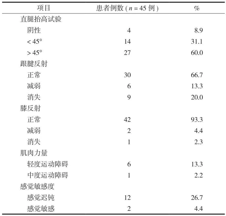
本组 45 例中,男 25 例,女 20 例;年龄 19~58 岁,平均 38.3 岁,病程 15~150 天,平均78.5 天;所有病例术前均伴有不同程度的腰痛和单侧坐骨神经痛,经保守治疗无效,行 X 线平片、MRI 检查,诊断为 L4~5或 L5~S1单节段非椎间孔型软性腰椎间盘突出症。突出节段:L5~S137 例、L4~58 例;突出类型:包含型 40 例,其中侧方型28 例、旁中央型 12 例;非包含型 5 例,其中脱出型 3 例,游离型 2 例;在 MRI 正中矢状位 T2WI 图像上,椎间盘退变分级采用 Pfirrmann 分级[8]:II 级9 例、III 级 18 例、IV 级 12 例、V 级 6 例。7 例术前伴有轻度或中度运动障碍;14 例术前伴有感觉缺陷。对患者直腿抬高试验、跟腱反射、膝反射、肌肉力量及感觉敏感度等进行术前检查 ( 表 1 )。所有手术均为同一术者完成,手术步骤参考本研究前期文献 [3]。

表1 术前临床检查结果Tab.1 Clinical Findings in the preoperative examination
二、观察指标及功能评定
患者腿、腰痛,采用视觉模拟评分 ( visual analoge scale,VAS ) 评定。腰椎功能采用 Oswestry 功能障碍指数 ( oswestry disability index,ODI ) 评估。采用问卷调查的方式评价患者主观满意度[9]:( 1 ) 是否认为自己的手术取得成功?( 2 ) 是否愿意推荐自己的朋友做这项手术 ( 如果你朋友不幸患上和你一样的疾病,你是否推荐你朋友做这项手术?)。参照改良 MacNab 分级标准[10]对患者临床疗效分级评估:症状完全消失,恢复原来的工作和生活视为优;有轻微症状,活动轻度受限,对工作生活无影响视为良;症状减轻,活动受限,影响正常工作和生活视为可;治疗前后无差别,甚至加重视为差。
三、统计学分析
数据分析采用统计学软件包 SPSS 17.0,变量VAS 评分、ODI 指数采用配对 t 检验,分类变量采用 χ2检验,非参数检验采用秩和检验,设检验水准α=0.05。
结 果
所有病例手术均顺利完成,手术时间 35~11 min,平均 62.4 min;住院时间 3~10 天,平均5.2 天;2 例术中出现硬膜小裂口,未予处理;无神经根损伤及术后出血等并发症发生。
所有患者均获门诊随访,时间术后 48~66 个月,平均 54.8 个月。腿痛 VAS 评分:术前、术后3 个月、术后 1 年和末次随访时分别为 ( 7.6±1.6 )分、( 1.2±0.8 ) 分、( 1.1±0.9 ) 分和 ( 1.6±1.2 ) 分,且术后 3 个月、术后 1 年、末次随访均较术前小,差异有统计学意义 ( P<0.01 );末次随访与术后1 年比较,差异无统计学意义 ( P>0.05 )。腰痛 VAS评分:术前、术后 3 个月、术后 1 年和末次随访分别为 ( 3.1±2.2 ) 分、( 1.8±1.5 ) 分、( 1.6±1.4 ) 分和( 1.7±0.9 ) 分,且术后 3 个月、术后 1 年均较术前小,差异有统计学意义 ( P<0.01 );末次随访与术后1 年比较,差异无统计学意义 ( P>0.05 )。腰椎 ODI指数:术前、术后 3 个月、术后 1 年和末次随访分别为 ( 69.5±10.5 )、( 19.3±6.5 )、( 15.6±5.9 ) 和( 21.8±7.0 ),且术后 3 个月、术后 1 年较术前明显减少,差异有统计学意义 ( P<0.01 );末次随访与术后 1 年比较,差异无统计学意义 ( P>0.05 )。
问卷调查主观满意度 93.3% ( 42 / 45 )。改良MacNab 临床疗效评价:优 16 例 ( 35.6% ),良 24 例( 53.3% ),可 5 例 ( 11.1% ),无差评,总体优良率为88.9% ( 40 / 45 )。单变量分析显示性别、年龄、突出类型及术前腰痛与改良 MacNab 结果无相关性 ( P>0.05 );而术前 Pfirrmann 分级、感觉缺陷及手术节段与改良 MacNab 结果显著相关 ( P<0.05 )。术前椎间盘轻、中度退变 ( ≤ III 级 ) 患者临床疗效优于椎间盘重度退变 ( >III 级 ) 患者,术前无感觉缺陷患者优于感觉缺陷患者,L5~S1节段手术患者临床疗效优于 L4~5节段 ( 表 2 )。

表2 患者术前临床资料与改良 MacNab 分类相关性分析Tab.2 Correlativity analysis of the preoperative clinical data and the outcomes according to the Modifed Macnab Classifcation
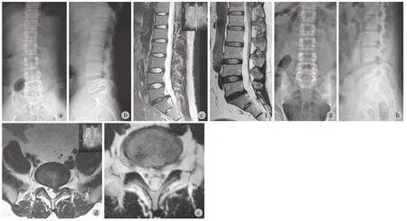
1 例 L4~5节段患者术后 1 周发生下肢麻木加重,予以脱水、激素治疗等保守治疗后 2 周症状缓解,末次随访时上述麻木症状明显缓解;1 例 L5~S1节段患者术后 4 个月出现手术节段椎间盘再次突出,经保守治疗无效,行小切口开放手术翻修。末次随访时,2 例轻度运动障碍,6 例感觉异常,但较术前均显著改善。典型病例如图 1。
讨 论
本组应用 FE 技术外科治疗 45 例腰椎间盘突出症中期随访结果显示,在完全内镜下通过 8 mm 微小通道进行操作,不仅可以获得安全、有效的椎间盘髓核摘除和神经根减压效果,而且能够最大限度地减少手术对脊柱后方结构破坏,从而减轻患者术后腰、腿痛状况。随着随访时间的延长,腰、腿痛VAS 评分略有上升,可能与部分患者随年龄增长、椎间盘继续发生退变及腰椎生理功能减退引发新的腰腿痛症状有关[11-12]。但总体来讲,末次随访与术后 1 年比较,差异无统计学意义 ( P>0.05 ),表明术后临床疗效可以得到良好的维持。根据美国 FDA标准,手术前后 ODI 指数变化 ≥15 分具有临床意义[13],本组腰椎 ODI 指数在术后 3 个月、术后 1 年和末次随访时均较术前明显下降 ( P<0.01 )。在上述时间点有效率分别为 95.6% ( 43 / 45 )、97.8% ( 44 / 45 )、和 91.1% ( 41 / 45 ),与 Ruetten 等[4]应用 FE 经椎板间隙入路 2 年随访报道结果一致,且优于传统开放手术的中期随访结果[14-15],进一步表明 FE 经椎板间隙入路技术能够促进患者早期康复并避免术后因慢性腰痛而造成的活动障碍。

图 1 患者,男,31 岁,左下肢放射痛、麻木 6 个月 a ~ b:术前 X 线正侧位片上示 L5 ~ S1 椎板窗 10 mm × 9 mm;c ~ d:术前MRI 示 L5 ~ S1 椎间盘突出压迫左侧 S1 神经根,Pfrrmann 分级 IV级;e ~ f:应用 FE 技术行 L5 ~ S1 髓核摘除后 3 个月复查 MRI 显示髓核摘除彻底,神经根完全松解;g ~ h:末次随访时,X 线正侧位片示腰椎序列维持良好Fig.1 A 31-year-old man had radiating pain and numbness in the left lower extremity for 6 months a-b: The preoperative anteroposterior and lateral X-ray films showed L5-S1 lamina window area was 10 mm×9 mm; c-d: The preoperative MRI showed the patient had L5-S1 disc herniation with the left S1 nerve root compressed, and the Pfrrmann grading was IV; e-f: The postoperative MRI showed the left S1 nerve root was released completely, with no recurrent disc herniation at 3 months after the surgery; g-h: In the latest follow-up, the anteroposterior and lateral X-ray flm showed a good sequence of the lumbar.
本组末次随访主观满意度达 93.3% ( 42 / 45 ),总体优良率 88.9% ( 40 / 45 ) 与传统开放手术的75%~95% 优良率一致[11]。我们通过单变量分析发现,患者性别、年龄、突出类型及术前腰痛与改良 MacNab 结果差异无统计学意义 ( P>0.05 ),而与术前 Pfirrmann 分级、感觉缺陷及手术节段与改良MacNab 结果呈显著相关性 ( P<0.05 )。术前椎间盘轻、中度退变 ( ≤ III 级 ) 患者临床疗效优于椎间盘重度退变 ( >III 级 ) 患者,术前无感觉缺陷患者优于感觉缺陷患者。此外,L5~S1节段手术患者临床疗效优于 L4~5节段。分析认为,无论开放手术还是FE 技术,明显的腰椎间盘退变和神经根损害等术前病理状况均会同等程度对患者术后临床疗效造成影响。但对于 FE 技术而言,手术节段的不同也会进一步影响患者术后疗效。为安全有效摘除髓核组织,FE 技术首先需要对黄韧带进行微小开窗,在显露椎管及内容物后进行减压操作。研究表明[16],深层黄韧带在不完全性切开后,由于弹性作用,会出现皱褶挛缩并向椎管内移位。在 L5~S1节段,由于椎板间骨窗、侧隐窝和椎间孔较大,神经根起点较高,减压后水肿的神经根有一定空间避让黄韧带断端挤压。但由于 L4~5节段的椎板窗较小且不规则、侧隐窝狭小且 L5神经根起点低、根囊角大[17],减压后黄韧带断端回缩、神经根水肿、局部血肿或积液等因素形成软性压迫,造成神经根出口继发狭窄。本组1 例术后发生下肢麻木加重,可能与此病理现象有关,从而对 L4~5节段患者术后疗效评估结果造成影响。当然,技术上可以通过改进 FE 下黄韧带开窗方式,并在髓核摘除减压后预防性扩大神经根出口来提高在 L4~5节段中远期结果,但受到 FE 下操作器械的限制,其可行性和有效性仍有待研究。
腰椎间盘突出症手术治疗后的复发问题一直是关注焦点。对传统手术而言,肌肉广泛去神经化支配、后方结构破坏、髓核大量摘除造成的医源性不稳往往加速椎间盘退变,可导致中远期随访复发。Wera 等[18]报道 259 例开放手术后复发率为 4.5% ( 12 / 259 ),平均复发时间 6.7 年。Soliman 等[19]报道采用开放手术治疗 54 例腰椎间盘突出症,术后7.2 年复发率为 11.1% ( 6 / 54 ),其中术后 1 年复发率仅为 3.7% ( 2 / 54 )。FE 下经椎板间隙入路技术治疗腰椎间盘突出症术后复发时间与开放手术有所不同,大部分出现在术后早期。Ruetten 等[4]报道232 例应用 FE 技术治疗腰椎间盘突出症,2 年随访期内复发率 6% ( 14 / 232 )。Chumnanvej 等[7]报道60 例 FE 技术治疗的腰椎间盘突出症患者中 2 例发生复发 ( 3.3% ),均发生在术后数个月内。本组 1 例Pfirrmann 分级为 IV 级 L5~S1节段患者,术后 4 个月发生同节段椎间盘突出复发。可能的原因是,FE下通过 8 mm 微小通道进行操作,虽然能够有效减少手术对脊柱后方结构破坏,避免医源性远期腰椎不稳,但细小的镜下显微器械和操作角度仅能对髓核进行有限摘除,对腰椎间盘退变程度较重、突出偏中央、巨大型或脱垂游离者,尤其对初期开展者,操作时容易遗漏盘内部分游离髓核,从而造成术后短期复发。另外,外科手术后椎间盘突出的复发原因与纤维环裂口过大和软骨终板处理不当有关。Ruetten 等[6]报道 3 例纤维环裂隙过大者发生复发,75% 的复发物为终板组织。因此,为提高中远期疗效、避免短期复发问题,建议在开展 FE 椎板间隙入路手术前,须根据 CT 和 MRI 详细评估椎板骨窗大小、判断椎间盘退变程度,以及突出的髓核类型、大小、位置和神经根的对应关系,术中采用髓核摘除靶向技术或对椎间盘进行亚甲兰染色,防止术中盘内髓核遗漏[20],并避免随意扩大纤维环裂隙,在内窥镜引导下处理椎间隙与终板。
总之,FE 经椎板间隙入路手术治疗腰椎间盘突出症具有创伤小、出血量和并发症少、术后恢复快等优点,中期临床疗效满意。然而,由于下腰椎解剖因素限制和技术本身存在的问题,FE 经椎板间隙入路手术治疗腰椎间盘突出症有其严格的适应证,尚需技术上的改进。
[1]Ruetten S, Komp M, Godolias G. Lumbar discectomy with the full-endoscopic interlaminar approach using newly-developed optical systems and instruments. WSJ, 2006, 1(3):148-156.
[2]王冰, 吕国华, 李晶, 等. 完全内镜下经椎板间入路治疗腰椎间盘突出症的对比研究. 中华外科杂志, 2011, 49(1):74-78.
[3]吕国华, 王冰, 刘伟东, 等. 完全内窥镜下经椎板间入路手术治疗腰椎间盘突出症. 中国脊柱脊髓杂志, 2010, 20(6): 448-452.
[4]Ruetten S, Komp M, Merk H, et al. Full-endoscopic interlaminar and transforaminal lumbar discectomy versus conventional microsurgical technique: a prospective, randomized, controlled study. Spine, 2008, 33(9):931-939.
[5]Kuonsongtum V, Paiboonsirijit S, Kesornsak W, et al. Result of full endoscopic uniportal lumbar discectomy:preliminary report. J Med Assoc Thai, 2009, 92(6):776-780.
[6]Ruetten S, Komp M, Merk H, et al. Use of newly developed instruments and endoscopes: full-endoscopic resection of lumbar disc herniations via the interlaminar and lateral transforaminal approach. J Neurosurg Spine, 2007, 6(6):521-530.
[7]Chumnanvej S, Kesornsak W, Sarnvivad P, et al. Full endoscopic lumbar discectomy via interlaminar approach: 2-year results in Ramathibodi Hospital. J Med Assoc Thai, 2011, 94(12):1465-1470.
[8]Pfrrmann CW, Metzdorf A, Zanetti M, et al. Magnetic resonance classifcation of lumbar intervertebral disc degeneration. Spine, 2001, 26(17):1873-1878.
[9]Weiner BK, Walker M, Brower RS, et al. Microdecompression for lumbar spinal canal stenosis. Spine, 1999, 24(21): 2268-2272.
[10]Macnab I. Negative disc exploration. An analysis of the causes of nerve-root involvement in sixty-eight patients. J Bone Joint Surg Am, 1971, 53(5):891-903.
[11]Casal-Moro R, Castro-Menéndez M, Hernández-Blanco M, et al. Long-term outcome after microendoscopic diskectomy for lumbar disk herniation: a prospective clinical study with a 5-year follow-up. Neurosurgery, 2011, 68(6):1568-1575.
[12]McGirt MJ, Ambrossi GL, Datoo G, et al. Recurrent disc herniation and long-term back pain after primary lumbar discectomy: review of outcomes reported for limited versus aggressive disc removal. Neurosurgery, 2009, 64(2):338-344.
[13]Roland M, Fairbank J. The roland-morris disability questionnaire and the oswestry disability questionnaire. Spine, 2000, 25(24):3115-3124.
[14]Atlas SJ, Keller RB, Chang Y, et al. Surgical and nonsurgical management of sciatica secondary to a lumbar disc herniation: fve-year outcomes from the Maine Lumbar Spine Study. Spine, 2001, 26(10):1179-1187.
[15]Wilson DH, Harbaugh R. Microsurgical and standard removal of the protruded lumbar disc: a comparative study. Neurosurgery, 1981, 8(4):422-427.
[16]Olszewski AD, Yaszemski MJ, White AA 3rd. The anatomy of the human lumbar ligamentum favum. New observations and their surgical importance. Spine, 1996, 21(20):2307-2312.
[17]李光, 王冰, 吕国华, 等. 成人下腰椎神经根与椎板骨窗之间CT容积再现技术成像特点与意义. 中国脊柱脊髓杂志, 2013, 23(1):42-46.
[18]Wera GD, Dean CL, Ahn UM, et al. Reherniation and failure after lumbar discectomy: a comparison of fragment excision alone versus subtotal discectomy. J Spinal Disord Tech, 2008, 21(5):316-319.
[19]Soliman J, Harvey A, Howes G, et al. Limited microdiscectomy for lumbar disk herniation: a retrospective long-term outcome analysis. J Spinal Disord Tech, 2014, 27(1):E8-E13.
[20]李振宙, 侯树勋, 宋科冉, 等. 经椎板间隙入路完全内窥镜下椎间盘摘除术治疗L5~S1非包含型椎间盘突出症. 中国脊柱脊髓杂志, 2013, 23(9):771-777.
( 本文编辑:代琴 )
Mid-term clinical outcomes of full-endoscopic interlaminar approach for lumbar disc herniation
WANG Bing,LÜ Guo-hua, LI Ya-wei, LI Lei, KUANG Lei, ZHOU Qian, LI Peng-zhi. Department of Spinal Surgery, the second Xiangya Hospital of Central South University, Changsha, Hunan, 410011, PRC
ObjectiveTo evaluate the mid-term clinical outcomes of full-endoscopic ( FE ) interlaminar approach for lumbar disc herniation.MethodsA retrospective study of 45 patients with lumbar disc hernation undergoing FE interlaminar approach from August 2008 to February 2010 was carried out. There were 25 males and 20 females, whose mean age was 38.3 years old ( range: 19-58 years ). The Visual Analogue Scale ( VAS ) of low back pain and leg pain and Oswestry Disability Index ( ODI ) were evaluated preoperatively, at 3 months and 1 year after the operation and in the latest follow-up. The modifed Macnab criteria was used to assess the clinical outcomes, and all the data was statistically analyzed.ResultsThe operation was successfully completed in all the patients. The mean operation time was 62.4 min, and the mean length of hospital stay was 5.2 d. Small dural tears were noticed during the operation in 2 patients, which were not handled. There were no nerve root injuries or postoperative blood loss. All the patients were followed up from 48 to 66 months. The VAS score of leg pain was ( 7.6±1.6 ) points preoperatively, which was reduced to ( 1.2±0.8 ), ( 1.1±0.9 ) and ( 1.6±1.2 ) points at 3 months and 1 year after the operation and in the latest follow-up respectively ( P<0.01 ). No statistically signifcant differences existed between the scores in the latest follow-up and that at 1 year after the operation ( P>0.05 ). The VAS score of low back pain was ( 3.1±2.2 ) points preoperatively, which was reduced to ( 1.8±1.5 ), ( 1.6±1.4 ) and ( 1.7±0.9 ) points at 3 months and 1 year after the operation and in the latest follow-up respectively ( P<0.01 ). No statistically signifcant differences existed between the scores in the latest follow-up and that at 1 year after the operation ( P>0.05 ). The ODI score was ( 69.5±10.5 ) pointspreoperatively, which was reduced to ( 19.3±6.5 ), ( 15.6±5.9 ) and ( 21.8±7.0 ) points at 3 months and 1 year after the operation and in the latest follow-up respectively ( P<0.01 ). No signifcant differences existed between the scores in the latest follow-up and that at 1 year after the operation ( P>0.05 ). In the latest follow-up, good subjective satisfaction was noticed in 93.3% of the patients, and the clinical excellent and good rate was 88.9%. There was no correlation between the results based on the modifed Macnab criteria and the gender, age, type of disc herniation and preoperative low back pain ( P>0.05 ). However, significant correlation was found between the results based on the modified Macnab criteria and the preoperative Pfrrmann grading, sensation defcits and surgical segments ( P<0.05 ). A patient with L4-5disc herniation had worsened lower limb numbness at 1 week after the operation, who underwent dehydration and hormonotherapy and got relieved at 2 weeks after the traditional treatment. Reherniation of the surgical disc was detected at 4 months after the operation in 1 patient with L5-S1disc herniation, who then received small-incision open surgery. In the latest follow-up, mild dyskinesia occurred to 2 patients and paresthesia to 6 patients, which were obviously improved when compared with the preoperative symptoms.ConclusionsSatisfactory mid-term clinical outcomes can be achieved by FE interlaminar approach for lumbar disc herniation, with the advantages of less trauma, blood loss and complications and rapid postoperative recovery.
Intervertebral disc displacement; Endoscopy; Laminectomy; Lumbar vertebrae; Surgical procedures, minimally invasive
10.3969/j.issn.2095-252X.2014.08.007
Th776.1, R681.5
410011 长沙,中南大学湘雅二医院脊柱外科
2014-06-13 )
