视神经管和颈内动脉与鞍区结构位置关系的CT研究
2013-11-02燕程也张斯文
彭 燕程 也张斯文
1.长春大学校医院医技科,吉林 长春 130022;2.吉林大学白求恩医学院;吉林 长春 130022
视神经管和颈内动脉与鞍区结构位置关系的CT研究
彭 燕1程 也2张斯文2
1.长春大学校医院医技科,吉林 长春 130022;2.吉林大学白求恩医学院;吉林 长春 130022
目的:通过CT对视神经管和颈内动脉在鞍区的位置进行研究并为神经外科医生进行经鼻蝶的鞍区手术提供指导。方法:观察并测量120个成人颅脑CT图像,并通过多平面重建技术进行冠状面、矢状面和轴面的测量。结果:①视神经管内侧壁与经鞍底正中矢状线的距离为平面1处:13.66±0.84mm,平面2处:11.14±0.98mm,平面3处:7.99±1.02mm;②BO与BM1的夹角为平面1处:89.61±6.38°,平面2处:82.34±7.45°,平面3处:83.99±5.90°;③颈内动脉内侧壁与经鞍底正中矢状线的距离为在平面4处:10.23±2.26mm,平面5处:8.82±3.15mm,平面6处:8.85±1.58mm;④BA2与BM2的夹角为74.99±5.21°;BA1与BM1的夹角为97.12±12.05°;⑤视神经管与水平线的夹角为10.14±4.99°。结论:视神经管、颈内动脉在鞍区的位置的测量,可为二者的位置的解剖学研究和内窥镜下经鼻蝶进行鞍底的手术提供指导。
颈内动脉;视神经管;蝶窦;鞍区;经鼻蝶手术
视神经管和颈内动脉与鞍区结构关系 (如蝶窦、鞍底)比较复杂,视神经管位于视神经管内而颈内动脉位于海绵窦内,称为颈内动脉海绵窦段(cavernous segment of the internal carotid artery,CSICA)[1-2]。视神经管和颈内动脉都可以突入蝶窦并且在蝶窦腔内形成隆突[3-4]。因此它们在颈内动脉瘤切除、垂体瘤切除等经蝶窦的手术中需要被特殊保护[5]。如果打开蝶窦侧壁时未进行限制会造成视神经和颈内动脉的损伤[6],因此,对视神经管和颈内动脉进行准确的定位十分重要。已有很多文献报道以视神经隆突和颈内动脉隆突作为蝶窦侧壁开口的标志,但这些隆突在不同人所形成的程度不同,并由很大一部分缺如的情况,因次,CT的测量提供相应的数据是必要的。
1 材料和方法
观测120例健康成年人的蝶窦CTA影像,男69例,女51例,所选取的成年人样本年龄为19~76岁(平均48.3岁),图像不含蝶窦畸形,垂体腺瘤、颈内动脉畸形的患者。测量地点为吉林大学中日联谊医院,仪器为东芝320排容积CT(片层0.5mm)。所有图像进行多平面重建(multiplanar reconstruction,MPR)。
测量平面的选择:选取6个冠状平面 (图1)和一个垂直平面 (图4),各个平面的位置见表1,选取的测量线段的位置见表2。
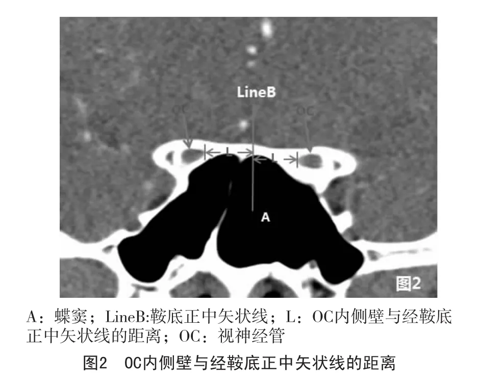
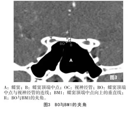
①在平面1、2、3测量OC内侧壁与经鞍底正中矢状线的距离以及线BO和BM1之间的夹角(图2,图3);
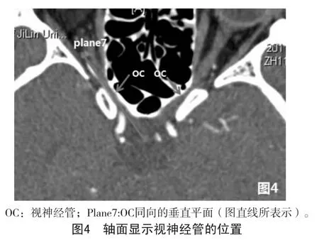
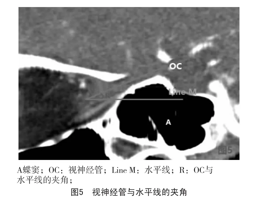
②在平面7测量OC和水平线的夹角(图5);
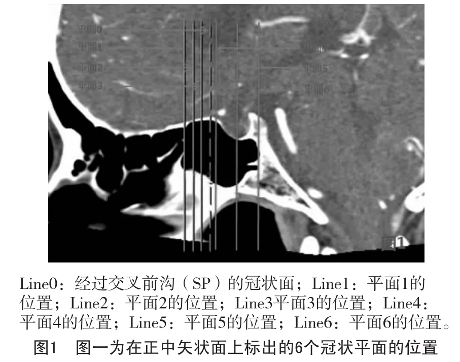
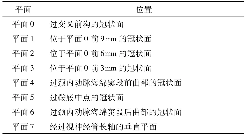
表1 测量平面
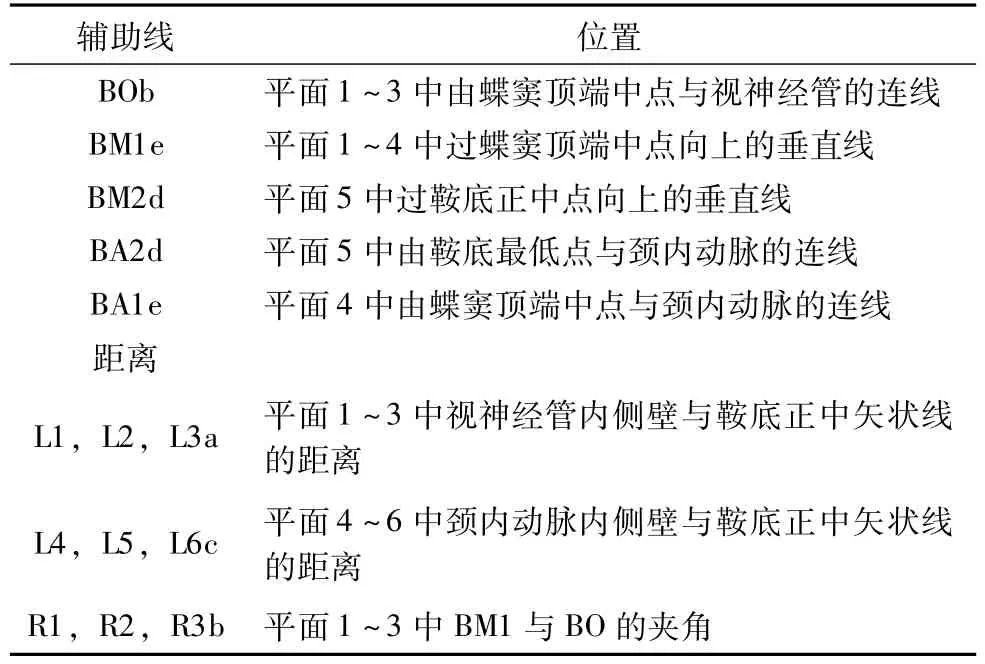
表2 测量所用辅助线与测量距离
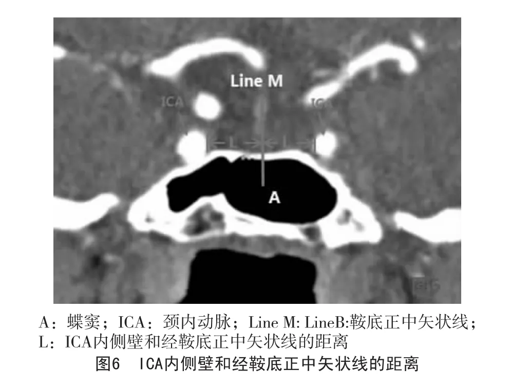
③在平面4、5、6测量ICA内侧壁和经鞍底正中矢状线的距离(图6),在平面5测量BA2和BM2的距离;
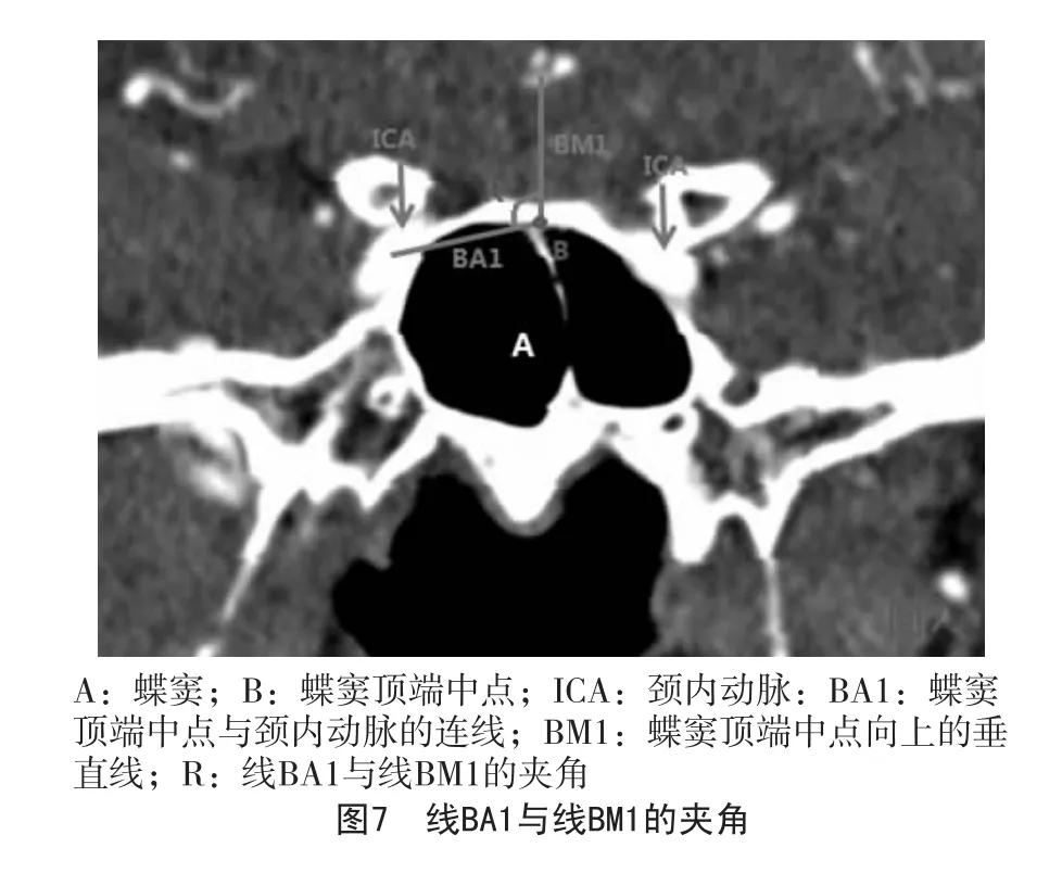
④在平面4进行线BA1与线BM1夹角的测量(图7)
统计结果采用SPSS15.0软件进行统计。
2 结果
在CTA上可以在冠状面、矢状面和轴面清楚看到OC和ICA的位置及走形。OC在冠状面上类似“蝶眼状”在矢状面上类似 “轨道状”。视神经管呈相对低密度儿颈内动脉呈相对高密度。测量结果表明视神经管和境内动脉都有很好的对称性。
2.1 视神经管与鞍区结构的关系
OC内侧壁与鞍底正中矢状线的距离见表3。
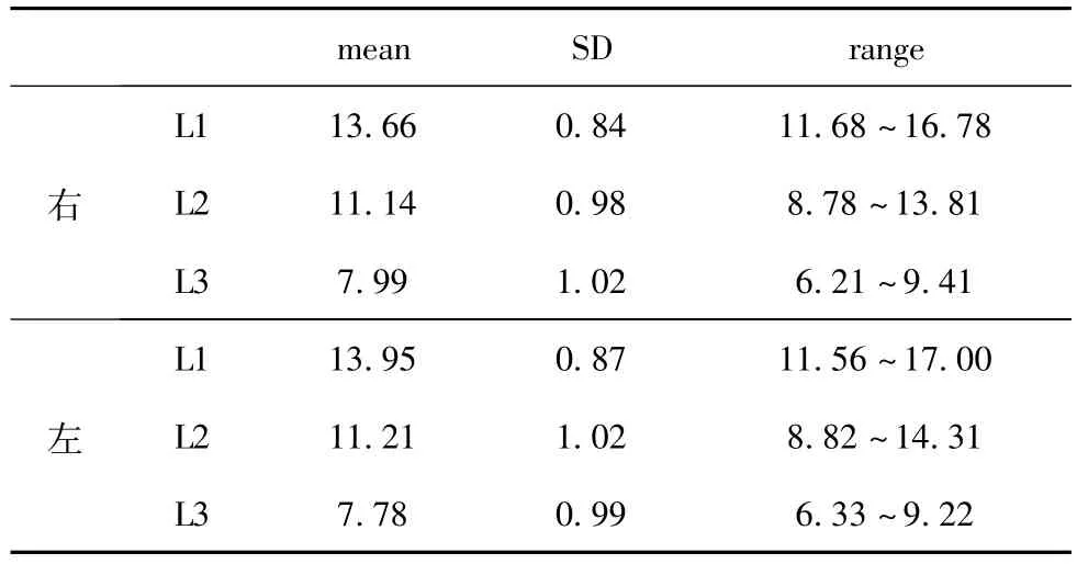
表3 OC内侧壁与鞍底正中矢状线的距离(mm)
经统计学ANOVA分析,在L1、L2和L3之间存在显著差异。经Student-t检验,L1与L2之间以及L2与L3之间存在显著差异。且男女之间无显著差异(P>0.05)。BO与BM1的夹角见表4。统计结果t检验表明R1与R2间无显著差异,但R3与R1之间以及R3与R2之间存在显著差异,表明R3与R1和R2存在差异,且数据显示R3更接近90°,因此视神经管在平面3位置与蝶窦上缘几乎平齐。
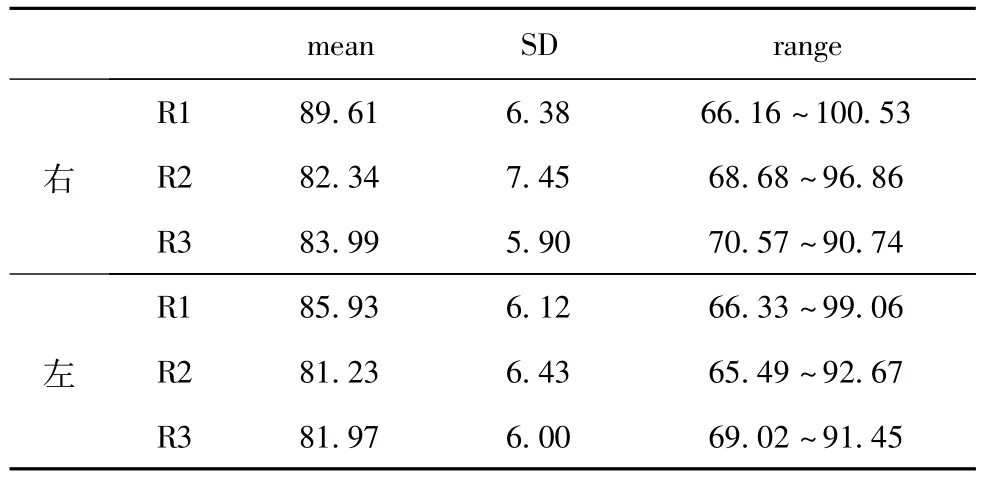
表4 BO与BM1的夹角
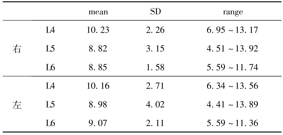
表5 颈内动脉与鞍底正中矢状线的距离 (mm)
视神经管与水平线的夹角为右侧10.14±4.99°;左侧9.99±5.03°
2.2 颈内动脉与鞍区结构的关系 颈内动脉与鞍底正中矢状线的距离见表5,结果在平面4相对较大在平面6相对较小,与WEI[7]以及XUE[8]的结果一致。线BA2与BM2的夹角为右侧74.99±5.21°,左侧76.02±5.98°;线BA1与BM1的夹角为右侧97.12±12.05°,左侧98.00±11.02°。
3 讨论
随着内窥镜技术的发展,内镜下经鼻蝶手术可以在较小创伤的情况下进行垂体瘤颈内动脉瘤的手术,而较小的创伤意味着更小的手术成本,更短的恢复时间,更容易控制的手术并发症而达到更好的外科治疗效果[6,9-11]。对于肿瘤的切除来说,完整地暴露病灶是安全手术的保障[12],而对于经蝶入路来说,对蝶骨侧壁的破坏增加了损伤视神经管和颈内动脉的危险性,而OC与ICA形态的多样性意义蝶窦气化的程度差异也增加了OC和ICA损伤的危险性[13]。虽然视神经隆突和颈内动脉隆突可作为手术的重要标志,但它们在很大一部分人中会缺如[3],从而又增加了手术的难度。因此寻找视神经管和颈内动脉和鞍区其它固定结构的位置关系变得更加重要。
3.1 OC的位置以及和鞍区结构的关系 视神经损伤依然是经蝶手术的主要并发症之一。视神经隆突被确定为是进行蝶窦开窗的最外界限,但除了视神经隆突缺如的可能性外 (86.6%可见[14]),视神经的近侧段和远侧段也无法形成隆突。因此CT的测量可在无法参考视神经隆突的情况下提供帮助。
同时,结节隐窝(Tubercular recess,TR)是视神经管的起始部位也是视神经进入视神经管的部位[3],TR唯一交叉前沟的正下方,平面1、平面2、平面3的位置可以根据TR的位置而确定。而对OC的测量结果提示:为了避免对视神经的损伤,在TR前3mm处,开窗的宽度应该小于7.80±1.23mm(右侧),7.92±1.14mm(左侧);在TR前6mm处,开窗的宽度应该小于11.35±1.08mm(右侧)11.09±1.12mm(左侧);在TR前9mm处,开窗的宽度应该小于13.97±0.78mm(右侧)14.01±0.87(左侧)。
由于蝶窦的顶端很容易确定,因此线BO与BM1的角度可以通过蝶窦顶端寻找OC的位置。
3.2 ICA的位置以及和鞍区结构的关系 当颈内动脉隆突缺如时,鞍底正中点,蝶窦顶端可以作为寻找颈内动脉的标志。由于双侧颈内动脉的高度有时不一致,因此在冠状面上进行颈内动脉内侧壁距离鞍底中线距离的测量优与轴面 (图6)。颈内动脉内侧壁与鞍底正中矢状线之间的距离可以视为蝶窦侧壁开窗的最大距离。因此,在颈内动脉前曲部,开窗的宽度应该小于10.23±2.26mm(右侧),10.12±2.21mm(左侧);在鞍底中点,开窗宽度应该小于8.82±1.15mm(右侧),8.76±1.23mm(左侧);在颈内动脉后曲部,开窗宽度应该小于8.85±1.58mm(右侧),8.45±1.61mm(左侧)。
鞍底最中点也是垂体窝的最低点,它已经被研究和定位[15]。根据线BA2和BM2的夹角和鞍底最低点的位置可以确定ICA的位置。同时,在颈内动脉的前曲段,根据线BA1和BM1的夹角以及蝶窦顶部中点可以确定颈内动脉的位置。
4 结论
由于OC和ICA在CTA上较易发现,因此我们可以根据影像学的定位进行术前定位。同时,精确的数据测量也是必不可少的。研究表明,OC和ICA与鞍区结构之间有一定的关系,因此OC和ICA的位置可以根据这些结果进行确定。
[1]Bouthillier A,van Loveren HR,Keller JT:Segments of the internal carotid artery:a new classification.Neurosurgery,1996,38:425-33.
[2]Fischer E.Die lageabweichungen der vorderen hirnarterie imgefaβblid[J].Zentralbl Neurochir,1938,3:300-313.
[3]Yilmazlar S,Saraydaroglu O,Korfali E.Anatomical aspects in the transsphenoidal-transethmoidal approach to the optic canal:An anatomic-cadaveric study.JCraniomaxillofac Surg.2012,40:198-205.
[4]ZHU Xi-wen,SUN Jia-qi,LIYue-feng:The correlation between the volume of the sphenoid and the bulge of the internal carotid artery in the sphenoid:CT study.Chinese journal of clinical anatomy,2010,28:551-553.
[5]Cavallo LM,Messina A,Cappabianca P,Esposito F,de Divitiis E,Gardner P,et al:Endoscopic endonasal surgery of themidline skull base:anatomical study and clinical considerations.Neurosurg Focus,2005,19(1):E2.
[6]Ossama Al-Mefty,Svetlana Pravdenkova,Cristian Gragnaniello:A technical note on endonasal combined microscopic endoscopic with free head navigation technique of removal of pituitary adenomas.Neurosurg Rev2010,33:243-249.
[7]WEIYukui,KANG Jun,WANG Renzhi:Microscopic and endoscopic anatomy of the cavernous segment of the internal carotid artery.CMINSJ,2008,13:64-67.
[8]XUE Liang,JING Jun-Jie:Microscopic anatomy of cavernous segmentof internal carotid artery via extended transnasal approach.J Fourth M il M ed Univ,2009,30:229-231.
[9]Berker M,Hazer DB,Yücel T,Gürlek A,Cila A,Aldur M,Onerci M:Complications of endoscopic surgery of the pituitary adenomas:analysis of 570 patients and review of the literature.Pituitary.2012;15(3):288-300.
[10]Raithatha R,McCoul ED,Woodworth GF,Schwartz TH,Anand VK:Endoscopic endonasal approaches to the cavernous sinus.Int Forum Allergy Rhinol,2012,2:9-15.
[11]Chamoun R,CouldwellWT:Practical and technical aspects of trans-sphenoidal surgery.JNeurosurg Sci,2011,55:265-75.
[12]Yazhuo Zhang,Zhongcheng Wang,Yejian Liu,Xuyi Zong,Ming Song,Ao Pei,Peng Zhao,Pengfei Zhang and Mingxue Piao:Endoscopic transsphenoidal treatment of pituitary adenomas.Neurological Research,2008,30:581-586.
[13]LocatelliM,Bertani G,Carrabba G,Rampini P,Zavanone M,Caroli M,Sala E,Ferrante E,Gaini SM,Spada A,Mantovani G,Lania A:The transsphenoidal resection of pituitary adenomas in elderly patients and surgical risk.Pituitary 2012 Apr 10.[Epub ahead of print].
[14]Cappabianca P,Cavallo LM,Esposito F,De Divitiis O,Messina A,De Divitiis E:Extended endoscopic endonasal approach to themidline skull base:the evolving role of transsphenoidal surgery.Adv Tech Stand Neurosurg,2008,33:151.
[15]Nikolaos Lazaridis,Konstantinos Natsis,Juergen Koebke et al:Nasal,Sellar,and Sphenoid Sinus Measurements in Relation to Pituitary Surgery.Clinical Anatomy,2010,23:629-636.
The position of optic canal and internal carotid artery in sella region:CT study
Yan Peng1,Ye Cheng2,Siwen Zhang2
1.Changchun University Hospital,Medical department;2.Norman Bethune Medical School of Jilin University
Background:The position of Optic canal(OC)and internal carotid artery(ICA)are relatively complex in sella region,they can be injured in the transsphenoidal surgery and result in severe complications such as haemorrhage and optic nerve damage.Purpose:the aim of our study is to provide new specific and comprehensive data about the location of OC and ICA in sella region in order to guide surgeons through the difficulties in the approaches.Material and method:Computer topographic angiography(CTA)images of 120 sphenoid sinuses in adultswere reviewed.Themeasurementwas on coronal,sagittal and axis planes aftermultiplanar reconstruction(MPR).We selected 6 coronal planes(plane1 to plane6)tomake themeasurement.Results:The distance between themedial wall of OC and the sagittalmidline of sella bottom were 13.66±0.84mm in plane1,11.14±0.98mm in plane2,7.99±1.02mm in plane3.The angle between line BO and BM1 were 89.6±6.38 degree in plane1,82.34±7.45 degree in plane2,83.99±5.90 degree in plane3.The distance between ICA and the sagittalmidline of sella bottom were 10.23±2.26mm in plane4,8.82±1.15mm in plane5,8.85±1.58mm in plane6.The angle between line BA2 and BM2 was74.99±5.21 degree;the angle between line BA1 and BM1 was 97.12±12.05 degree.The angle between OC and the horizontal line was 97.12±12.05 degree.Conclusion:After finding the relationships among OC,ICA and the structures in sella region,we can have amore comprehensive anatomical understanding of them as well as the clinical importance of the location of them.
ICA;OC;Sphenoid sinus;Sella region;Transsphenoidal approach
R651.1
A
1007-8517(2013)11-0060-04
2013.03.19)