大鼠脑缺血/再灌注后bFGF和GAP-43的表达与神经再生
2013-03-30石旺清,郑关毅,陈晓东等
(正文见第65页)

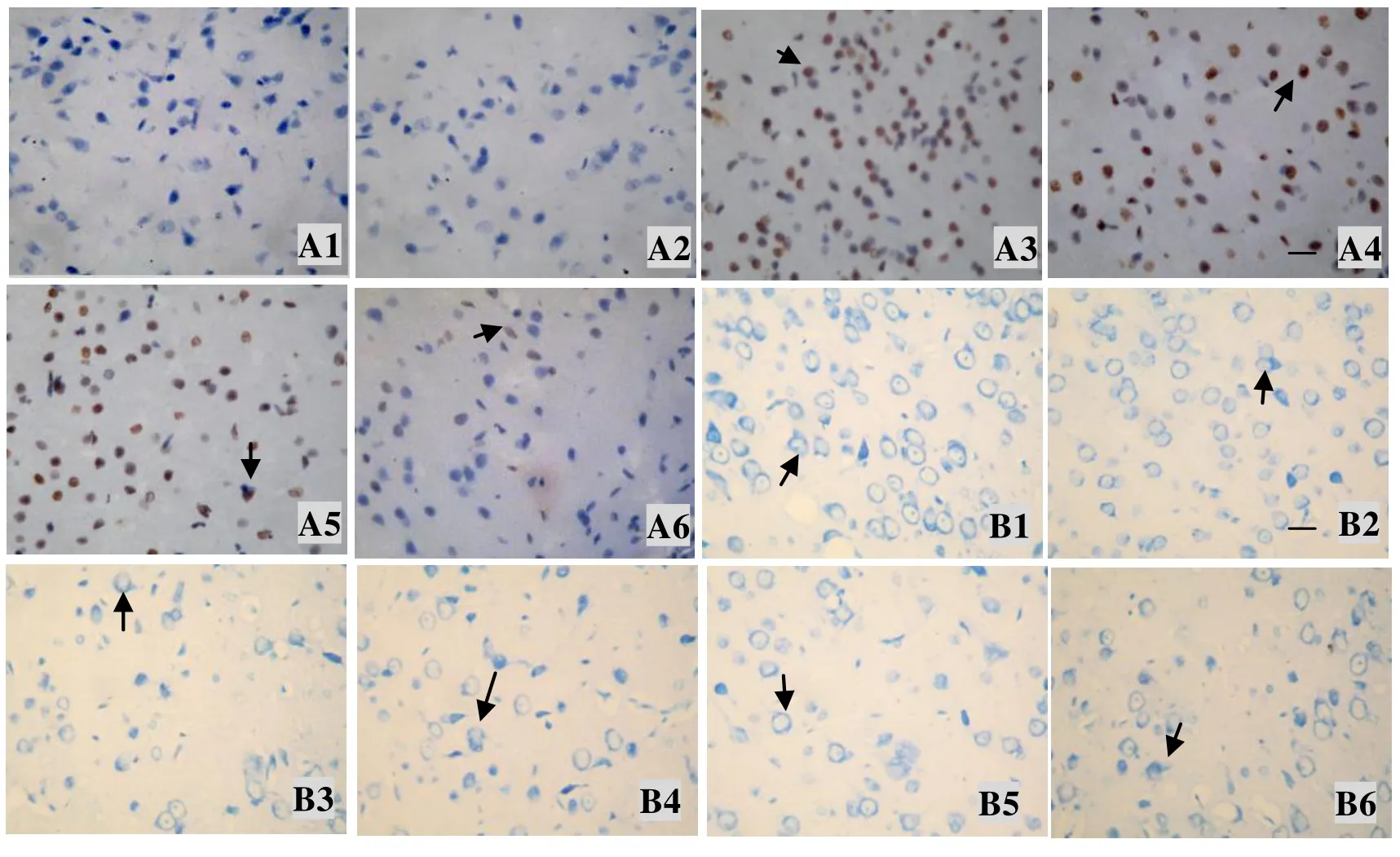
Fig. 1 Neuronal apoptosis after cerebral ischemia/reperfusion(Scale bar: 25 μm ×400)The coronal brain sections were stained with TUNEL(A)and Nissl(B). The arrows head point to the positive neuron A1, B1:Control;A2, B2:Sham;A3−A6, B3−B6:3 d, 7 d, 14 d and 28 d

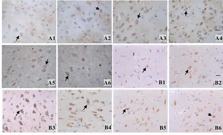
Fig. 4 Immunohistochemistry of bFGF(A)and GAP-43(B)after cerebral ischemia/reperfusion(Scale bar: 25 μm ×400)The arrows head point to the positive neuronA1, B1:Control;A2, B2:Sham;A3−A6, B3−B6:3 d, 7 d, 14 d and 28 d
(正文见第65页)

(正文见第65页)

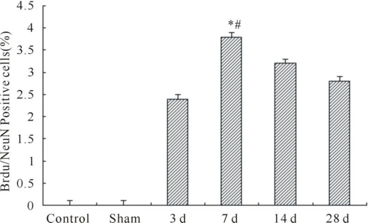
Fig. 5 Immunofluorescence staining with BrdU(red)and NeuN(green)on rat ischemic cortex in cerebralischemia/reperfusion. Arrowheads indicate double-stained cellsA: Control;B: Sham;C, D, E, F: 3 d, 7 d, 14 d and 28 d;G: Quantitative analysis of Brdu/NeuN positive cells* P<0.05 vs 3 d;#P<0.05 vs 28 d
