Assessment of tumor vascularization with functional computed tomography perfusion imaging in patients with cirrhotic liver disease
2011-07-07JinPingLiDeLiZhaoHuiJieJiangYaHuaHuangDaQingLiYongWanXinDingLiuandJinWang
Jin-Ping Li, De-Li Zhao, Hui-Jie Jiang, Ya-Hua Huang, Da-Qing Li, Yong Wan, Xin-Ding Liu and Jin-E Wang
Harbin, China
Assessment of tumor vascularization with functional computed tomography perfusion imaging in patients with cirrhotic liver disease
Jin-Ping Li, De-Li Zhao, Hui-Jie Jiang, Ya-Hua Huang, Da-Qing Li, Yong Wan, Xin-Ding Liu and Jin-E Wang
Harbin, China
BACKGROUND:Hepatocellular carcinoma (HCC) is a common malignant tumor in China, and early diagnosis is critical for patient outcome. In patients with HCC, it is mostly based on liver cirrhosis, developing from benign regenerative nodules and dysplastic nodules to HCC lesions, and a better understanding of its vascular supply and the hemodynamic changes may lead to early tumor detection. Angiogenesis is essential for the growth of primary and metastatic tumors due to changes in vascular perfusion, blood volume and permeability. These hemodynamic and physiological properties can be measured serially using functional computed tomography perfusion (CTP) imaging and can be used to assess the growth of HCC. This study aimed to clarify the physiological characteristics of tumor angiogenesis in cirrhotic liver disease by this fast imaging method.
METHODS:CTP was performed in 30 volunteers without liver disease (control subjects) and 49 patients with liver disease (experimental subjects: 27 with HCC and 22 with cirrhosis). All subjects were also evaluated by physical examination, laboratory screening and Doppler ultrasonography of the liver. The diagnosis of HCC was made according to the EASL criteria. All patients underwent contrast-enhanced ultrasonography, pre- and post-contrast triple-phase CT and CTP study. A mathematical deconvolution model was applied to provide hepatic blood flow (HBF), hepatic blood volume (HBV), mean transit time (MTT), permeability of capillary vessel surface (PS), hepatic arterial index (HAI), hepatic arterial perfusion (HAP) and hepatic portal perfusion (HPP) data. The Mann-WhitneyUtest was used to determine differences in perfusion parameters between the background cirrhotic liver parenchyma and HCC and between the cirrhotic liver parenchyma with HCC and that without HCC.
RESULTS:In normal liver, the HAP/HVP ratio was about 1/4. HCC had significantly higher HAP and HAI and lower HPP than background liver parenchyma adjacent to the HCC. The value of HBF at the tumor rim was significantly higher than that in the controls. HBF, HBV, HAI, HAP and HPP, but not MTT and PS, were significantly higher in the cirrhotic liver parenchyma involved with HCC than those of the controls. Perfusion parameters were not significantly different between the controls and the cirrhotic liver parenchyma not involved with HCC.
CONCLUSIONS:CTP can clearly distinguish tumor from cirrhotic liver parenchyma and controls and can provide quantitative information about tumor-related angiogenesis, which can be used to assess tumor vascularization in cirrhotic liver disease.
(Hepatobiliary Pancreat Dis Int 2011; 10: 43-49)
liver cirrhosis; hepatocellular carcinoma; perfusion imaging; computed tomography
Introduction
Hepatocellular carcinoma (HCC) is a common malignant tumor in China, and early diagnosis is critical for patient survival.[1]In patients with liver cirrhosis, HCC lesions are known to develop from benign regenerative and dysplastic nodules, and the differences in their respective blood supplies can assist in tumor detection and characterization.[2]
Regenerative nodules receive most of their blood supply from the portal vein, and the evolution from a low-grade dysplastic nodules to HCC is associated with increased arterial blood supply, mainly due to tumorrelated arterial neovascularization.[3,4]These hemodynamic and physiological properties can be measured serially using the functional computed tomography perfusion(CTP) technique and multi-parameter imaging maps. A mathematical deconvolution method is used to calculate perfusion parameters.[5]The time-attenuation curve takes advantage of the linear relationship between iodine concentration and CT attenuation numbers.[6]Thus, perfusion parameters can improve the sensitivity and specificity of diagnostic liver imaging and give a true assessment of the hemodynamic status of the liver tissue.[7]
The present study aimed to use CTP in the quantitative assessment of tumor-related angiogenesis in patients with cirrhotic liver disease and HCC versus normal controls and to clarify the physiological characteristics of tumor angiogenesis in cirrhotic liver disease.
Methods
Patients
Informed consent was obtained from all subjects before the study and after the nature of the procedure had been fully explained to them in accordance with the regulations of the institutional review board. Thirty subjects who were free of liver disease (8 women and 22 men; average age 54.5 years, range 30-78 years) underwent CTP examination of the abdomen for unrelated causes. The experimental subjects consisted of an additional 49 patients (12 women and 37 men; average age 48.2 years, range 35-61 years) of whom, 12 had alcoholic cirrhosis, 2 had primary biliary cirrhosis, 8 had post-hepatic cirrhosis, and 27 had HCC. Both control and experimental subjects were also evaluated by physical examination, laboratory screening and Doppler ultrasonography of the liver, and a thorough medical history was taken from each subject. The diagnosis of HCC was made according to the European Association for the Study of the Liver (EASL) criteria.[8,9]
Contrast-enhanced ultrasonography (CEUS)
CEUS was performed using a Voluson E8 ultrasound scanner (GE Healthcare, Austria) equipped with a convex 3-5 MHz probe and agent detection imaging software. Contrast-enhanced sequences were obtained using dedicated low-mechanical index (low-MI) contrastimaging software (MI<0.2). Standard pre-settings were used, with the possibility to adjust them to the individual patient. After baseline evaluation, a second generation contrast agent (SonoVue; Bracco, Milan, Italy) was injected intravenously as a bolus of 2.4 ml, followed by a flush of 5 ml normal saline. All images were obtained with the probe focused on the region of interest and part of the surrounding liver parenchyma.
Conventional contrast CT and CTP protocol
In the 79 subjects (49 patients and 30 controls), CT was performed using spiral CT (Lightspeed 64-slice VCT; GE Healthcare, Milwaukee, WI, USA), with both pre- and post-contrast enhanced imaging. Contrastenhanced triple-phase scans (arterial, portal venous, and equilibrium phases) were obtained after an intravenous bolus injection of 100 ml of nonionic iodinated contrast material (OmnipaqueTM(iohexol); GE Healthcare) at a rate of 4.5 ml/sec via a 20-gauge intravenous catheter in the antecubital fossa. Single level monitoring low-dose scanning (120 kVp, 60 mA) was initiated after injection of the contrast material. Contrast material enhancement was automatically calculated by placing the region of interest cursor over the abdominal aorta, and the trigger threshold was set at 120 Hounsfield units. After the threshold had been reached, the arterial phase scanning began automatically. The mean scanning time delay of the arterial phases was 15-20 seconds. The portal venous and equilibrium phases were acquired at 45 and 120 seconds, respectively, after the threshold had been reached.
A plain scan was first performed to localize the central slice of the tumor, and a perfusion scan was performed on this slice. The CT parameters for perfusion imaging were as follows: scanning was carried out using a low radiation dose (120 kV, 60 mA), cinescan mode, volume coverage up to 40 mm, 50 seconds of continuous scanning time set at 5 seconds after the injection of contrast material, 1 second per 360° revolution, 5 mm slice thickness image reconstruction, and a matrix size of 512×512 pixels. The rate of injection of contrast medium (OmnipaqueTM(iohexol); GE Healthcare) was 4-5 ml/sec in all studies, with a dose of 1.0 ml/kg body weight. A bolus of contrast agent was injected 5 seconds after the beginning of acquisition. Images were reconstructed continuously over 50 seconds, extrapolating to a total of 396 slices.
Data acquisition and perfusion images
Computed tomography images were transferred to a workstation (Advantage Windows 4.3; GE Medical Systems Ltd., Milwaukee, WI, USA). Liver tissue was considered a double-input system (hepatic artery and portal vein) to take into account the dual hepatic perfusion, with a single output (hepatic veins). The deconvolution algorithm allowed for the calculation of seven parameters quantifying perfusion for each tissue region of interest (ROI). Hepatic blood flow (HBF) was expressed as ml/(min/100 mg of body weight), hepatic blood volume (HBV) as ml/100 mg, mean transit time (MTT) in seconds, the permeability of capillary vesselsurface (PS) as ml/(min/100 mg), and hepatic arterial index (HAI) as the percentage of total blood flow that was of arterial origin. Hepatic arterial perfusion (HAP) and hepatic portal perfusion (HPP) were expressed as ml/(min/100 mg). The HBF was calculated using the following equations:
HAP+HPP=HBF, where HAP=HBF×HAI and HPP= HBF×(1-HAI).
The ROIs were placed on a color map (Fig. 1). Perfusion parameters were measured three times at each time point for each ROI and the mean of the three measurements was used in the analysis. An ROI of 20-30 mm2was placed in the aorta, portal vein and liver parenchyma. For liver tissue surrounding the tumor, an equivalent ROI was placed in an area of liver parenchyma surrounding the tumor which did not show any abnormality on conventional contrast-enhanced CT. In each of the control subjects, two or three ROIs were placed randomly in the liver parenchyma. To evaluate the tumor rim in scans containing HCC, two or three ROIs were placed in the areas seen as ring enhancement on contrast-enhanced CT. For cirrhotic liver parenchyma without HCC, two or three ROIs were placed randomly in the liver parenchyma.
Statistical analysis
All data were expressed as mean±SD. All analyses were carried out using SPSS 11.5 for Windows (SPSS Inc., Chicago, IL, USA). The Mann-Whitney U test was used to determine differences in perfusion parameters between background cirrhotic liver parenchyma and HCC and between cirrhotic liver parenchyma with HCC or without HCC. A P value less than 0.05 was considered statistically significant.
Results
On the functional perfusion maps of HBF, HBV and HAI, the red region of tumor tissue indicated an area of high blood perfusion. The mean values of perfusion parameters were also calculated for controls, tumor rim and background liver tissues surrounding the tumor.
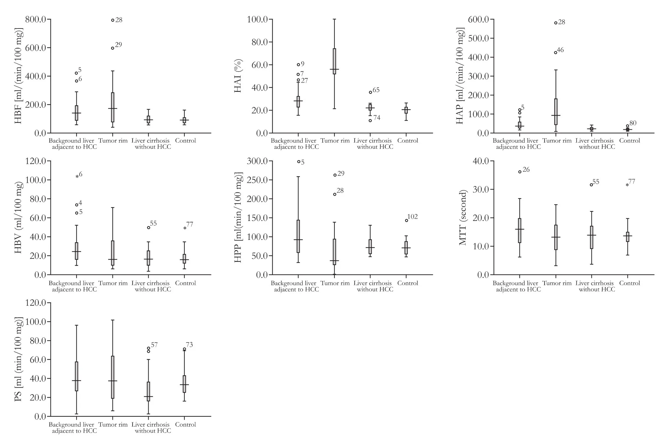
In experimental subjects, HCC showed nodular or ring-like enhancement on contrast-enhanced CT. The contour of the tumor was clearly delineated as a hyperparametric area on HBF and HAI maps compared to the background liver parenchyma adjacent to HCC (Fig. 1). The results of ROI analysis are summarized in Tables 1 and 2. With regards to the controls, the ratio of HAP/HVP was about 1/4, which is consistent with the physiological percentages of hepatic arterial blood flow relative to portal venous blood flow. At the tumor rim,the perfusion parameters (HAI and HAP) were higher (P<0.05) and HPP values were lower (P<0.05) than both the controls and background cirrhotic liver parenchyma adjacent to HCC. In our series, HBF was higher at the tumor rim than in the controls (P<0.05), but there was no difference in HBF between background cirrhotic liver parenchyma adjacent to HCC and tumor rim (P>0.05) (Table 1 and Fig. 2).

Table 1. Values obtained by CTP in different regions of interest in background liver parenchyma adjacent to HCC (n=27), tumor rims (n=27) and the controls (n=30)

Fig. 1. CTP study performed in liver cirrhosis and focal lesion of HCC in the right hepatic lobe. A: Raw data from 64-slice VCT scan image showing ROIs placed on the tumor rim (3, 4, 5), aorta (1) and portal vein (2). B: HAI perfusion color map. ROIs are also shown on the tumor rim (3, 4, 5), aorta (1) and portal vein (2). C: Time-density curves derived from analyses of ROIs (aorta and portal vein) for production of perfusion parameters. The first aortic peak was sharp and high. The aortic peak was followed by a lower broader portal peak.

Fig. 2. Box plots of group perfusion parameters. Errors bar below and above boxes indicate minimum and maximum values, respectively. For the tumor rim, values of the perfusion parameters HAI and HAP were higher (P<0.05) and HPP values were lower (P<0.05) than the controls and background liver parenchyma adjacent to HCC. The value of HBF in the tumor rim was higher than the controls (P<0.05). For cirrhotic liver parenchyma, HBF, HPP, HAI, HAP and HBV were higher (P<0.05) in cirrhotic liver parenchyma containing HCC compared with the controls.There were no differences between the controls and cirrhotic liver without HCC (P>0.05).

Table 2. Values obtained by CTP in different regions of interest in the controls (n=30), cirrhotic liver parenchyma with HCC (n=27) and without HCC (n=22)
In cirrhotic liver parenchyma, the HBF, HPP, HAI, HAP and HBV, but not MTT and PS, were higher in cirrhotic liver parenchyma containing HCC than in the controls (P<0.05). All of parameters showed no differences between the controls and cirrhotic liver without HCC (P>0.05, Table 2 and Fig. 2).
Discussion
A long-standing goal for most researchers in the field of cirrhosis has been to develop a simple, noninvasive imaging method that can quantify the changes in arterial and portal venous blood flow in the cirrhotic liver. More importantly, changes in the hepatic microcirculation in cirrhosis influence the progression of the disease. The development of HCC occurs in conjunction with the formation of new arterial vessels (and not portal venous branches). When cirrhotic nodules evolve from low to high-grade dysplastic nodules and eventually to HCC, the number of arterial vessels increases to the greatest number. Therefore, animaging technique is needed that can quantify multiple perfusion parameters in both normal and pathologic tissues.
Various methods exist for the determination of hepatic microcirculation in clinical practice.[10-15]Among these, nuclear medicine techniques, specifically positron emission tomography and single photon emission tomography, have the longest history and are regarded as the gold standards for blood flow determination.[16]A limitation of nuclear techniques, however, is the requirement for radioisotopes and expensive specialized equipment. The application of magnetic resonance imaging (MRI) is also limited in the quantification of blood flow, because the signal enhancement of MRI does not show a linear correlation with the concentration of the contrast medium.[17]The new CTP technique using the central volume principle with a deconvolution algorithm is more practical and much safer than other methods, and requires an injection rate of only 3-5 ml/sec, a normal rate in routine imaging.[5]
In the present study, tumor rims were used as ROIs for the assessment of tumor angiogenesis. These rims were seen as "ring" enhancement on conventional contrast-enhanced CT, where angiogenesis is typically most intense and correlates with a high level of peripheral perfusion, blood volume or permeability. Also, we investigated the role of several tissue perfusion parameters obtained with functional CT in the quantitative assessment of HCC-related angiogenesis. Our results showed that the values of the perfusion parameters were significantly different in HCC tissue at the tumor rims compared to both controls and cirrhotic liver parenchyma adjacent to HCC.
In our study, the HAP/HVP ratio of approximately 1/4 was a good approximation to the ratio of relative blood supply from the hepatic artery compared to the portal veins. The result is consistent with normal liver physiology, suggesting that CTP can accurately assess the microcirculation in normal liver.
The values of HAI and HAP for the tumor rims were significantly higher and HPP values were significantly lower than the controls and background liver parenchyma adjacent to HCC. This result may be secondary to an increase in arterial perfusion caused by liver arterialization. Thus, HAI is an important parameter reflecting the status of the hepatic artery and portal vein, and can characterize liver tumors in radiological practice. Compared to the controls, the HBF was also increased in the background liver parenchyma adjacent to HCC, but no significant difference was found between the surrounding liver parenchyma adjacent to HCC and the tumor rims. These findings indicate that, in the surrounding liver parenchyma adjacent to HCC, the blood flow is largely influenced by HCC growth. These findings are also comparable to results from earlier experimental and clinical studies.[18-21]
Previously, we also correlated CTP results with histologic microvessel density counts of angiogenesis and concluded that functional CT has the potential to evaluate angiogenesis.[22]The high PS found in the tumor rims may indicate a high level of vascular permeability, allowing the tumor volume to increase with subsequently higher HBV. The MTT reflects the period during which the contrast medium passes through the blood capillaries. The high PS or increased tumor-induced vascular permeability also resulted in fast MTT. However, in the tumor rim, MTT, PS and HBV were not significantly different compared to either the controls or background liver parenchyma adjacent to HCC and these results should be interpreted cautiously.
In cirrhotic liver parenchyma containing HCC, the perfusion parameters HBF, HPP, HAI, HAP and HBV, but not MTT and PS, were significantly higher than the controls. This indicates that perfusion changes due to HCC growth involve cirrhotic liver parenchyma. Not only was blood flow increased, but other perfusion parameters (HPP, HAI, HAP and HBV) were also increased. Our findings demonstrated that CTP is an important imaging technique that can assess the physiological changes of liver cirrhosis and HCC, not visualized by conventional CT imaging.
Interestingly in the present study, the perfusion parameters HBF, HPP, HAI, and HAP were increased in cirrhotic liver parenchyma without HCC compared to the controls; however, the differences were not significant. Also, PS and HBV were lower in cirrhotic liver without HCC compared to the controls. These results imply that the physiological changes associated with cirrhotic liver can be detected by CTP, even when the changes are not obvious. These findings highlight the potential use of CTP to evaluate changes in arterial and portal blood flow (as liver cirrhosis develops into HCC) as well as tumor-related angiogenesis in cirrhotic liver disease.
Our results are in line with recently reported experimental studies where CTP has been used to assess the response of HCC to treatment by evaluating perfusion changes.[23,24]As is well known, therapeutic evaluation of liver tumors has largely relied on morphologic features such as the presence or change in size of a mass.[25,26]However, some malignant tumors may remain unchanged in size in spite of successful treatment. CTP has the potential to evaluate the therapeutic response by evaluating perfusion changes,thereby showing conditions associated with malignant tumors, such as changes in arterial and portal perfusion.
Compared to previous studies, which used a maximum width of 20 mm scanned in cine mode and an examination range limited to the porta hepatica, the 64-slice VCT used in our study offers a wide volumetric coverage (up to 40 mm), suitable not only for a shorter scan time and improved spatial resolution, but also encompassing a larger volume per single rotation. Scanning time is also reduced compared to 16-slice spiral CT as it takes less than 10 seconds to scan the entire liver. The CTP technique thus allows assessment of more lesions and lesions smaller than those previously evaluated.
In conclusion, based on the deconvolution algorithm, CTP is a safe and accurate imaging technique for measurements of the hepatic microcirculation. In addition, it provides quantitative information about tumor-related angiogenesis in cirrhotic liver parenchyma with and without HCC. Further useful information regarding differentiating between benign and malignant nodules may be provided by various perfusion parameters. These findings underscore the importance of CTP as a noninvasive tool to quantify hepatic angiogenesis parameters in the cirrhotic liver.
Funding:This work was supported by grants from the Natural Science Foundation of Heilongjiang Province (No. D2009-05) and the Educational Committee of Heilongjiang Province (No. 11541166).
Ethical approval:Not needed.
Contributors:LJP and ZDL contributed equally to the article. JHJ proposed the study. LJP, ZDL and JHJ wrote the first draft. LJP analyzed the data. All authors contributed to the design and interpretation of the study and to further drafts. JHJ is the guarantor.
Competing interest:No benefits in any form have been received or will be received from a commercial party related directly or indirectly to the subject of this article.
1 Yang L, Parkin DM, Ferlay J, Li L, Chen Y. Estimates of cancer incidence in China for 2000 and projections for 2005.Cancer Epidemiol Biomarkers Prev 2005;14:243-250.
2 Itai Y, Matsui O. Blood flow and liver imaging. Radiology 1997;202:306-314.
3 Miles KA. Functional computed tomography in oncology. Eur J Cancer 2002;38:2079-2084.
4 Efremidis SC, Hytiroglou P. The multistep process of hepatocarcinogenesis in cirrhosis with imaging correlation. Eur Radiol 2002;12:753-764.
5 Bisdas S, Baghi M, Wagenblast J, Knecht R, Thng CH, Koh TS, et al. Differentiation of benign and malignant parotid tumors using deconvolution-based perfusion CT imaging: feasibility of the method and initial results. Eur J Radiol 2007;64:258-265.
6 Miyazaki M, Tsushima Y, Miyazaki A, Paudyal B, Amanuma M, Endo K. Quantification of hepatic arterial and portal perfusion with dynamic computed tomography: comparison of maximum-slope and dual-input one-compartment model methods. Jpn J Radiol 2009;27:143-150.
7 Pandharipande PV, Krinsky GA, Rusinek H, Lee VS. Perfusion imaging of the liver: current challenges and future goals. Radiology 2005;234:661-673.
8 Bruix J, Sherman M, Llovet JM, Beaugrand M, Lencioni R, Burroughs AK, et al. Clinical management of hepatocellular carcinoma. Conclusions of the Barcelona-2000 EASL conference. European Association for the Study of the Liver. J Hepatol 2001;35:421-430.
9 Bruix J, Sherman M; Practice Guidelines Committee, American Association for the Study of Liver Diseases. Management of hepatocellular carcinoma. Hepatology 2005; 42:1208-1236.
10 Goetti R, Leschka S, Desbiolles L, Klotz E, Samaras P, von Boehmer L, et al. Quantitative computed tomography liver perfusion imaging using dynamic spiral scanning with variable pitch: feasibility and initial results in patients with cancer metastases. Invest Radiol 2010;45:419-426.
11 Zapletal C, Jahnke C, Mehrabi A, Hess T, Mihm D, Angelescu M, et al. Quantification of liver perfusion by dynamic magnetic resonance imaging: experimental evaluation and clinical pilot study. Liver Transpl 2009;15:693-700.
12 Johnson DJ, Muhlbacher F, Wilmore DW. Measurement of hepatic blood flow. J Surg Res 1985;39:470-481.
把酒临风:科技创新不是一个急功近利的问题,在中国你成功之后别人可能会抄袭,但在法治国家不行,你抄袭就重罚你,谁都不能随便侵犯他人,如果真做到这一点,我们的科技创新就能产出更多成果。也就是说完善的财产保护制度,才能让大家看到技术创新暴富的可能性。
13 Zeeh J, Lange H, Bosch J, Pohl S, Loesgen H, Eggers R, et al. Steady-state extrarenal sorbitol clearance as a measure of hepatic plasma flow. Gastroenterology 1988;95:749-759.
14 Miles KA, Hayball MP, Dixon AK. Functional images of hepatic perfusion obtained with dynamic CT. Radiology 1993;188:405-411.
15 Taourel P, Blanc P, Dauzat M, Chabre M, Pradel J, Gallix B, et al. Doppler study of mesenteric, hepatic, and portal circulation in alcoholic cirrhosis: relationship between quantitative Doppler measurements and the severity of portal hypertension and hepatic failure. Hepatology 1998;28:932-936.
16 Frackowiak RS, Lenzi GL, Jones T, Heather JD. Quantitative measurement of regional cerebral blood flow and oxygen metabolism in man using 15O and positron emission tomography: theory, procedure, and normal values. J Comput Assist Tomogr 1980;4:727-736.
17 Brasch RC, Weinmann HJ, Wesbey GE. Contrast-enhanced NMR imaging: animal studies using gadolinium-DTPA complex. AJR Am J Roentgenol 1984;142:625-630.
18 Tsushima Y, Funabasama S, Aoki J, Sanada S, Endo K. Quantitative perfusion map of malignant liver tumors, created from dynamic computed tomography data. Acad Radiol 2004;11:215-223.
20 Fournier LS, Cuenod CA, de Bazelaire C, Siauve N, Rosty C, Tran PL, et al. Early modifications of hepatic perfusion measured by functional CT in a rat model of hepatocellularcarcinoma using a blood pool contrast agent. Eur Radiol 2004;14:2125-2133.
21 Sahani DV, Holalkere NS, Mueller PR, Zhu AX. Advanced hepatocellular carcinoma: CT perfusion of liver and tumor tissue--initial experience. Radiology 2007;243:736-743.
22 Jiang HJ, Zhang ZR, Shen BZ, Wan Y, Guo H, Li JP. Quantification of angiogenesis by CT perfusion imaging in liver tumor of rabbit. Hepatobiliary Pancreat Dis Int 2009;8: 168-173.
23 Kan Z, Kobayashi S, Phongkitkarun S, Charnsangavej C. Functional CT quantification of tumor perfusion after transhepatic arterial embolization in a rat model. Radiology 2005; 237:144-150.
24 Kan Z, Phongkitkarun S, Kobayashi S, Tang Y, Ellis LM, Lee TY, et al. Functional CT for quantifying tumor perfusion in antiangiogenic therapy in a rat model. Radiology 2005;237: 151-158.
25 Miles KA, Charnsangavej C, Lee FT, Fishman EK, Horton K, Lee TY. Application of CT in the investigation of angiogenesis in oncology. Acad Radiol 2000;7:840-850.
26 Katyal S, Oliver JH, Peterson MS, Chang PJ, Baron RL, Carr BI. Prognostic significance of arterial phase CT for prediction of response to transcatheter arterial chemoembolization in unresectable hepatocellular carcinoma: a retrospective analysis. AJR Am J Roentgenol 2000;175:1665-1672.
Accepted after revision December 13, 2010
A gentleman is open-minded and optimistic; a small person is narrow-minded and pessimistic.
–the Analects
August 16, 2010
Author Affiliations: Department of Radiology, Second Affiliated Hospital, Harbin Medical University, Harbin 150086, China (Li JP, Zhao DL, Jiang HJ, Huang YH, Li DQ, Wan Y, Liu XD and Wang JE)
Hui-Jie Jiang, PhD, Department of Radiology, Second Affiliated Hospital, Harbin Medical University, Harbin 150086, China (Tel: 86-451-86605576; Email: jhj68323@yahoo.com.cn)
© 2011, Hepatobiliary Pancreat Dis Int. All rights reserved.
猜你喜欢
杂志排行
Hepatobiliary & Pancreatic Diseases International的其它文章
- Hepatobiliary & Pancreatic Diseases International (HBPD INT)
- Primary hepatic carcinosarcoma
- Aberrant methylation frequency of TNFRSF10C promoter in pancreatic cancer cell lines
- Protective effects of glutamine preconditioning on ischemia-reperfusion injury in rats
- Effects of suppressing glucose transporter-1 by an antisense oligodeoxynucleotide on the growth of human hepatocellular carcinoma cells
- Peroxisome proliferator-activated receptor gamma inhibits hepatic fibrosis in rats
