Akt1和Akt2基因转染对人胃黏膜上皮细胞GES-1生长的影响
2010-04-06谢甲贝张庆瑜康春生韩静王涛张洁
谢甲贝,张庆瑜,康春生,韩静,王涛,张洁
Akt1和Akt2基因转染对人胃黏膜上皮细胞GES-1生长的影响
谢甲贝,张庆瑜,康春生,韩静,王涛,张洁
【摘要】
目的观察 Akt1 和 Akt2 基因转染人胃黏膜上皮 GES-1细胞后对其生长的影响。
方法采用脂质体法将分别含 Akt1 和 Akt2 基因全长cDNA 序列的 p-LXSN-Akt1(Akt1 组)和 p-LXSN-Akt2(Akt2 组)质粒转染人胃黏膜上皮 GES-1 细胞,以转染p-LXSN 空质粒的细胞作为空载组,正常未转染细胞作为对照组,并经抗生素 G418 筛选抗性克隆。提取各组细胞的总蛋白,蛋白质印迹法检测转染后细胞 Akt1、Akt2 蛋白及细胞周期蛋白 D1(Cyclin D1)的表达水平;采用 MTT 法检测转染后细胞的增殖活性;应用流式细胞仪检测 Akt1 和Akt2 基因转染对细胞周期的影响。
结果Akt1 和 Akt2 组均获得了稳定表达 Akt1 和 Akt2基因的 GES-1 细胞模型。Akt1 组:① Akt1 蛋白相对表达量(0.96 ± 0.05)分别是空载组(0.47 ± 0.02)和对照组(0.64 ± 0.03)的 2.03 ± 0.22 和 1.48 ± 0.19 倍(均 P <0.01),Cyclin D1 蛋白相对表达量(0.74 ± 0.01)分别是空载组(0.29 ± 0.01)和对照组(0.22 ± 0.01)的 2.57 ± 0.05 和3.44 ± 0.04 倍(均 P < 0.01);②吸光度(A490)值于第 1、2 天分别与空载组和对照组比较差异无统计学意义,第 3 ~6 天差异均有统计学意义(均 P < 0.01);③ S 期细胞数与空载组和对照组比较分别增多约 20.00% 和 21.10%,G2期细胞数减少约 20.80% 和 15.48%(均 P < 0.01)。Akt2组:① Akt2 蛋白相对表达量(0.95 ± 0.01)分别是空载组(0.62 ± 0.02)和对照组(0.56 ± 0.01)的 1.53 ± 0.04 和1.69 ± 0.01 倍(均 P < 0.01),Cyclin D1 蛋白相对表达量(0.31 ± 0.02)与空载组(0.29 ± 0.01)和对照组(0.22 ± 0.01)比较差异均无统计学意义;② A490值于第 1 ~ 6 天分别与空载组和对照组比较差别均无统计学意义;③ S 期细胞数与空载组和对照组比较差异均无统计学意义。
结论Akt1 基因可使 GES-1 细胞 S 期细胞数增多,并提高细胞的增殖能力;而 Akt2 基因对 GES-1 细胞的细胞周期和增殖能力的影响不明显。
【关键词】基因转移技术; 细胞周期蛋白 D1; 胃黏膜
www.cmbp.net.cn 中国医药生物技术, 2010, 5(6):423-428
胃癌的发生是一个多步骤、多因素的发展过程。Akt 信号通路与肿瘤发生发展密切相关,在促进其生长增殖和侵袭转移方面发挥着重要作用[1],并且 Akt 信号通路与胃癌的发生发展亦密切相关[2]。Akt 亚型 Akt1、Akt2 对细胞增殖周期的作用不尽相同,在不同细胞系中对增殖和转移的影响甚至相反[3-4]。为此我们将外源性 Akt1、Akt2 基因导入人胃黏膜上皮 GES-1 细胞中,分析观察两者对 GES-1 细胞的增殖和细胞周期的影响,旨在探索 Akt1、Akt2 基因是否通过延长细胞生存时间为胃黏膜上皮细胞恶性转化提供可能性。
1 材料与方法
1.1 材料
1.1.1 细胞株与质粒 人胃黏膜上皮 GES-1 细胞购自北京百和营科技发展有限责任公司;p-LXSN质粒以及分别含 Akt1 和 Akt2 基因全长 cDNA序列的 p-LXSN-Akt1、p-LXSN-Akt2 质粒由美国南佛罗里达大学 Dr. Jin Q. Cheng 惠赠。
1.1.2 主要试剂与仪器 质粒抽提及纯化试剂盒、总蛋白提取试剂盒购自天根生化科技(北京)有限公司;脂质体 LipofectamineTM2000、DMEM培养液、小牛血清、MTT、抗生素 G418 购自北京索莱宝科技有限公司;兔抗人 Akt1、Akt2、细胞周期蛋白 D1(Cyclin D1)单抗购自美国 Santa Cruz公司;抗 GAPDH 单克隆抗体、HRP 标记的羊抗兔 IgG 抗体购自北京中杉金桥生物技术有限公司;ECL 化学发光试剂盒和化学发光检测系统购自美国 Pierce 公司(Super Signal West Pico 34080);FACS Calibur 流式细胞仪为美国 BD 公司产品;荧光相差倒置显微镜 IX-71 为日本 Olympus 公司产品。
1.2 方法
1.2.1 基因转染和克隆筛选 人胃黏膜上皮GES-1 细胞置于含 10% 小牛血清的 DMEM 完全培养液中,于 37 ℃、5% CO2、湿润条件下进行体外培养。当细胞生长到 60% ~ 70% 融合时,参照脂质体 LipofectamineTM2000 说明书分别转染p-LXSN-Akt1(Akt1 组)、p-LXSN-Akt2 质粒(Akt2组),48 h 后换用含浓度为 400 μg/ml G418 的完全培养液筛选,3 周后选出抗性克隆株继续扩大培养。以正常未转染的 GES-1 细胞作为对照组,以转染 p-LXSN 空质粒的 GES-1 细胞作为空载组。
1.2.2 转染后细胞蛋白表达检测 采用蛋白质印迹法。待各组细胞培养至约 5 × 10⁷时,按总蛋白提取试剂盒说明书分别提取各组待测细胞的总蛋白,并检测样品蛋白浓度。将各组细胞总蛋白按比例与 5 倍上样缓冲液混合,置 100 ℃ 沸水中煮15 min。取等量蛋白样品溶液及蛋白 Marker 上样进行 SDS-PAGE 凝胶电泳,然后将凝胶电转移至PVDF 膜上,置于膜封闭液中封闭过夜。依次加入一抗为兔抗人 Akt1、Akt2、Cyclin D1 单抗(1∶1000),二抗为 HRP 标记的羊抗兔 IgG 抗体(1∶1000)。用增强化学发光法(ECL)显色,化学发光检测系统进行检测。洗膜后以抗 GAPDH单克隆抗体二次杂交检测作为内对照。实验重复3 次。
1.2.3 转染后细胞增殖活性检测 采用 MTT法。取处于对数生长期的各组细胞,常规 0.25% 胰酶消化,以 200 μl/孔接种于 96 孔培养板中继续培养,以加入 200 μl DMEM 完全培养液作为空白对照,每组各设 8 孔。每孔加入 20 μl MTT 溶液(5 mg/ml),37 ℃ 继续孵育 4 h,小心吸弃孔内培养上清液。每孔加入 150 μl DMSO,振荡 10 min,使结晶物充分溶解。在酶联免疫检测仪上测定各孔波长 490 nm 处吸光度(A490)值,记录结果并绘制细胞生长曲线。连续检测 6 d,实验每天重复3 次。
1.2.4 转染后细胞周期检测 取处于对数生长时期的各组细胞,常规 0.25% 胰酶消化,离心收集细胞,冷 PBS 洗涤 2 次。加入预冷(4 ℃)的 75%乙醇溶液固定,RNA 酶 A(RNase A)消化,碘化丙啶染色 30 min 后,应用 FACS Calibur 流式细胞仪(激发波长 488 nm)检测各组细胞的细胞周期。实验重复 3 次。
1.3 统计学处理
应用 SPSS16.0 统计学软件进行数据处理,检测数据以±s表示,组间数据的比较采用单因素方差分析和 χ2检验,以 P < 0.05 为差异有统计学意义。
2 结果
2.1 转染 Akt1、Akt2 基因的 GES-1 细胞筛选
p-LXSN-Akt1、p-LXSN-Akt2 质粒分别转染GES-1 细胞后,在抗生素 G418(400 μg/ml)作用下大部分细胞死亡,培养约 10 d 后可见剩余细胞成岛状生长;经过 3 周抗生素 G418 抗性筛选,获得生长良好的抗性克隆株,而正常未转染 GES-1细胞在约 10 d 后完全死亡。
2.2 转染 Akt1、Akt2 基因的 GES-1 细胞蛋白表达
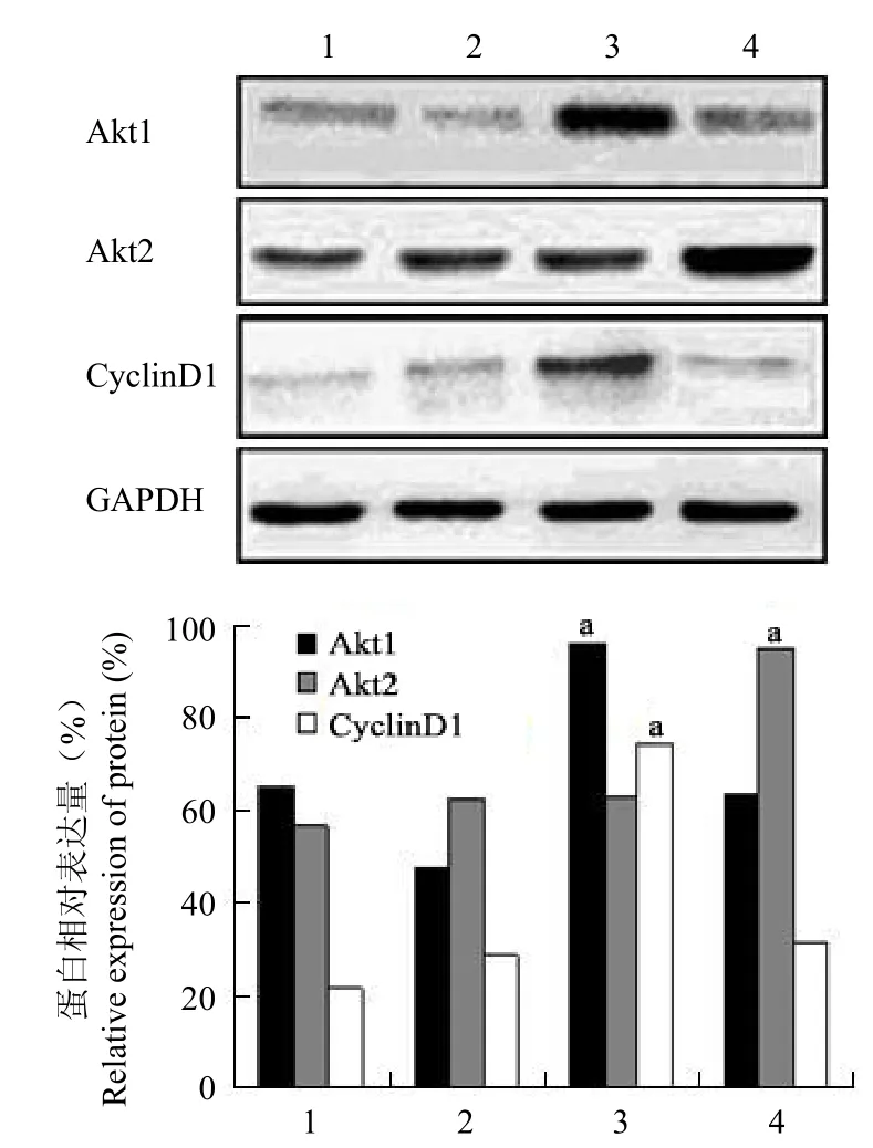
图 1 转染 Akt1 和 Akt2 基因的 GES-1 细胞蛋白表达的蛋白质印迹检测(a分别与对照组和空载组比较,均 P <0.01)Figure 1 The expression of protein in the GES-1 cells transfected by Akt1 and Akt2 genes by Western blot.aCompare with the control group and blank vector group respectively, all P < 0.01.
蛋白质印迹分析显示,Akt1 组 Akt1 蛋白的表达水平高于空载组和对照组,灰度值(Akt1/GAPDH)分析表明其 Akt1 蛋白相对表达量(0.96 ± 0.05)分别是空载组(0.47 ± 0.02)和对照组(0.64 ± 0.03)的 2.03 ± 0.22 和 1.48 ± 0.19倍,差异均有统计学意义(均 P < 0.01);同时 Akt1组 Cyclin D1 蛋白相对表达量(0.74 ± 0.01)分别是空载组(0.29± 0.01)和对照组(0.22 ± 0.01)的2.57 ± 0.05 和 3.44 ± 0.04 倍,差异也均有统计学意义(均 P < 0.01)。Akt2 组 Akt2 蛋白的表达水平高于空载组和对照组,灰度值(Akt2/GAPDH)分析表明其 Akt2 蛋白相对表达量(0.95 ± 0.01)分别是空载组(0.62 ± 0.02)和对照组(0.56 ± 0.01)的 1.53 ± 0.04 和 1.69 ± 0.01 倍,差异均有统计学意义(均 P < 0.01);而 Akt2 组 Cyclin D1 蛋白相对表达量(0.31 ± 0.02)与空载组(0.29 ± 0.01)和对照组(0.22 ± 0.01)比较无明显升高,差异均无统计学意义(均 P > 0.05)(图 1)。表明 p-LXSNAkt1 和 p-LXSN-Akt2 质粒可以在 GES-1 内稳定表达。
表 1 MTT 法测定转染 Akt1 和 Akt2 基因的 GES-1 细胞的 A490值(±s)Table 1 The A490values of the GES-1 cells transfected by Akt1 and Akt2 genes by MTT method (±s)

表 1 MTT 法测定转染 Akt1 和 Akt2 基因的 GES-1 细胞的 A490值(±s)Table 1 The A490values of the GES-1 cells transfected by Akt1 and Akt2 genes by MTT method (±s)
注:a分别与对照组和空载组比较,均 P < 0.01Note:aCompared with the control group and blank vector group respectively, all P < 0.01
细胞培养时间(d)Cells culture time (d)对照组Control group空载组Blank vector group Akt1组Akt1 group Akt2 组Akt2 group 1 0.254 ± 0.055 0.221 ± 0.008 0.269 ± 0.118 0.243 ± 0.009 2 0.233 ± 0.114 0.239 ± 0.126 0.440 ± 0.365 0.323 ± 0.369 3 0.474 ± 0.217 0.455 ± 0.404 0.714 ± 0.313a 0.535 ± 0.249 4 0.569 ± 0.216 0.566 ± 0.611 0.998 ± 0.914a 0.639 ± 0.355 5 0.805 ± 0.993 0.821 ± 0.351 1.438 ± 0.844a 0.950 ± 0.608 6 1.065 ± 0.514 1.113 ± 0.134 1.723 ± 0.912a 1.087 ± 0.175
2.3 Akt1、Akt2 基因对 GES-1 细胞增殖能力的影响
MTT 法检测结果表明,Akt1 组吸光度(A490)值于第 1、2 天分别与空载组和对照组比较差异无统计学意义(均 P > 0.05),第 3 ~ 6 天差异均有统计学意义(均 P < 0.01)。而 Akt2 组吸光度(A490)值于第 1 ~ 6 天分别与空载组和对照组比较差异均无统计学意义(均 P > 0.05)(表 1、图 2)。表明转染 Akt1 基因后 GES-1 细胞增殖能力增强。
2.4 Akt1、Akt2 基因对 GES-1 细胞细胞周期的影响

图 2 转染 Akt1 和 Akt2 基因的 GES-1 细胞生长曲线Figure 2 The cells growth curves of the GES-1 cells transfected by Akt1 and Akt2 genes

图 3 转染 Akt1 和 Akt2 基因的 GES-1 细胞细胞周期的流式细胞术分析(A:对照组;B:空载组;C:Akt1 组;D:Akt2 组)Figure 3 The cells cycle distribution of the GES-1 cells transfected by Akt1 and Akt2 genes by flow cytometry. A: Control group; B: Blank vector group; C: Akt1 group; D: Akt2 group.
相对于空载组和对照组,Akt1 组 S 期细胞数分别增多约 20.00% 和 21.10%,G2 期细胞数相应减少约 20.80% 和 15.48%,差异均有统计学意义(均 P < 0.01);而与空载组和对照组比较,Akt2 组S 期细胞数仅增多 1% ~ 2%,差异均无统计学意义(P > 0.05)(图 3、表 2)。

表 2 Akt1、Akt2 基因转染对 GES-1 细胞细胞周期的影响Table 2 The effect of Akt1 and Akt2 genes transfection on the cells cycle of the GES-1 cells
3 讨论
Akt/PKB 信号通路是细胞内信号传导的重要环节,参与细胞代谢、生存、凋亡、分化和增殖等过程。哺乳动物的 Akt/PKB 由 3 个亚型组成,分别是 Akt1/PKBα、Akt2/PKBβ 和 Akt3/PKBγ,它们分别受 3 个完全不同的基因编码调节,主要表现为位于激酶区域的第 308 号位点苏氨酸(Thr308)和位于 C 末端的第 473 号位点丝氨酸(Ser473)的磷酸化,只有这 2 个位点的氨基酸都被激活,才能充分发挥其激酶功能。3 个亚型都具有相似的蛋白质结构,对底物表现相似的特异性。动物实验证明,敲低 Akt1 基因的小鼠生长发育迟缓,细胞凋亡增加,表明 Akt1 基因在细胞存活过程中具有关键作用;敲低 Akt2 基因的小鼠表现为糖代谢障碍;而敲低 Akt3 基因的小鼠有脑损伤表现,实际上 Akt 各亚型特别是 Akt1 和 Akt2,有着极其复杂的作用,并且相互重叠[5-7]。在不同的组织细胞,Akt 各亚型的表达水平不一,Akt1 基因扩增见于胃癌,其高表达见于乳腺癌;Akt2 基因扩增见于卵巢癌和胰腺癌,其高表达见于肝癌和结肠癌[8]。Koseoglu 等[9]发现 Akt 这 3 种亚型依赖性细胞的存活具有细胞系特异性,在人乳腺癌ZR-75 细胞中,Akt1 基因对细胞增殖和存活起主导作用;而对于人卵巢腺癌 IGROV1 细胞,Akt2基因则对细胞增殖起主导作用,但其存活非依赖单一 Akt 亚型。研究显示 Akt1 基因及磷酸化 Akt1(p-Akt1)基因在胃腺癌组织中的阳性表达率高于癌旁组织和正常胃黏膜组织,并且其表达水平也同肿瘤分级和 TNM 分期相关;利用磷酸肌醇 3-激酶(phosphatidylinositide 3-kinase,PI-3K)抑制剂LY-294002 抑制 p-Akt1 基因,尤其对于 Akt1 激活的细胞,可使胃癌细胞增殖受阻,并诱导 G1 期停滞,表明 Akt1 基因作为一个激酶与胃癌的进展和细胞增殖密切相关[10-11]。
Akt1、Akt2 基因在细胞周期进程中的作用不尽相同。Yun 等[12]研究发现,Akt1 基因在细胞周期 G1/S 期过度和增殖中具有重要作用。细胞缺少Akt1 基因则表现为增殖伴 G1/S 期过度减弱,缺少 Akt2 基因的细胞该进程不受影响;而 Akt1 基因对增殖和 G1/S 期过度的作用完全可以通过Akt2 基因交换 PH 区域来废止,这表明 PH 区域是 Akt1 基因完全激活并促使 G1 到 S 期过度的关键结构域。Héron-Milhavet 等[3]发现敲低非转化哺乳动物细胞的 Akt1 基因而不是 Akt2 基因,可导致 Cyclin A 水平下降,细胞进入 S 期减弱;而Akt2 基因的过表达则通过增加核内 p21 蛋白使细胞周期阻滞在 M-G1 期。这同样证明了 Akt1基因对细胞增殖的关键作用,而 Akt2 基因则促进细胞周期阻滞。本实验与以上研究结果基本一致,其机制可能是 Akt 基因通过其下游雷帕霉素靶点(mTOR)将有丝分裂信号传递给 p70S6K1,使细胞周期主要蛋白如细胞周期蛋白的翻译上调,以及抑制 p21CIP1、p27KIP1的表达,促进 G1 期进展,使细胞周期加速。
本实验以脂质体介导的基因转染法将含有Akt1-cDNA、Akt2-cDNA 的真核表达载体 p-LXSN导入人胃黏膜上皮 GES-1 细胞,成功地建立了稳定表达 Akt1 及 Akt2 蛋白的 GES-1-Akt1 和GES-1-Akt2。进一步的 MTT 生长曲线测定和流式细胞周期分析实验表明,转染 Akt1 基因的 GES-1细胞,增殖能力提高,细胞周期 S 期明显增多,提示 Akt1 基因可能通过延长细胞生存时间为人胃黏膜上皮细胞的恶性转化提供可能,而 Akt2 基因对 GES-1 细胞的影响不明显。下一步我们需要探讨的是 Akt 通路下游,如 mTOR 等一系列与细胞增殖相关靶基因的变化,并从细胞形态及迁移能力方面进行对比分析,期待能为抑制 GES-1 细胞恶性转化提供新的途径或方法。
参考文献
[1] Samuels Y, Ericson K. Oncogenic PI3K and its role in cancer. Curr Opin Oncol, 2006, 18(1):77-82.
[2] Han Z, Wu K, Shen H, et al. Akt1/protein kinase B alpha is invoved in gastric cancer progression and cell proliferation. Dig Dis Sci, 2008, 53(7):1801-1810.
[3] Héron-Milhavet L, Franckhauser C, Rana V, et al. Only Akt1 is required for proliferation, while Akt2 promotes cell cycle exit through p21 binding. Mol Cell Biol, 2006, 26(22):8267-8280.
[4] Toker A, Yoeli-Lerner M. Akt signaling and cancer: surviving but not moving on. Cancer Res, 2006, 66(8):3963-3966.
[5] Chen WS, Xu PZ, Gottlob K, et al. Growth retardation and increased apoptosis in mice with homozygous disruption of the Akt1 gene. Genes Dev, 2001, 15(17):2203-2208.
[6] Garofalo RS, Orena SJ, Rafidi K, et al. Severe diabetes, age-dependent loss of adipose tissue, and mild growth deficiency in mice lacking Akt2/PKBbeta. J Clin Invest, 2003, 112(2):197-208.
[7] Tschopp O, Yang ZZ, Brodbeck D, et al. Essential role of protein kinase B gamma (PKB gamma/Akt3) in postnatal brain development but not in glucose homeostasis. Development, 2005, 132(13):2943-2954.
[8] Gonzalez E, McGraw TE. The Akt kinases: isoform specificity in metabolism and cancer. Cell Cycle, 2009, 8(16):2502-2508.
[9] Koseoglu S, Lu Z, Kumar C, et al. AKT1, AKT2 and AKT3-dependent cell survival is cell line-specific and knockdown of all three isoforms selectively induces apoptosis in 20 human tumor cell lines. Cancer Biol Ther, 2007, 6(5):755-762.
[10] Cui XW, Zhang QY, Kang CS, et al. Protein expression of Akt1, MMP2 and PTEN in gastric adenocarcinoma and the correlations among these indices. Chin J Clin Oncol, 2009, 36(5):258-261.(in Chinese)崔小伟, 张庆瑜, 康春生, 等. 利用组织芯片技术研究 Akt1、MMP2和 PTEN在胃腺癌中的表达及其相关性. 中国肿瘤临床, 2009, 36(5):258-261.
[11] Han Z, Wu K, Shen H, et al. Akt1/protein kinase B alpha is involved in gastric cancer progression and cell proliferation. Dig Dis Sci, 2008, 53(7):1801-1810.
[12] Yun SJ, Tucker DF, Kim EK, et al. Differential regulation of Akt/protein kinase B isoforms during cell cycle progression. FEBS Lett, 2009, 583(4):685-690.
ObjectiveTo observe the effects of Akt1 and Akt2 genes transfection on the growth of human gastric epithelial GES-1 cells.
MethodsThe plasmids of p-LXSN-Akt1 which contained the whole Akt1 gene sequence (Akt1 group) and p-LXSN-Akt2 which contained the whole Akt2 gene sequence (Akt2 group) were used to transfect human gastric epithelial GES-1 cells with lipofectamine, the GES-1 cells which were transfected by the empty vector p-LXSN were used as the black vector group, and the no transfected ones were used as the control group. Then the positive cell clones were selected with antibiotics G418. The protein expressions of Akt1, Akt2 and Cyclin D1 in the GES-1 cells were measured by Western blotting analysis; MTT method was applied to detect the proliferation activity of GES-1 cells; the effect of Akt1 and Akt2 genes transfection on the cells cycle of the GES-1 cells was tested by flow cytometry (FCM).
Results Cell modes which expressed Akt1 gene (Akt1 group) and which expressed Akt2 gene (Akt2 group) were obtained. Akt1 group: ①The relative expressions of Akt1 protein (0.96 ± 0.05) were respectively 2.03 ± 0.22 times of the black vector group (0.47 ± 0.02) and 1.48 ± 0.19 times of the control group (0.64 ± 0.03) (all P < 0.01), the relative expressions of Cyclin D1 protein (0.74 ± 0.01) were respectively 2.57 ± 0.05 times of black vector group (0.29 ± 0.01) and 3.44 ± 0.04 times of the control group (0.22 ± 0.01) (all P < 0.01); ②Compared with the black vector group and control group, the absorbance (A490) values in the first 1, 2 days were no significant differences, but there were statistically significant differences (all P < 0.01) in the first 3 to 6 days; ③Compared with the black vector group and control group, S phase cells were increased by approximately 20.00% and 21.10%, G2 phase cells were reduced by approximately 20.80% and 15.48% (all P < 0.01). Akt2 group: ①The relative expression of Akt2 protein (0.95 ± 0.01) were 1.53 ± 0.04 times of the black vector group (0.62 ± 0.02) and 1.69 ± 0.01 times of the control group (0.56 ± 0.01) (all P < 0.01), compared with the black vector group (0.29 ± 0.01) and the control group (0.22 ± 0.01), the relative expression of Cyclin D1 protein (0.31 ± 0.02) was no significant differences; ②Compared with the black vector group and the control group, the A490values of the first 1 to 6 days were no significant differences; ③Compared with the black vector group and control group, S phase cells were no significant differences.
ConclusionAkt1 gene transfection could increase the proportion of S-phase cells and enhance the proliferation ability of the GES-1 cells, but the effects of Akt2 gene was not obvious.
【Key words】Gene transfer techniques; Cyclin D1; Gastric mucosa
Author Affiliation:Department of Gastroenterology, Tianjin Medical University General Hospital, Tianjin 300052, China (XIE Jia-bei, ZHANG Qing-yu, HAN Jing, WANG Tao, ZHANG Jie); Laboratory of Neuro-Oncology, Tianjin Neurological Institute, Tianjin 300052, China (KANG Chun-sheng)
Chin Med Biotechnol, 2010, 5(6):423-428
Corresponding Author:ZHANG Qing-yu, Email: zhangqy@tijmu.edu.cn www.cmbp.net.cn
作者单位:300052 天津医科大学总医院消化内科(谢甲贝、张庆瑜、韩静、王涛、张洁);300052 天津市神经病学研究所神经肿瘤研究室(康春生)
通讯作者:张庆瑜,Email:zhangqy@tijmu.edu.cn
收稿日期:2010-08-25
DOI:10.3969/cmba.j.issn.1673-713X.2010.06.005
The effect of Akt1 and Akt2 gene transfection on growth of human gastric epithelial cell GES-1
XIE Jia-bei, ZHANG Qing-yu, KANG Chun-sheng, HAN Jing, WANG Tao, ZHANG Jie
【Abstract】