Fractal Study on the Evolution ofMicro-Pores in Concrete Under Freeze-Thaw
2024-04-10SUNHaoranZOUChunxiaXUDeruGUOXiaosongHUANGKun
SUN Haoran, ZOU Chunxia*, XU Deru, GUO Xiaosong, HUANG Kun,2
(1.College of Water Conservancy and Civil Engineering, Inner Mongolia Agricultural University, Hohhot 010018, China; 2.Xi’an HaiTang Vocational College, Xi’an 710038, China)
Abstract: After exposure to freeze-thaw cycles, scanning electron microscopy (SEM) and nuclear magnetic resonance (NMR) were used to test the four mixtures.The microstructure is qualitatively analyzed from the 2D SEM image and the 3D pore distribution curve before and after freezing and thawing.The fractal dimension is utilized to characterize the two-dimensional topography image and the three-dimensional pore distribution, quantitatively.The results reveal that the surface porosity and volume porosity increase as the freeze-thaw action increases.Self-similarity characteristics exist in micro-damage inside the concrete.In the fractal dimension, it is possible to characterize pore evolution quantitatively.The fractal dimension correlates with pore damage evolution.The fractal dimension effectively quantitatively characterizes micro-damage features at various scales from the local to the global level.
Key words: fractal dimension; freeze-thaw cycle; concrete; SEM; NMR
1 Introduction
Freezing-thawing has always been one of the severe damage of channel concrete in the cold area of North China, which leads to its mechanical property and durability deterioration.In order to prevent this serious damage, research has been intensified.A study revealed that under freezing and thawing, the internal structure of concrete is loose and the pores gradually deteriorate[1].The freezing point and amount of freezing in the pores are affected by porosity, pore size, and pore distribution[2].Therefore, controlling parameters like air content and porosity in concrete can help to increase its frost resistance[3].At present, research focuses on phenomenological research to characterize the deterioration of macro-mechanical characteristics and anticipate concrete’s service life based on peak stress and relative dynamic modulus of elasticity damage[4,5].However, presenting the evolution process at the pore structure is hard.In the study of frost-resisting durability[6,7], it is critical to quantitatively describe the pore structure, which will reveal the microscopic damage mechanism of concrete.Hence, researchers focus on the damage mechanisms at the microscopic scale during freezing and thawing[8-10].Concrete’s resistance to freeze-thaw cycles was simulated by the solution of the poromechanical problm for studying the impacts of pore space and entrained air void size distribution on concrete’s durability.The fractal dimension was regarded as a numerical index that describes the complexity of a pore structure.Scanning electron microscopy(SEM) may be used to observe the surface’s irregular cracks and nonlinear damage[11], and through nuclear magnetic resonance (NMR) measurements the complex pore structure is tested[12,13].However, the quantitative characterization of pore structure has mainly focused on a single experiment but ignored the dimensional relationship in fractal objects.
The fractal theory is compatible with concrete’s complex pore structure changes as a subject for studying irregularities and complicated matter processes.It may quantitatively define the micro-damage of concrete by the fractal dimension of 2D micro-topography and 3D pore structures.Furthermore, appropriate fractal research will contribute to determining the dimensional relationship between the topography and pore structure in freeze-thaw cycles, which reveals the micro-damage appeared quantitatively experiencing freezing-thawing.
The primary objective of this article is to investigate the fractal characteristics of pore evolution at various scales.For this purpose, scanning electron microscopy (SEM) and nuclear magnetic resonance (NMR)measurements were combined with fractal geometry to analyze the internal pore structure in concrete, resulting in a powerful method for evaluating the spatial properties of the microstructure.The statistical analysis of pore damage was conducted on two scales: 2D images and 3D structures.The relationship between the pore damage evolution and the freeze-thaw was assessed based on fractal dimension characteristics.Additionally, this research targets to provide theoretical and experimental support as well as an essential reference for the micro-damage and macro-durability property evaluation of freeze-thaw action.
2 Experimental
2.1 Materials
The Mengxi P·O 42.5R Portland cement was preferred for the concrete mixtures.Fly ash (3.6% in fineness) is from Baotou Power Plant, and silica fume(321.36 m2/kg in specific surface area, and 94.51% in SiO2content) is solid waste from the jade processing plant in Urad Front Banner, which are used to partially replace the cement at different ratios.YE-NGX naphthalene series was preferred as a superplasticizer additive.Concrete mixing water is ordinary water and freeze-thaw test water is obtained from the Yellow River.The fine aggregate (2 586 kg/m³ in apparent density and 1 631 kg/m³ in bulk density) and coarse aggregate(5-19 mm in particle sizes) are chosen from UradFront Banner.The mix proportions are shown in Table 1.

Table 1 Mix ratio of concrete/(kg/m³)
2.2 Test Method
2.2.1 Freeze-thaw cycle test
The specimen size for the freeze-thaw test was 100×100×400 mm.According to the climate characteristics and the laws of agricultural irrigation[14]of Hetao Irrigation District in Inner Mongolia, 25 freeze-thaw cycles have been assumed.During the freezing process,temperatures were lowered from 5 to -18 degrees Celsius.The temperature was rose from -18 to 5 degrees Celsius throughout the thawing process.
2.2.2 Scanning electron microscope test
A Phenom LE scanning electron microscope tested the microstructure and morphology of the concrete samples.The samples under the action of freezing and thawing were taken from the same position inside the concrete.Then the samples in the transition area of the interface were put into ethanol and natural drying after ultrasonic dispersion.In order to make the SEM image of the microstructure appearance clearer, spraying treatment is required before the test.The electron microscope scanning test was carried out on the samples under the action of freeze-thaw cycles, and the quantitative analysis of the microscopic damage was transformed from a qualitative analysis of the microstructure and morphology.
2.2.3 Nuclear magnetic resonance test
The nuclear magnetic resonance (NMR) test was performed using the MesoMR23-60 nuclear magnetic resonance (NMR) tester of Suzhou Niumei Company.The MesoMR23-60 nuclear magnetic resonance tester is a permanent magnetic field with a magnetic field strength of 0.55 T, a magnet temperature of 32°C, and an H proton resonance frequency of 23.32 MHz.Based on the response of hydrogen nuclei, the nuclear magnetic resonance instrument was used to test the concrete test block in the saturated state to determine the relaxation characteristics of the H element in the internal pore structure.Pores of different sizes in saturated concrete correspond to different water contents.Each type of pore corresponds to a certain spin-echo decay magnitude.After inversion of the spin-echo attenuation amplitude, the T2spectrum was obtained from a set of attenuation constants.Before the test, the concrete core was sampled with a diamond coring machine.The specimens were then placed in a vacuum-saturated device of 0.1 MPa and evacuated for 24 h.NMR tests were carried out on concrete with 50, 100, and 150freeze-thaw cycles, respectively.The evolution law of fractal damage was explored by the characteristic parameters of pore structure of concrete.
3 Theoretical methods
3.1 SEM fractal dimension
The damage characteristics in the surface can be obtained from a microscopic image of the concrete taken with a scanning electron microscope (SEM).Fractal dimension is related to damage characteristics complexity and disorder, and more complex image surfaces have a higher fractal dimension.When the fractal dimension transforms the qualitative description into quantitative analysis, the damage evolution will be found out quantitatively.The fractal dimension of SEM images can be calculated using a variety of approaches[15,16].In this experiment the box-counting method was used to conduct fractal analysis on SEM images under freeze-thaw.The SEM images were processed into binary images using MATLAB.A diagram of lnN(ε)–ln(ε)was determined by the least square method[17], and the fractal dimensionDScan be defined as:
whereDSis the fractal dimension of SEM images.
The image was binarized for fractal dimension calculation.The target image consisted of only black and white parts, then a box with side lengthεwas taken to divide the target image.The number of regions containing black (white) parts, was denoted asN(ε), (ε= 1,2, 4,..., 2i(i= 0, 1, 2, 3,...)).1, 2, 4,..., 2ipixel size was taken as edge length and finallyN(1),N(2),N(4),...,N(2i), the number of boxes was obtained.With a smaller side length, more boxes are obtained; when the side length of the boxεtends to 0, the number of boxesN(ε)will tend to infinity.Fig.2 shows the fractal calculation process after SEM image binarization.
3.2 NMR fractal dimension
The microscopic image of the concrete acquired with a scanning electron microscope (SEM) reveals the damage characteristics on the surface.
The damage evolution law will make quantitative sense when the fractal dimension transforms qualitative description into quantitative analysis.The amount of pores with pore size are suggested to follow the fractal scaling law as[18].Therefore, the cumulative pore volumeSVin the NMR measurements can be expressed as:
whereris the pore radius,rminandrmaxare the smallest and largest pore sizes, respectively.DVis the pore fractal dimension.
The fractal formula of the NMRT2spectrum is shown in Eq.(3):
whereSVis the percentage of the cumulative pore volume (the lateral relaxation time is less thanT2) to the total pore volume.
Therefore, the fractal dimensions can be calculated from the slope (3-D) of the best fitting line by plottingSVand the correspondingT2in an lg–lg plot as shown in Eq.(4):
4 Results and discussion
4.1 Microstructure
The SEM micro-morphology images of concrete under freeze-thaw cycles are shown in Fig.1.The scale of the SEM image is 10 μm and the magnification is 5 000×.In Fig.1(a), the microstructure appears uniformly and scaly with some hydration products.After freeze-thaw exposure (Fig.1(b)), the scaly products disappear, and the acid salt from the Yellow River gradually etch the granular fly ash, followed by the formation of cracks gradually.After 150 freeze-thaw cycles,the fibrous product becomes thin and partially fractures due to irreversible damage or micro-damage on the concrete.The images of F15S4, F10S8 and F20S4 are selected because we comprehensively considered the SEM and NMR tests.F15S4 has the smallest fractal dimension and porosity, F20S4 is the largest, and F10S8 is in the middle.The three groups of mix ratios represent different damage characteristics.
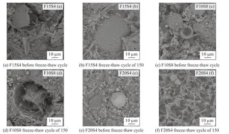
Fig.1 SEM images before and after freezing and thawing

Fig.2 Fractal calculation process after SEM image binarization
The surface porosity of the SEM image was calculated using Image J to evaluate the evolution of microscopic pore structure.2D(SP) in Table 4 is the SEM surface porosity value of mix ratio in concrete.2D(SP) in Table 4 is the SEM microscope surface porosity value of mix ratios of concrete.As shown in Fig.3, convert the image to 8-bit, select the analysis area,and calculate the pore area of the chosen area.Surface porosity equals to pore area/total area.
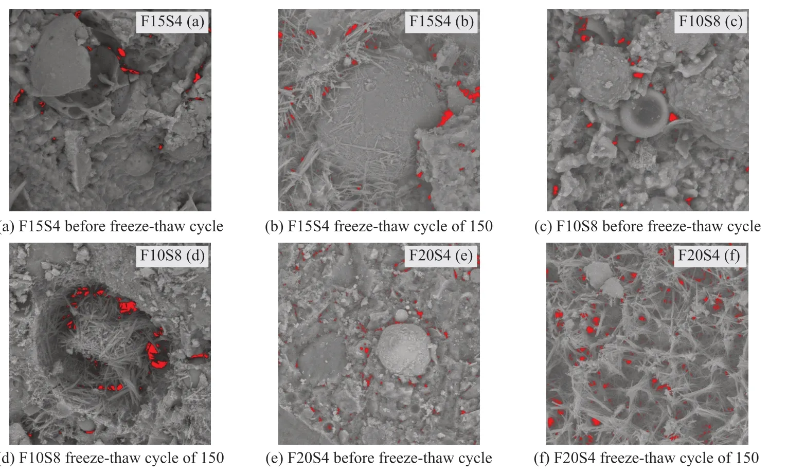
Fig.3 Pore distribution of SEM image calculated by Image J
4.2 Pore structure of concrete
The NMR test obtains the evolution of the pore structure under freeze-thaw action (Fig.4).The relaxation time correlates with the pore radius[19], and the relaxation timeT2is related to the pore size.This means that the shorter the relaxation time, the smaller the pore radii; also, the longer the relaxation time, the larger the pore radii[20,21].The surface relaxation determines the relaxation time, as shown in Eq.(5):
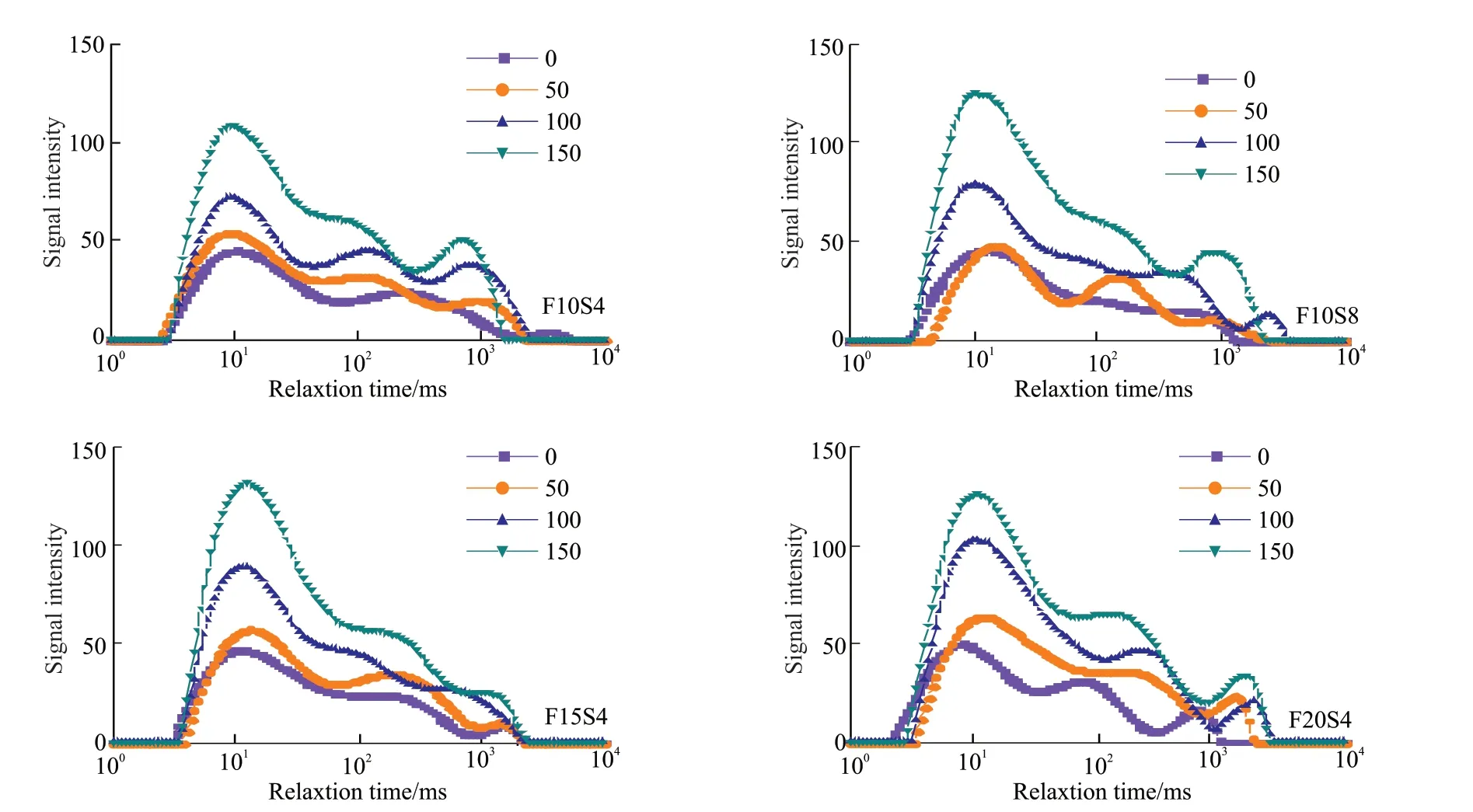
Fig.4 T2 spectrum distribution of pore structure via NMR measurements
whereρis the lateral surface relaxation strength,μm/s;Sis the surface area of the pore;Vis the pore volume.
NMRT2spectra give nearly the entire pore body size distribution in Fig.4.TheT2response spectrum reveals the presence of three populations of pore sizes that gradually increased with freeze-thaw cycles.In addition, shortT2components have a significant elevation of small pores, while longT2components correspond to large pores, gradually occupying a certain proportion under freeze-thaw cycles.The longT2components of F15S4 were relatively small, indicating fewer large pores after 150 cycles.Combined with the SEM image analysis, the evolution of microscopic morphology and pore structure is inherently consistent.
4.3 Fractal characteristics of pore structure
4.3.1.Fractal dimension via SEM image
Table 2 calculates the binarization threshold and fractal dimension of the SEM images.As the fibrous product begins to break after 150 freeze-thaw cycles,the fractal dimension allowing quantitative analysis of its morphological damage characteristics gradually grew.The fractal dimension can explain the damage in the microscopic morphology by the box-counting method.Under the same freeze-thaw cycles, F15S4 and F10S8 detect smaller fractal dimensions and lower damage degrees.For the F15S4, compared with before freezing and thawing, the fractal dimension increased from 3.62%, 4.6%, to 7.99%.At this point, the development of damage from the fractal dimension is not linear as considering the morphological characteristics of the SEM image, and the higher the fractal dimension, the more complex of the cracks, pores, and structural bedding.After freeze-thaw cycles, the fractal dimension varies with microscopic appearance and pore structure evolution.The fractal dimension quantitatively characterizes the damage of concrete, and the fractal dimension increases as the pore structure increases.Therefore, it means that the damage degree of concrete gradually deteriorates and its durability decreases.

Table 2 Results of SEM fractal dimension
4.3.2 Fractal dimension via NMR technology
The relationship betweenSVandT2is shown in Eq.(4).However, two straight sectional lines substitute for the flowing curve, which is a plot bylgSVagainstlgT2for all the samples in Fig.5, where the fractal curves divide into two segments at theT2cutoffvalue.From the slopes (k1, k2) of the relationship betweenlgSVandlgT2in Eq.(4), obtain the fractal dimensionsDVminandDVmaxare obtained.Related results are shown in Table 3.The fractal dimension increases continuously as the complicacy of the pore increases[22].As aresult, fractal sizes reaching 3.0 revealed a heterogeneous pore structure, with values ranging from 2.699 to 2.822, with a mean of 2.765, indicating pore structures of moderate to high complexity and heterogeneity.The fractal dimension and the irreducible water content vary proportionally, and the fractal dimension can determine the irreducible water content in concrete.The rate of increase of fractal dimension for F15S4 ranged from 1.56% to 2.48% to 3.89%.The ranges of values were consistent with the fractal size of the SEM images, indicating that the calculation results obtained with the different test methods are only the size difference.
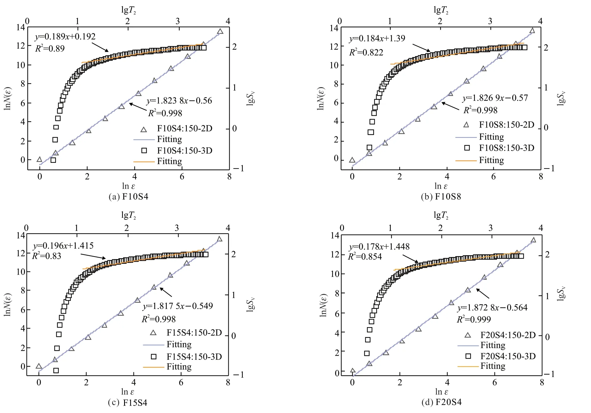
Fig.5 Fractal dimension of pore structure in concrete with SEM image and NMR technology

Table 3 Fractal dimension calculated based on nuclear magnetic resonance
The rate of increase of fractal dimension from 1.25% to 1.21% reduced to 1.12% under freeze and thaw, showing that the degree of concrete damage is progressively approaching the limit.Therefore, the fractal dimension can evaluate the heterogeneity, which has advantages in the characterization of the pore structures of the concrete.
4.4 Fractal dimension fitting results of 2D SEM image and 3D NMR technology
The straight line is calculated by the box-counting methods of the SEM image, whose absolute slope value is the fractal dimension (Fig.5 (2D)).The graph with the correlation coefficientR2> 0.99 obtain a better fitting result.ThelgSV-lgT2plot (Fig.5(d)) divide into two segments atT2≤T2cutoff.The linear trendline with the correlation coefficientR2> 0.82 support the large pore area corresponding to the longT2components and is essentially fractal.Small-scale pores (shortT2components) find that the range of the slope is from 3.315 to 4.55 through linear regression, while large-scale pores(longT2components) have the range of the slope from 0.189 to 0.3.By linear regression, a fractal dimension ranges from 2.699 to 2.822.
The 2D images and 3D structures demonstrate similar properties of fractal bodies at different scales based on the fitting curves (Fig.5).The fractal dimensions of 2D images or 3D structures can accurately reflect pore structure evolution.Therefore, it is possible to quantitatively characterize damage development with the fractal dimension.
Fractal dimension is related to surface morphological complexity and disorder, and more complex structures have a higher fractal dimension.Fig.5 depicts the fractal dimension of pore growth in 2D images and 3D structures.Overall, as the number of freeze-thaw cycles increases, the fractal dimension of 2D images and 3D structures grows, as does the abundance of pores.The average fractal dimension of concrete in the 2D image is 1.697 before freeze-thaw action, and then it increases to 1.752, 1.771, and 1.824 after freeze-thaw action.The variation trend of fractal dimension in the 3D structure is similar to that in the 2D image.The average fractal dimensions of different mix ratios calculated by 3D structure were 2.715, 2.749, 2.782, and 2.813 from before freeze-thaw to after 150 freeze-thaw cycles,which increases by 1.25%, 2.48%, and 3.63%, respectively.Notably, the fractal dimension of 3D structures is higher than the fractal dimension from 2D images by approximately 1.0.
In the concept of Euclidean geometry to fractal geometry, the intersection ofn-dimensional on the cross-section isn-1 dimensional.DL,DS,DVcan be used to express the fractal dimensions from the section line, plane, and space[23], where the mathematical relationship could be expressed as follows:
Eq.(6) obtains the dimensional relationship between the two-dimensional topography image and the three-dimensional pore structure, which describes the degree of roughness of the pore surface and the complex pore area structure.Fig.6 shows the dimensional relationship.
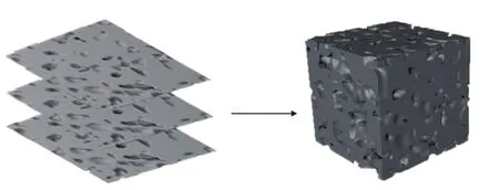
Fig.6 Dimensional relationship of 2D image and 3D structure
Fig.7 illustrates the 2D and 3D porosity of concrete, including the 3D pore structure and 2D SEM image.As mentioned before, the fractal dimension reflects information about pore space regulation.More pore damage evolves in fractal space as the freeze-thaw process increases.Two crucial indicators, surface porosity(SP) and volume porosity (VP) describe the fractal properties of micro-damage.Surface porosity reflects the distribution characteristics of a 2D SEM image,whereas volume porosity from a 3D structure extendsinto pore space.

Fig.7 Porosity of 2D SEM image and 3D pore structure
From Table 4, the porosity (SP and VP) increases with increasing fractal dimension, similar to 2D images.The porosity of a 2D image is greater than that of a 3D structure under the same freeze-thaw action, which could be because 3D structures include more information regarding pore space.It’s also clear that the fractal dimension has a linear relationship with the porosity of damage evolution, implying that fractal pore structure indicators can represent micro-damage and durability.

Table 4 Indicators of the pore evolution under freezing and thawing/%
5 Conclusions
a) The interior micro-pore structure damage mechanism can be qualitatively captured by SEM images and NMR measurements.The microporous structures in concrete is a fractal objects with self-similar properties.The fractal theory effectively analyzes the self-similarity of concrete by accurate coverage and intelligent calculation.
b) The fractal dimension allows for quantitative identification of concrete microstructure damage under freezing and thawing.By the SEM experiment in different freeze-thaw cycles, the value of fractal dimension increases from 1.683 6 to 1.8282, with increments of 3.73%, 1.11%, and 2.9%, respectively, and the average value is 1.761 6.In addition, by the fractal dimension of the NMR experiment, the value ranges from 2.699 to 2.822, with the rate of increase from 1.25%,1.21%, to 1.12%, and the average value is 2.765.
c) The fractal dimension can comprehensively describe the freeze-thaw damage characteristics of the pore structure by utilizing the 2D SEM image and 3D pore structure.Based on the dimensional relationship of fractal dimension, it can provide a theoretical basis for exploring concrete’s comprehensive pore damage evolution under freeze-thaw action through different experiments.
Conflict of interest
All authors declare that there are no competing interests.
杂志排行
Journal of Wuhan University of Technology(Materials Science Edition)的其它文章
- Effect of VEGF/GREDVY Modified Surface on Vascular Cells Behavior
- Evolution of Biofilm and Its Effect on Microstructure of Mortar Surfaces in Simulated Seawater
- Synthesis of Organic-Inorganic Hybrid Aluminum Hypophosphite Microspheres Flame Retardant and Its Flame Retardant Research on Thermoplastic Polyurethane
- Surface Metallization of Glass Fiber (GF) /Polyetheretherketone (PEEK) Composite with Cu Coatings Deposited by Magnetron Sputtering and Electroplating
- Effect of Size Change on Mechanical Properties ofMonolayer Arsenene
- Effects of Sinusoidal Vibration of Crystallization Roller on Composite Microstructure of Ti/Al Laminated Composites by Twin-Roll Casting
