Development status of novel spectral imaging techniques and application to traditional Chinese medicine
2023-12-14QiWngYongZhngBofengYng
Qi Wng ,Yong Zhng ,Bofeng Yng
a Department of Medicinal Chemistry and Natural Medicinal Chemistry, College of Pharmacy, Harbin Medical University, Harbin,150081, China
b Department of Pharmacology (The State-Province Key Laboratories of Biomedicine-Pharmaceutics of China, Key Laboratory of Cardiovascular Research,Ministry of Education), College of Pharmacy, Harbin Medical University, Harbin,150081, China
c Research Unit of Noninfectious Chronic Diseases in Frigid Zone, Chinese Academy of Medical Sciences, Harbin,150081, China
d Institute of Metabolic Disease, Heilongjiang Academy of Medical Science, Harbin,150086, China
e Department of Pharmacology and Therapeutics,Melbourne School of Biomedical Sciences,Faculty of Medicine,Dentistry and Health Sciences University of Melbourne, Melbourne, VIC, 3010, Australia
Keywords:Chinese medicine Spectral imaging Fluorescence spectroscopy Photoacoustic imaging
ABSTRACT Traditional Chinese medicine (TCM) is a treasure of the Chinese nation,providing effective solutions to current medical requisites.Various spectral techniques are undergoing continuous development and provide new and reliable means for evaluating the efficacy and quality of TCM.Because spectral techniques are noninvasive,convenient,and sensitive,they have been widely applied to in vitro and in vivo TCM evaluation systems.In this paper,previous achievements and current progress in the research on spectral technologies (including fluorescence spectroscopy,photoacoustic imaging,infrared thermal imaging,laser-induced breakdown spectroscopy,hyperspectral imaging,and surface enhanced Raman spectroscopy) are discussed.The advantages and disadvantages of each technology are also presented.Moreover,the future applications of spectral imaging to identify the origins,components,and pesticide residues of TCM in vitro are elucidated.Subsequently,the evaluation of the efficacy of TCM in vivo is presented.Identifying future applications of spectral imaging is anticipated to promote medical research as well as scientific and technological explorations.
1.Introduction
Throughout China's long history,traditional Chinese medicine(TCM) has been an alternative for the Chinese nation.With rapid advancements in science and technology,the modernization of TCM has been of considerable importance in satisfying the medical needs of people and adapting to the development of modern society.Modern TCM can be divided into two main categories: nondrug,technology-focused therapy (represented by acupuncture and moxibustion),and traditional therapy (represented by the preparation of Chinese herbal compounds or the application of active ingredients).
Accordingly,properly evaluating the efficacy and quality of TCM is critical.In recent years,optical spectral imaging has been widely applied to physical and biological analyses;new spectroscopic technologies have been developed in this regard.This paper provides a comprehensive review of different types of spectroscopies utilized in real-life applications and their significance in the medical field.A range of spectral technologies offering both high precision and sensitivity is highlighted,focusing on their diagnostic advantages.Additionally,distinctive outcomes obtained through various spectroscopic analyses are examined.
2.Fluorescence spectroscopy technology
The concept and mechanism of fluorescence generation were proposed and explained by Stokes in 1852 [1].Fluorescence spectral imaging was first developed in 1966 [2].Fluorescence spectroscopy technology utilizes the principles of fluorescence to determine and analyze substances,which measures the absorption and emission spectra of substances in order to analyze and identify their composition and characteristics [3].Fluorescence spectral imaging technology is based on the fluorescence effect of substances combined with fluorescence analysis and space imaging technology.The technology is used to analyze measured substances quantitatively and qualitatively with the advantages of tracing and highly sensitive,nondestructive,and real-time dynamic detection.Recently,techniques derived from fluorescence spectroscopy(such as fluorescence lifetime imaging [4],fluorescence in situ hybridization [5],fluorescence resonance energy transfer [6],and threedimensional (3D) fluorescence spectroscopy [7,8]) have been emerged.Since 1990,fluorescence spectroscopy has been widely applied to the assessment of pathophysiology and pharmacodynamics[9],determination of drug composition and content in vivo[10],study of drug pharmacological mechanisms[11],optimization of drug dosage forms,and quality evaluation and identification of TCM (Fig.1) [3,9-13].
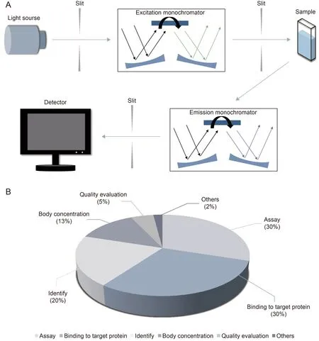
Fig.1.Fluorescence spectroscopy technology.(A) Principle and (B) applications of fluorescence spectroscopy technology.Reprinted from Refs.[3,9-13] with permission.
Fluorescence spectroscopy is a potential tool for analyzing,diagnosing,and predicting the pathological progression of diseases.For example,in the current coronavirus disease 2019 (COVID-19)pandemic,developing a sensitive method for assessing the prognosis of the disease among patients is critical.The increased concentration of collagen degradation products in plasma leads to a shortened plasma fluorescence lifetime because of the formation of numerous collagen scars during pulmonary fibrosis induced by COVID-19 [14].Wybranowski et al.[15] assessed the degree of pulmonary fibrosis in patients with COVID-19 by measuring the plasma fluorescence lifetime.The detection of the plasma fluorescence lifetime is a new method for determining the prognosis of the disease among those infected [16].Spectroscopy combined with machine learning methods has been used to detect and analyze abnormal metabolic indicators of porphyrin derivatives and bilirubin in urine for diagnosing hepatocellular carcinoma and liver cirrhosis;the overall diagnostic accuracy is as high as 83.42% [17].Fluorescence spectroscopy is a new method for the noninvasive and simple screening of these two diseases [17].The tears of healthy subjects and patients with glaucoma were used to collect 3D synchronous fluorescence data that were processed into second-derivative spectral data to achieve a sensitive and rapid diagnosis of glaucoma [18].Xi et al.[19] designed a gold nanocluster fluorescent probe encapsulated in bovine serum albumin to detect heavy ions in blood.Three different concentrations of dopamine were used as nonspecific receptors to quench the fluorescent probe by forming polydopamine.Heavy metal ions prevented fluorescence quenching by interfering with the formation of polydopamine.Therefore,heavy metals can be conveniently detected using fluorescent probes that react with heavy metal ions[19].Accordingly,fluorescence spectral imaging is a potential approach for the direct and rapid quantitative detection of pathological changes in diseases.However,presently,only a few disease indicators can be detected by fluorescence spectroscopy,and corresponding research is insufficient.Accordingly,further development of disease spectrum and detection indicators is necessary.
Some studies have shown that fluorescence spectroscopy is a sensitive and rapid detection method [20].Matrix equipotential synchronous fluorescence comprising 3D fluorescence spectroscopy combined with first-derivative technology was used to determine diflunisal and salicylic acid in human serum,providing a simple and sensitive method for the determination of two antiinflammatory drugs in serum [10].Similarly,a direct and rapid quantitative study of curcumin and dimethoxycurcumin in human plasma samples was performed by Zhai et al.[21].They used excitation emission 3D fluorescence spectroscopy combined with an alternate trilinear decomposition second-order correction method.The same method was used by Yang et al.[22]to study the cohosh content and recovery of cimifugin in plasma quantitatively.Fluorescence spectroscopy is a new method used for the more sensitive and convenient detection of drug content in vivo;however,its effectiveness is limited by whether the drug contains fluorophore.
Owing to the characteristics of fluorophores in many active ingredients of TCM,fluorescence spectroscopy has also been used to explore the action mechanism in vivo.Based on the variations in the fluorescence spectral properties of low-density lipoprotein and oxidation products obtained using 3D fluorescence spectroscopy,Zhang et al.[23] found that the non-oil components of cloves inhibited the oxidation of these protein molecules.Serum albumin,the most abundant and important carrier and target molecule in the blood,is widely involved in the transport,distribution,metabolism,and elimination of vital substances in the body.In this regard,3D fluorescence spectroscopy is used to determine the binding effect between serum albumin and the active components of TCM (e.g.,galangin,pachymic acid,juglone,and pharmacochemically modified rhaponticin) [24-27].Drugs exert pharmacological effects by binding to DNA and affecting the expression of corresponding genes.The interaction between quercetin and DNA is investigated using fluorescence spectroscopy [28].Resveratrol is a non-flavonoid polyphenolic compound with several biological and pharmacological effects.Polyphenolic compounds can interact with pepsin,thereby affecting its pharmacodynamic properties.The interaction between resveratrol and pepsin was confirmed using fluorescence spectroscopy [29].Fluorescence spectroscopy is a new method that is simple and convenient for the pharmacokinetic and pharmacodynamic study of active pharmaceutical ingredients.However,the application of this method remains limited by whether the ingredients contain fluorophore.The authors are committed to the study of the pharmacological action and mechanism of berberine (BBR).BBR has been previously verified to prevent postoperative intestinal adhesion and inflammation by downregulating the intercellular adhesion molecule-1 in rats[27].It also prevents primary peritoneal adhesion by directly suppressing the tissue inhibitors of matrix metalloprotease [28].BBR can be detected at a maximum excitation of 365 nm and emission of 409 nm.Accordingly,fluorescence spectroscopy was used to detect its distribution in the heart,liver,spleen,kidney,brain,and serum of mice after administrating 130 mg/kg BBR for 2 h and to analyze its absorption and metabolism.Flow cytometry showed that the number of drug particles in the serum peaked when the fluorescence signal was approximately 103.Results showed distinct fluorescence at the edges of the tissues in the heart,liver,spleen,kidney,and brain.BBR was found to be more distributed in these areas and accumulated the most in the heart (Fig.2).Compared with traditional pharmacokinetic analysis,fluorescence scanning technology has the advantage of rapid,sensitive,and accurate characterization of BBR absorption distribution.
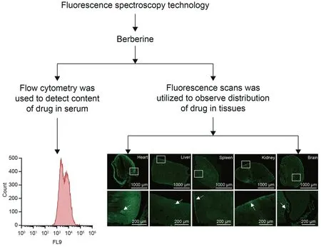
Fig.2.Fluorescence analysis of berberine (BBR)after administration for 2 h in mice.Flow cytometry was used to detect the content of BBR in serum,and fluorescence scans were utilized to observe the distribution of BBR in the heart,liver,spleen,kidney,and brain.
For drug dosage form optimization,fluorescence spectroscopy was used to explore the combination and action relationship between drugs and carriers.Zakaria et al.[30] found that curcumin binds to polylactic-co-glycolic acid through hydrogen bonds and van der Waals forces.It also combines with polydiallyldimethylammonium chloride through hydrophobic interactions,providing a means for validating the binding relationship between the active ingredients of TCM and drug delivery systems.Li et al.[31] verified that the combination of resveratrol and phosvitin significantly increased the solubility of resveratrol using fluorescence spectroscopy.This was based on the fact that drug combination was an effective and reliable method for promoting the full utilization of the pharmacodynamic value of drugs.In addition to assisting in vivo studies,fluorescence spectroscopy plays an important role in content determination and quality control of active ingredients in TCM in vitro.It can be used to describe the fluorescence spectral information of TCM comprehensively,provide complete fingerprint feature information,and analyze the complex components of TCM accurately with the advantages of high sensitivity,good selectivity,and high detection speed [32].
In summary,fluorescence spectroscopy has been widely used in TCM research in vivo and in vitro;it is noninvasive,convenient,sensitive,and rapid.Although the technique is constrained by the presence of fluorophores,this problem can be overcome by the artificial addition of fluorescent labels.However,despite the wide application of this technology,research remains insufficient,and the scope and quantity of detected samples must be further expanded (Table 1).

Table 1 Novel spectroscopic techniques.
3.Photoacoustic imaging technology
In 1880,Alexander Graham Bell discovered that the photoacoustic effect occurs when certain substances absorb light energy to produce specific acoustic signals.Subsequently,the photoacoustic effect was gradually applied to military communications,chemical industry,and other fields;however,its scope of application was limited[33].In the 1960s,with the vigorous development of detection,sensing,and light source technology,photoacoustic imaging technology combined the advantages of optical and ultrasonic imaging to obtain sufficiently high imaging resolution and contrast.Consequently,the technology has been widely used and has attracted attention in the biomedical field[34].The principle of photoacoustic imaging technology is the photoacoustic effect.After a pulse or modulated laser irradiates an object,the object absorbs light energy and converts it into heat energy,and this process is followed by thermal expansion,contraction,and the outward radiation of sound [35].The images collected by photoacoustic imaging technology contain a considerable amount of information,including the morphology and functional information of detected objects.More importantly,photoacoustic imaging technology compared with the use of ordinary light microscopes offers high sensitivity,novel imaging contrast,and high resolution.It is generally an all-optical imaging method that differs from other microscopic techniques[34].Presently,it is popular in the fields of physics,chemistry,biomedicine,and environmental protection;moreover,it has broad application prospects in the medical field(Fig.3)[33-35].Optical imaging interferes with the accuracy of the measurement results owing to the influence of reflected light.In contrast,photoacoustic imaging allows the detection of substances through sound waves and produces a clear image with high resolution,which is two levels higher than that of ordinary optical imaging [36].
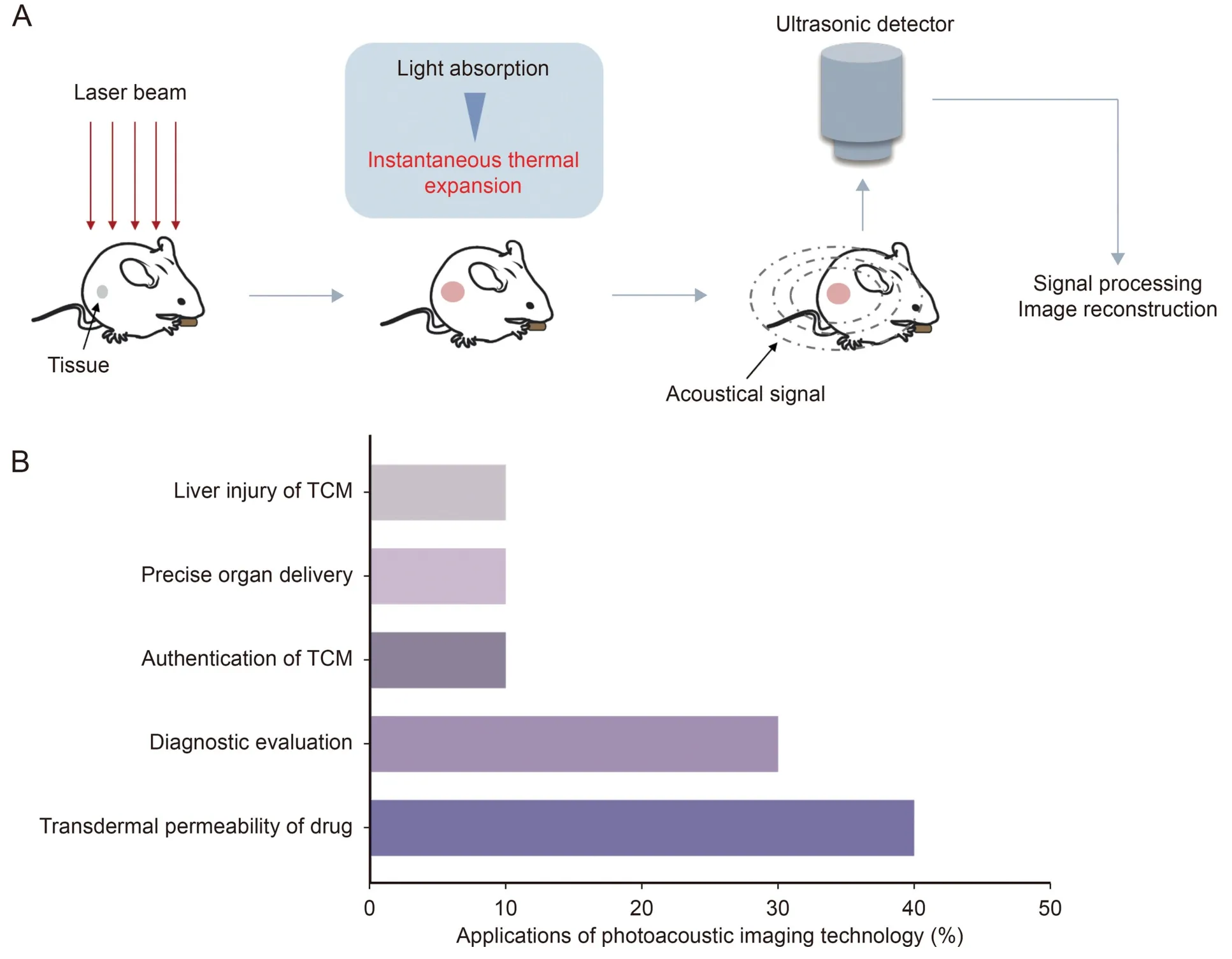
Fig.3.Photoacoustic imaging technology.(A)Principle and(B)applications of photoacoustic imaging technology.TCM:traditional Chinese medicine.Reprinted from Refs.[33-35]with permission.
Preliminary progress has been achieved in the photoacoustic imaging technology research for evaluating drug pharmacodynamics,particularly those of TCM extracts.This technology was used by Ames et al.[37] to assess the anti-inflammatory and percutaneous osmotic effects of fish oil.Mendes et al.[38]used the technology to evaluate the antifungal and nail penetration effects ofthe hydroalcoholic extract ofSapindus saponariaL.pericarps.They also utilized photoacoustic imaging to measure skin permeability with regard to emulsions and evaluated the antioxidant capacityMelochia arenosaextract[39].Photoacoustic imaging technology is also an effective method for evaluating the effect of TCM and active ingredients on wound healing.For example,the technology was applied to determine the effect of the crude extract ofStryphnodendron adstringens[40] andPoincianella pluviosa[41] on diabetic wound healing.Furthermore,the hemodynamic changes in the functional areas of the brains of mice after the administration of BBR could be detected by photoacoustic imaging technology.Further,the visualization of drug-induced cerebral hemodynamic changes was creatively realized [42].The effects of Astragali polysaccharide and curcumin on tumor vascular normalization and antihepatocellular carcinoma were visualized using photoacoustic imaging technology[43].
Optimizing new technology using photoacoustic imaging technology to satisfy the requirements of disease diagnosis and drug quality control is also one of the research topics that has currently attracted considerable attention.Li et al.[44] developed a nearinfrared fluorescent and photoacoustic dual-mode carbon monoxide probe (called QL-CO) that is sensitive and responsive to CO at inflammatory sites,thereby realizing the accurate diagnosis of inflammation.Sun et al.[45] designed a probe (called QY-N) that could be activated by nitric oxide.They combined optoacoustic imaging and near-infrared-II fluorescence imaging technology to detect nitric oxide in the liver for diagnosing herb-induced liver injury.Ouyang et al.[46] devised a novel nanoprobe that could generate infrared fluorescence and photoacoustic signals after activation.Additionally,the combined application of fluorescence spectroscopy and multispectral photoacoustic imaging technology was used to monitor tumor metastasis in mice with breast cancer and evaluate the effect of chemotherapy.Guo et al.[47]developed a dual-wavelength optically resolved photoacoustic laparoscope for precise in vivo drug delivery to achieve drug targeting.In line with other imaging techniques,photoacoustic imaging technology has also been used for enabling TCM to satisfy quality control requirements by identifying the typical photoacoustic spectral features of herbs [48].
In conclusion,photoacoustic imaging technology has facilitated some achievements in the research and application of TCM.This has allowed the evaluation of pharmacology in vivo and visualization of the in vitro quality of TCM,promoting its modernization and application (Table 1).
4.Infrared thermal imaging technology
German astronomer Sir William Herschel first discovered infrared radiation in 1800.Based on the principle of infrared radiation and using infrared thermal imaging technology,an object's thermal radiation infrared-specific band signal can be detected using photoelectric technology.Infrared thermal imaging technology utilizes an infrared detector and an optical imaging objective to capture data from the measured target,thereby obtaining infrared thermography that corresponds to the thermal distribution field of the object's surface [49].This signal can be visualized and used to reflect the temperature distribution of the measured object directly.By the 1960s,infrared thermal imaging was used in modern medicine [50].Infrared thermal imaging was regarded as a novel imaging technology in the 1970s.Unfortunately,owing to technical obstacles,the measurement information was unreliable and not reported to the public.Owing to the expanded application range of the thermal imaging technology,it was practically applied to large-scale temperature measurements during the COVID-19 pandemic.However,the practical applications of thermal imaging may extend beyond what is currently known[51].In the medical field,the infrared radiation signals of the body are collected by thermal imaging systems.These signals are used to assess temperature changes intuitively at different locations of the body and assist in the diagnosis and treatment of diseases [51].In recent decades,the application of infrared thermal imaging technology has gradually extended to the diagnosis,treatment systems,and pharmacodynamic evaluation of TCM [49,52-54] (Fig.4).
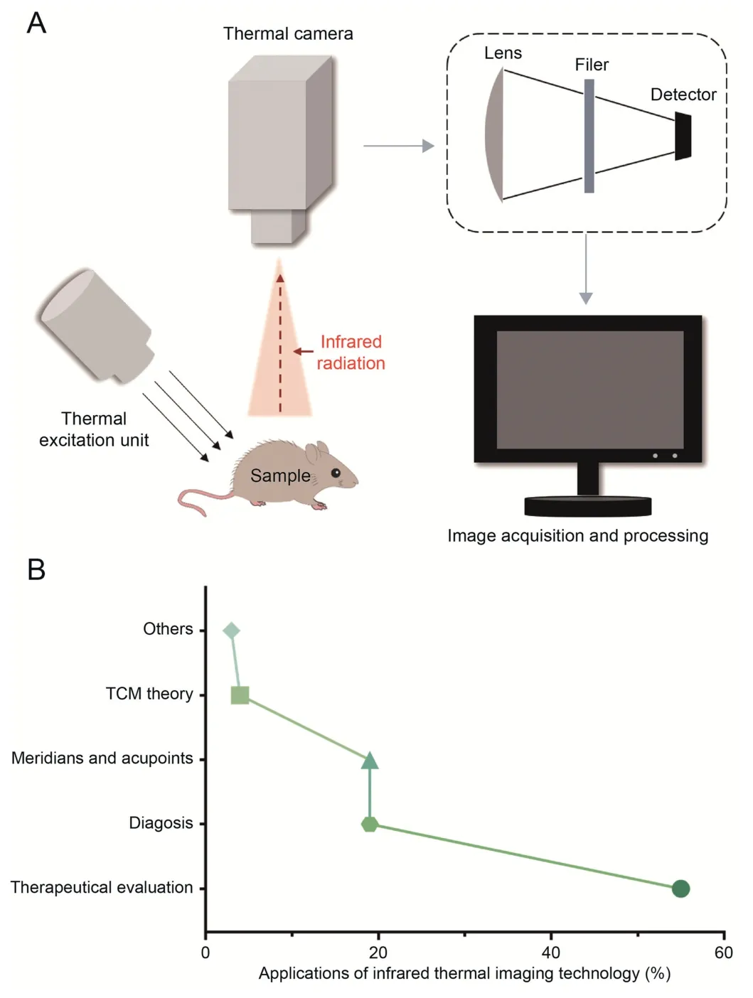
Fig.4.Infrared thermal imaging technology.(A) Principle and (B) applications of infrared thermal imaging technology.TCM: traditional Chinese medicine.
Infrared thermal imaging technology provides a visual strategy for evaluating the efficacy of TCM therapies,particularly acupuncture.The technology was used to clarify the co-localization relationship between acupuncture points and the perforation of cutaneous vessels.To a certain extent,this relationship explains the principle of acupuncture and moxibustion,providing clearer understanding[55].In principle,the effect of laser acupuncture on the small intestine meridian was verified,enabling the visualization of body energy changes caused by acupuncture [56].The same technology was used to explore the concatenative effects of acupuncture at different time points[57,58].Specifically,Cai et al.[59]monitored the thermal changes in the auditory region after acupuncture using an infrared thermal imaging instrument and clarified the mechanism of acupuncture in the treatment of tinnitus.In addition to acupuncture,infrared thermal imaging was used by Liu et al.[60]to monitor the effect of cupping therapy on local skin temperature to select appropriate negative pressure cupping conditions.Therefore,infrared thermal imaging technology links the acupuncture technology of TCM and the diagnosis and treatment evaluation systems of Western medicine.It plays a vital role in promoting the visual evaluation of the efficacy of acupuncture.
Infrared thermal imaging is typically used to evaluate the pharmacodynamic effects of TCM.Fundamentally,infrared thermal imaging technology has been used to determine the fundamental effects of TCM on body phenotypes.For example,Yin et al.[61]demonstrated thatEuodiae FructusandCoptidis Rhizomaaffected the body surface temperature of rats.Furthermore,Song et al.[62]monitored the temperature of pain areas in the shoulders and back of patients with myofascitis using infrared thermal imaging technology and found that Jiang's Huojing decoction could reduce the pain by improving the local blood supply.Similarly,the therapeutic effect of substituting tea for removing dampness and resolving turbidity in hyperuricemia and the improvement effect of Chinese medicine on nonalcoholic fatty liver diseases have been verified[63,64].Therefore,infrared thermal imaging technology provides a new means for evaluating the pharmacological effects of active ingredients and prescriptions in TCM.
Infrared thermal imaging also plays an important role in assisting clinical disease diagnosis and treatment.In recent years,the technology has also been applied to the development of new drugs and preoperative preparations for clinical surgery [65,66].According to one study,the accuracy and reliability of dynamic infrared thermography used in deep inferior epigastric perforator flap breast reconstruction were comparable to those of the well-recognized computed tomography angiography [67].Infrared thermal imaging has also been used to analyze skin tissues and identify tissues whose imaging is uneven to diagnose gynoid lipodystrophy [68].This technology has also promoted the development of drug carrier materials with thermal imaging properties [69].With this updated technology,digital infrared thermography,dynamic infrared thermography,and other derivative technologies were emerged during the development of infrared thermal imaging[66,70].
In summary,infrared thermal imaging technology is a noninvasive means for providing early warning of disease,is highly accurate,and has comprehensive coverage.This technology is anticipated to become one of the key technical means for quantitative TCM and modern medical research.Moreover,it is expected to become the standard for“TCM computed tomography” applied to modern drug research and clinical diagnosis and treatment(Table 1).
5.Laser-induced breakdown spectroscopy technology
Laser-induced breakdown spectroscopy (LIBS) was introduced in the 1960s[71].Since the first ruby laser emerged,the innovation of laser light source technology,optical detection technology,high time resolution measurement technology,imaging technology,and various spectral data processing technologies has continued.This technique uses a laser pulse as a source of excitation to induce the atomic emission spectrum of a laser plasma(Fig.5)[72].Moreover,the LIBS experimental equipment has been constantly updated and has since been applied to metallurgy,archaeology,and cultural relic identification.The LIBS technology was not used in the field of biomedicine until 2004.It has a broad application range because it has a fast detection speed and is capable of direct detection and simultaneous analysis of a variety of elements.Moreover,the LIBS equipment is small and simple to operate [73].The technology is rapid and allows microscopic and multi-element detection using a laser pulse as the excitation source to induce the atomic emission spectrum of laser plasma.Further,LIBS is characterized by fast multi-element detection;a single laser pulse is sufficient to predict the elemental composition of a sample within a few seconds.It is suitable for direct,in situ,online,and remote detection.Moreover,it can achieve rapid evaluation virtually without sample pretreatment [74].Traditional monopulse LIBS produces signals with a short lifetime and low signal-to-noise ratio.With continuous innovations in LIBS,such as double-pulse excitation,magnetic fieldconfined plasma,space-confined plasma,and discharge-enhanced plasma emission spectroscopy,sensitivity has considerably improved [75].With the development of laser and spectral detection technologies,the LIBS technology has become increasingly mature.Fiber coupling and portable and remote sensing instruments have become mainstream,rendering them more suitable for in situ,field,remote,and harsh environmental applications.The LIBS technology has been successfully applied to botany,space exploration,environmental pollution,and other fields.Therefore,it can be used not only in laboratory testing but also in industrial sites,geology,metallurgy,and pharmaceutical fields [76].
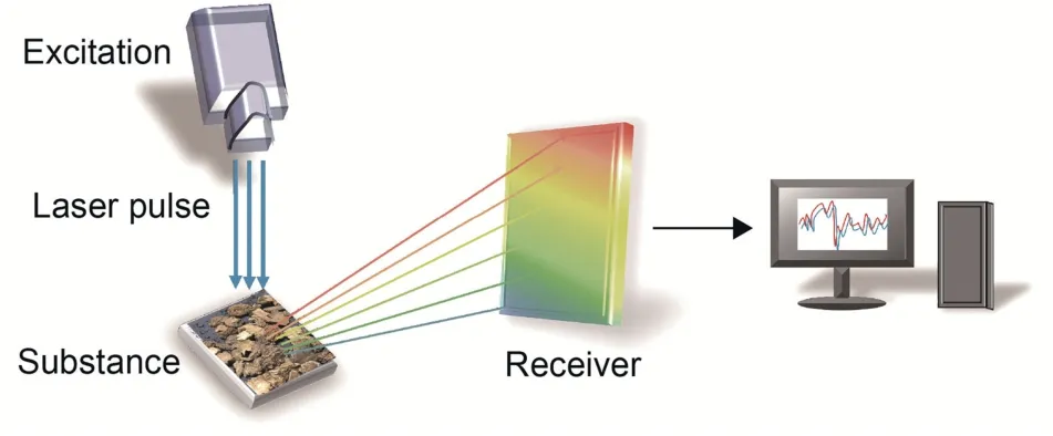
Fig.5.Principle of laser-induced breakdown spectroscopy technology.Reprinted from Ref.[72] with permission.
During the COVID-19 pandemic,LIBS was used to detect viruses and associated particles rapidly.Traditional viral infection detection methods are based on reverse transcription,e.g.,polymerase chain reaction or enzyme-linked immunosorbent assay;however,these techniques are limited because they require special reagents and detection conditions.Moreover,laser spectroscopy techniques,renowned for their noninvasive characteristics and their capacity to facilitate rapid,straightforward,and cost-effective analyses,have demonstrated notable improvements and innovations [75].Clinically,LIBS is currently being considered for detecting viral contents in infectious diseases [77] and identifying bacterial species [78].The detection of viral contents through LIBS can establish a corresponding relationship between different viral contents and characteristic line intensities to achieve quantitative or semiquantitative detection rapidly and effectively.Accordingly,LIBS is primarily used to detect the sources and elements in TCM.Liu et al.[79]used LIBS combined with chemometry to identify the origin of kudzu powder.Zhao et al.[80] identified the adulteration of Chinese yam powder,and Shen et al.[81] identified the nutritional elements ofPanax notoginseng.Therefore,the LIBS experimental method enabled the rapid analysis of the composition and content of TCM,providing a practical method for identifying the authenticity,content,and origin of medicine.The LIBS technology combined with neural networks has been employed to identify the origin ofginseng[82] andGentiana rigescensFranch [83].In addition,the compatibility stability of TCM was tested using LIBS,and the stability of a mixed system obtained by combining cinnabar and realgar in the An-Gong-Niu-Huang Wan treatment was determined by detecting variations in heavy metal elements (arsenic and mercury)[84].The spectral characteristics of heavy metal elements in Tibetan medicine were collected by Liu et al.[85] using LIBS,providing new ideas for the elemental analysis of ethnic medicine.This clearly demonstrates the advantages of the LIBS experimental method in terms of experimental analysis and quantitative identification,fully reflecting the stability and facility of this method.
In summary,with its advantages,LIBS has been widely used in sample element detection,quantitative analysis,medical treatment,cultural relic protection,industry,space exploration,and other related works.However,for LIBS to be applied to real life,it must be appropriately improved to promote the rapid conduct of LIBS experiments (Table 1).
6.Hyperspectral imaging technology
Multispectral imaging technology only allows the continuous imaging of a few bands.To overcome this limitation and satisfy the requirements of space exploration in the field of remote sensing,hyperspectral imaging (HSI) technology was developed in the 1970s.This technology enabled continuous-band imaging with a high resolution of hundreds of nanometers[86].In 1988,the advent of aerial imaging spectrometers marked the development of HSI technology.The technology allows the simultaneous measurement of the visual and spectral information of an object.It also has the advantages of atlas integration and good prediction accuracy.These advantages not only resolve the limitations of traditional technology but also provide more accurate and intuitive information[87].In 1999,HSI was applied to the content analysis of biological tissues[88].After the 20th century,HSI was gradually applied to pathological examinations,early disease screening [89],clinical treatment,and efficacy evaluations[90,91].In the field of medicine,HSI can only be used for initial tablet content analysis[92].Then,it can be used for the quality control and composition analysis of Chinese medicinal materials [93,94].Over the past two decades,HSI derivative technologies (such as quantitative hyperspectral reflectance imaging [95],hyperspectral confocal fluorescence imaging[96],excitation-scanning hyperspectral imaging microscopy [97],dark-field hyperspectral imaging [98],and bedside hyperspectral imaging [98]) have been developed (Fig.6).
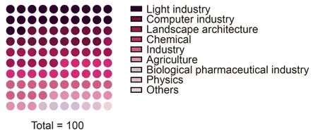
Fig.6.Applications of hyperspectral imaging technology.
The HSI has a wide application range,nondestructive nature,high precision,simple operation,and short consumption time.It is mainly used in TCM to determine the authenticity,origin,sulfur fuming,and content of medicinal materials [99].Xiao et al.[100]jointly applied the HSI and convolutional neural network to identify the origin ofRadix Astragali.The study onRhizome atractvlodis macrocephalaeof Ru et al.[101]and the study onGlycyrrhizae Radix et Rhizomaof Yin et al.[102]adopted the combined data processing technology of visible near-infrared and shortwave-infrared hyperspectral technology to establish a full 3D fusion strategy.The strategy included visible near-infrared and shortwave-infrared fusion,spectral and image fusion,and full data fusion,to identify the origin ofRhizoma Atractylodis Macrocephalae.Similarly,HSI was used to identify the origin of Dendrobium and detect the mannose and polysaccharides it contains[103].Zhang et al.[104]developed a visible near-infrared portable field imaging spectrometer based on the HSI technology to identify sulfur-fumed herbs rather than sun-dried herbs.Unfortunately,the effectiveness of HSI technology is not consistent.Wu et al.[105] attempted to describe the glycyrrhizic acid content in licorice using near-infrared spectroscopy.However,after testing 39 samples of different origins,they found that the technique was not suitable;the quality of the data was not consistent when testing TCM with complex components.As reported by Tankeu et al.[106],the HSI technology can be used to identify adulteratedStephania tetrandra,distinguishing it fromAristolochia fangchiwhen theAristolochia fanghiscontent exceeds 10%.Based on HSI,an original biomedical shortwave-infrared imaging acousto-optical tunable filter system was developed by Batshev et al.[107]to improve spectral sensitivity and imaging quality.The study showed that an identification model with high resolution and prediction accuracy could be established based on HSI.This indicates that HSI can be used as an effective means to provide technical support for the identification and content determination of medicinal materials in the field of TCM;however,certain technical problems must be further resolved.
Briefly,HSI,a technology that provides multidimensional imaging data,has several advantages,such as simple operation,high resolution,high prediction accuracy,wide application range,and atlas integration.Thus,it has excellent application prospects in TCM.However,as an emerging technology,HSI is confronted with considerable challenges in terms of equipment,data processing,and data analysis (Table 1).
7.Surface enhanced Raman spectroscopy (SERS) technology
Raman scattering was discovered in 1928[108].Owing to the low intensity of scattered light,Raman spectroscopy technology rapidly improved in 1960 with the development of laser technology [109].Raman spectroscopy is a molecular structure research and analysis method based on the principle of Raman scattering.It can be used to analyze the molecular vibration,rotation,and other information of a scattering spectrum,which differs from the frequency of incident light.It has been used for TCM identification,origin classification,and content determination.The technique has high reliability,speed,and sensitivity as well as enables real-time qualitative and quantitative analyses in addition to other advantages[110].
Raman spectroscopy has been used for raw material identification,crystal structure characterization,origin identification,content determination,and adulteration discrimination [111].Yin et al.[112] used Raman spectroscopy to monitor the content of liquiritin and glycyrrhizic acid in real time during the extraction of licorice formula particles.Liu et al.[113] employed this spectroscopy to identify the authenticity of pseudoginseng.Raman spectroscopy combined with inductively coupled plasma atomic emission spectroscopy was used by Feng et al.[114] to verify the authenticity of a pilose antler.Accordingly,Raman spectroscopy was also successfully used in identifying TCM materials.
However,the application of Raman spectroscopy is limited by its low detection sensitivity and severe fluorescence interference.In view of this,the SERS technology was derived from Raman spectroscopy.The SERS technology,developed in 1974,used the special surface of certain metals or semiconductors to adsorb relevant molecules or functional groups to enhance the intensity of the Raman spectrum[115].The Raman spectrometer consists of a laser source,a system of collection,a spectrophotometer,and a system of detection.Based on this,SERS is performed by adding gold,silver,and other molecules with resonance Raman effect to the collection system (Fig.7) [116].The SERS technology is one of the most remarkable spectral analysis techniques developed in recent years.It can provide rich chemical molecular structure information and has been widely used in biochemistry,physics,medicine,and other fields owing to its high sensitivity,high selectivity,ease of implementation,and minimal interference from water and fluorescence signals.However,this technology requires trained personnel to conduct disease analysis.Moreover,the required safety standards for disease analysis and equipment limit the application of SERS.
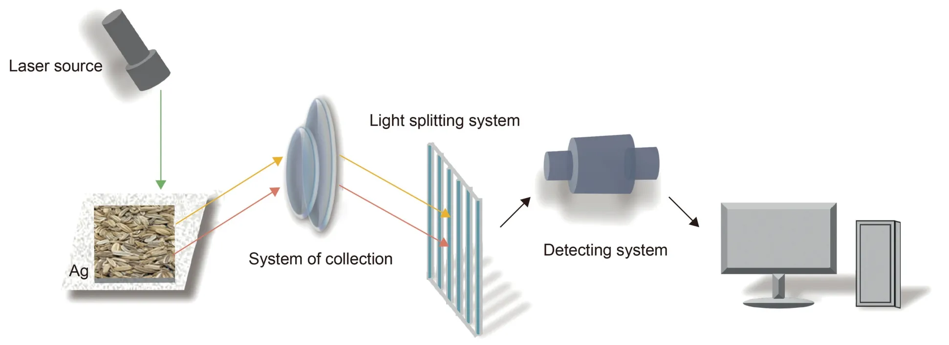
Fig.7.Principle of surface enhanced Raman spectroscopy (SERS).Reprinted from Ref.[116] with permission.
The SERS technology has been applied to the biomedical field since the 1990s[117].As early as 2009,Chen et al.[118]used SERS to analyze the spectrum ofRhizoma Atractylodis Macrocephalaedecoction.Further,they proposed that SERS may be used as a new technique for characterizing TCM decoctions.In 2014,Zhao et al.[119] used a silver nanosphere probe and SERS to detect BBR inCoptis RhizomaandPhellodendron amurense.They also determined the active component contents of TCM by SERS for the first time.Fan et al.[120] designed and synthesized a functional membrane substrate from a Si@Ag@PEI composite to enhance the Raman signal of sulfur dioxide inginseng,Salviae Miltiorrhizae Radix et Rhizoma,and bitter almonds.A 3D-printed headspace extraction device for the detection of sulfur dioxide was fabricated.The SERS technology has also been used to detect pesticide residues,heavy metals,fungomycin,and other pollutants in TCM [121].Ren et al.[122] used dynamic SERS to detect aflatoxin G1 contamination in Coicis Semen.Liu et al.[123]detected the organic pesticide residues inDioscoreae Rhizomausing SERS with a gold nanosol as an enhancement substrate.Sun [124] combined SERS and chemometrics to identify and classify the sources ofCordyceps sinensisandCordyceps militaris.For decades,coherent anti-Stokes Raman spectroscopy [125],Fourier transform Raman spectroscopy [126],tip-enhanced Raman spectroscopy [127],and other spectroscopic techniques have been developed and applied.Resonance Raman spectroscopy is a new Raman spectroscopy technique that has been used to study the interactions between small drug molecules and hemeproteins,such as sodium Danshensu and cytochrome C[128].Our research group used the SERS technology to analyze the origin ofCoptis Rhizoma,parts ofAstragali Radix,and components ofZanthoxyli Radix(Fig.8).Therefore,compared with Raman spectroscopy,SERS has more rapidly developed and covers a wider application range.
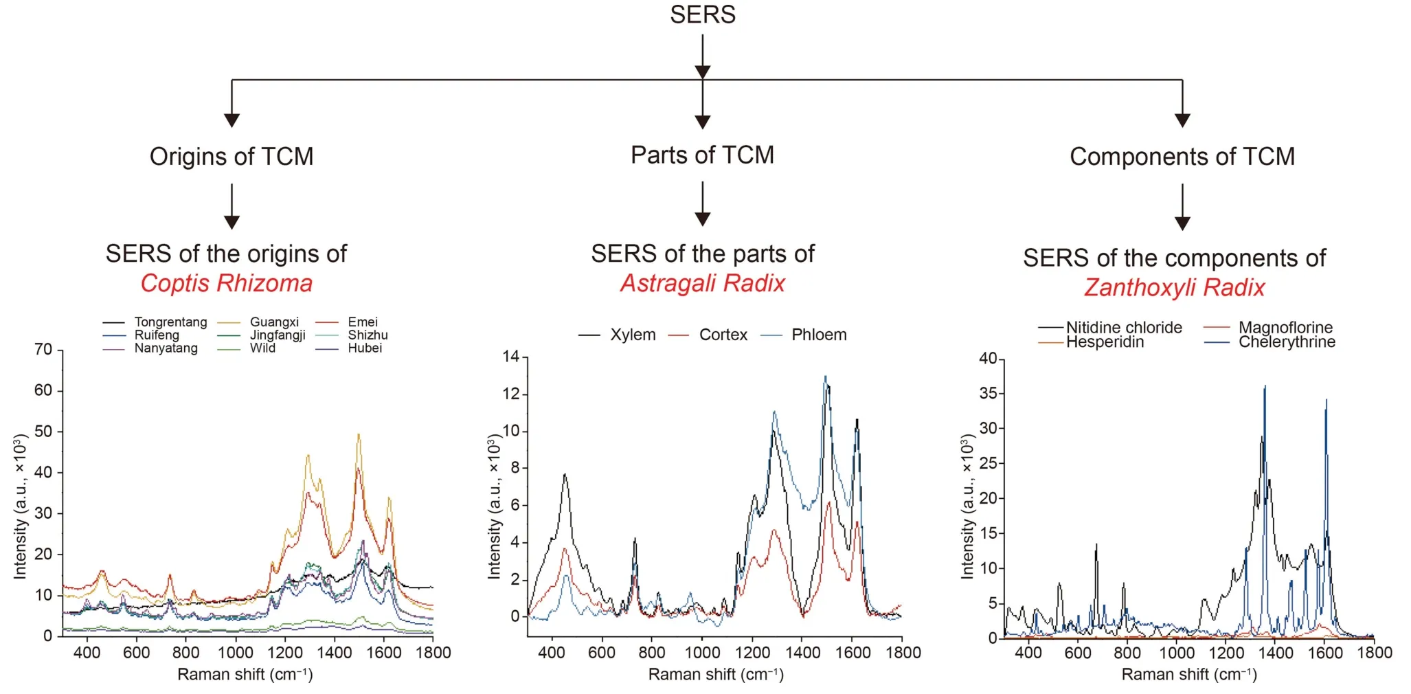
Fig.8.Origins,parts,and components of different types of traditional Chinese medicine (TCM) detected by surface enhanced Raman spectroscopy (SERS).
In summary,the SERS technology derived from Raman spectroscopy significantly promotes the in vitro quality control of TCM.In contrast,other derivative technologies with promising prospects remain in the developmental stage (Table 1).
8.Other spectral imaging techniques
Two-photon imaging has been applied to cell,tissue,and in vivo imaging in TCM studies to evaluate the effects of TCM or its active components on tissue cells and to explore corresponding mechanisms [129].Huang et al.[130] used two-photon time-lapse imaging to monitor the effect of the Chinese patent medicine Xueshuantong on the blood flow of mice with Alzheimer's disease.Two-photon laser scanning microscopy was also used by Liu et al.[131] to monitor the effect of the proprietary Chinese capsule medicine Tongxinluo on cerebral microcirculation in mice with ischemic stroke.In addition to blood flow monitoring,two-photon microscopy was used to image the vagus nerve.Two-photon microscopy was used by Huang et al.[132]to detect calcium currents in the gastroesophageal vagus nerve of mice and explore the antiemetic mechanism of 6-shogaol after administration.Therefore,two-photon spectroscopy is also a technique with considerable potential for evaluating the pharmacodynamic effects of TCM in vivo;evidently,its further development is advantageous.Magnetic resonance spectroscopy imaging(MRSI) is a noninvasive imaging technique that uses the magnetism of hydrogen nuclei to visualize tissue metabolism in vivo [133].Zhang et al.[134] used MRSI to detect the changes in the liver fat content in obese children.Using MRSI,Gu et al.[135]observed the changes in the levels of the brain metabolite(N-acetyl aspartate)during migraine;hence,MRSI is also an in vivo imaging technology.
Matrix-assisted laser desorption/ionization mass spectrometry imaging (MALDI-MSI) can detect the spatial distribution of compounds within tissue samples.It is the most employed MSI tool in clinical research,offering high resolution and sensitivity;moreover,it does not require labeling [136].Because of these characteristics,MALDI-MSI has been widely applied to various fields,such as pharmacokinetics,metabolomics,proteomics,and drug preparation [137].This visualization tool has also been employed in the study of TCM to uncover pharmacological mechanisms.Wang et al.[138] utilized MALDI-MSI to investigate the effects of oral BBR on the brain dopamine imaging intensity.They concluded that BBR acts as a tyrosine hydroxylase agonist inEnterococcus.In another study using MALDI-MSI,Genangeli et al.[139] conducted spatial analyses of the brain,liver,and intestines of rats treated with lentil extract.They discovered a synergistic cholesterol-lowering effect of the lentil extract.
In the future,more accurate and noninvasive imaging technologies will be developed to assist in clinical diagnosis and treatment.
9.Conclusions
The development of a new spectral technology promotes the accurate,noninvasive,and convenient diagnosis and treatment of diseases clinically.This is particularly true with the evaluation of the effectiveness and mechanistic exploration of TCM (such as acupuncture and moxibustion).Based on the research and development of drug use,the application of the new spectral technology promotes quality control,formulation optimization,and mechanistic exploration of Chinese medicinal materials.The nondestructiveness and high sensitivity of the spectral imaging technology in vivo and in vitro are evident advantages.Based on these,the application of spectral imaging is anticipated to promote future medical research as well as scientific and technological explorations.
CRediT author statement
Qi Wang:Investigation,Methodology,Validation,Writing -Original draft preparation;Yong Zhang:Formal analysis,Investigation,Resources,Methodology,Writing -Original draft preparation;Baofeng Yang:Conceptualization,Project administration,Supervision,Writing -Reviewing and Editing.
Declaration of competing interest
The authors declare that there are no conflicts of interest.
Acknowledgments
This work was supported by the National Key R&D Program of China (Grant No.: 2017YFC1702003) and the Chinese Academy of Medical Sciences Innovation Fund for Medical Sciences(Grant No.:2019-12M-5-078).
杂志排行
Journal of Pharmaceutical Analysis的其它文章
- Stage-specific treatment of colorectal cancer:A microRNA-nanocomposite approach
- Biosensors for waterborne virus detection: Challenges and strategies
- Oridonin restores hepatic lipid homeostasis in an LXRα-ATGL/EPT1 axis-dependent manner
- Ginsenoside Rg5 enhances the radiosensitivity of lung adenocarcinoma via reducing HSP90-CDC37 interaction and promoting client protein degradation
- Canonical transient receptor potential channel 1 aggravates myocardial ischemia-and-reperfusion injury by upregulating reactive oxygen species
- Nanoscale coordination polymer Fe-DMY downregulatingPoldip2-Nox4-H2O2 pathway and alleviating diabetic retinopathy
