Simultaneous determination of ten nucleosides and bases in Ganoderma by micellar electrokinetic chromatography
2022-11-28FeiyShengSongsongWngXioLuoJinboXioLinfengHuPengLi
Feiy Sheng, Songsong Wng, Xio Luo, Jinbo Xio, Linfeng Hu*, Peng Li,*
a State Key Laboratory of Quality Research in Chinese Medicine, Institute of Chinese Medical Sciences, University of Macau, Macao, 999078, China
b School of Basic Medical Sciences, Chengdu University, Chengdu 610106, China
c Chengdu Institute for Food and Drug Control, Chengdu, 610045, China
d Chongqing Key Laboratory of Medicinal Resources in the Three Gorges Reservoir Region, School of Biological and Chemical Engineering,Chongqing University of Education, Chongqing, 400067 China
Keywords:
Ganoderma
Nucleosides
Bases
Micellar electrokinetic chromatography
Quality assessment
A B S T R A C T
Ganoderma (lingzhi) is a famous herbal medicine and edible supplement in oriental countries for a long history. In this study, a simple micellar electrokinetic chromatography (MEKC) method was established for the analysis of nucleosides and bases, the major bioactive components in Ganoderma for the first time. By optimizing the borate concentration, the sodium dodecyl sulfate (SDS) concentration and the pH value of running buffer, 10 nucleosides and bases achieved an ideal separation. In real sample analysis, the developed method was successfully used to determine the 10 target analytes in 23 batches of Ganoderma samples from different regions. Results indicated that contents of 10 investigated analytes in each sample showed obvious variation. The principal components analysis (PCA) and hierarchical cluster analysis (HCA) analysis classified the samples into three groups, and the HCA tree visualized the relationships which was mainly contributed by geographical partition. The results indicated geographical origin to be an important factor that affect the accumulation of nucleosides and bases in Ganoderma. In summary, this study provides a simple and practical strategy for quality assessment and cultivation reference of Ganoderma.
1. Introduction
Ganoderma(lingzhi) is a famous herbal medicine and edible supplement in oriental countries for a long history [1,2]. According toChinese Pharmacopeia, the medicinal use ofGanodermainvolve invigorating Qi, easing the mind, relieving cough and asthma,alleviating neurasthenia and debility from prolonged illness, anorexia,dizziness, and insomnia. A number of pharmacological studies have reported various biological activities ofGanoderma[2,3], such as antitumor [4,5], anti-oxidation [6,7], anti-bacterial [8], anti-diabetes [9-11],inhibiting platelet aggregation [12], and immune-regulation [13,14].Clinically, it is mainly recognized as an alternative adjuvant for the treatment of leukemia, carcinoma, diabetes, hepatitis and bronchitis [2,8].Nowadays, cultivatedGanodermaand its related extracts have been processed to be commercial products as functional foods or nutraceuticals [15,16], such asGanodermatea,Ganodermacapsules,Ganodermabuccal tablets.
To date, a great deal of chemical analysis has clarified thatGanodermacontained a variety of constituents, including triterpenes,polysaccharides, nucleosides, etc. [17]. As fundamental parts ofGanoderma, nucleosides and bases were reported to possess various pharmacological activities [18-20]. For instance, uridine and uracil could lower the serum aldolase level of mice in myotonia experiment.Adenosine could inhibit platelet aggregation [12]. Meanwhile,adenosine has been generally used as a marker for quality control of another precious herb,Cordyceps[20]. Therefore, nucleosides and their bases are believed important toGanoderma, especially on quality assessment.
Previously, several analytical methods including high performance liquid chromatography (HPLC), ultra performance liquid chromatography (UPLC), liquid chromatography/mass spectrometry (LC/MS) and nuclear magnetic resonance (NMR) have been applied for the quality analysis ofGanoderma[21-26]. The LC-based methods were highly repeatable and sensitive; the MS and NMR related methods were extremely precise in identification and quantification of constituents. However, those approaches also showed some drawbacks, such as the large consumption of organic solvents or high demand of apparatus. Capillary electrophoresis (CE),characterized by high resolution, separation efficiency, and minimal solvents consumption, has been introduced to meet an environmental friendly and cost-effective alternative [27,28]. Micellar electrokinetic chromatography (MEKC) is a typical branch of CE, in which the surfactant above the critical micelle concentration is added to running buffer to form micelles. It is adequate to separate both charged and neutral ingredients in herbal medicine with high selectivity and micelle system stability [15,27].
In this work, the MEKC method was optimized via assessing the effects of buffer salt, sodium dodecyl sulfate (SDS), and buffer pH.Ten nucleosides and bases, including cytosine, hypoxanthine, adenine,guanine, guanosine, adenosine, uracil, thymine, cytidine and uridine in 23 batches ofGanodermasamples were simultaneously determined by the optimized method.
2. Materials and methods
2.1 Materials
Boric acid, SDS (purity ≥ 98% , AR grade), sodium hydroxide were purchased from Aladdin (Shanghai, China). All stock solutions were prepared and diluted using deionized water (18.2 MΩ·cm) from Milli-Q Advantage A10 system (Merck, Darmstadt, Germany).
Ten of analyte standards were purchased from Mansite Bio-Tech Co., Ltd (Chengdu, China). 23 batches ofGanodermawere purchased from different herbal companies or Hehuachi Chinese herbal medicine market (Chengdu, China). All theGanodermawere identified by Dr. Luo Xiao who comes from Chengdu Institute for Food and Drug Control. The two samples number 12 and 14 are identified asGanoderma sinenseZhao, Xu et Zhang, and the others samples belongs toGanoderma lucidum(Leyss. ex Fr.) Karst.
2.2 Apparatus
All separation experiments were performed on Beckman MDQ P/A CE MDQ electrophoresis system equipped with a photo diode array (PDA) detector (Beckman coulter, USA). Data were acquired by 32 Karat system version 8.0 (Beckman coulter, USA). Fusedsilica capillary were purchased from Yongnian Company (Hebei,China). The CE capillary column were lab made, with 75 cm total length (65 cm to detector) and 75 µm I.D. It was conditioned by rinsing with 0.1 mol/L NaOH, deionized water and running buffer sequentially before use.
Standards dissolution, sample extraction and degassing were carried out on ultrasonic cleaner (Branson, Danbury, USA).Lyophilized sample extracts were freeze-dried by ilShin TFD lyophilizer (ilShin, Korea).
2.3 Preparation of sample and standard solutions
EachGanodermasample was crushed to powder. An aliquot of 2.00 g powder was accurately weighed and mixed with 20 mL water. The suspension was soaked for 30 min at room temperature,and then ultrasonic extracted for 30 min. The extract was centrifuged at 3 000 r/min for 10 min. Then the supernatant was collected and frozen at –80oC for 12 h. After lyophilization, each dried sample was redissolved with 2 mL water, then centrifuged at 14 500 r/min for 10 min. The supernatant was filtered through 0.22 µm membrane for MEKC separation.
Individual standards were accurately weighted and firstly diluted to about 10 mg/mL in deionized water as stock solutions. The mixture of standards was prepared with appropriate amounts of individual standard solutions in water to desired concentration. All the solutions were kept in a refrigerator at 4oC.
2.4 Electrophoretic procedure
Capillary column was conditioned by sequential rinsing with water and running buffer for 5 min respectively before each separation. Both inlet and outlet running buffer were replaced per injection. In all experiments of this study, absorbance was monitored at 254 nm, separation voltage was set at 16 kV, capillary temperature was 25oC. Sample introduction was performed by pressure injection for 5 s under 0.5 psi. Buffers were filtered through 0.22 µm membrane and ultrasonic degassed before use.
3. Results and discussion
3.1 Optimization of separation conditions
A mixture solution of the 10 standards was used as sample for optimization of MEKC conditions. Many factors could affect MEKC separation, mainly including resolution, sensitivity, peak shape,and migration time. Firstly, we used phosphate buffer to define a preliminary pH for running buffer salt selection, which is pH 8.5.Based on this pH, several buffer salts, namely phosphate, borax,borate were tested to determine the most adequate running buffer.The results showed that borate was the best choice as the buffer salt since it had good interaction with molecules containing polyhydroxyl groups (such as guanosine, adenosine, cytidine and uridine) and it could bear a higher SDS concentration with a lower current comparing with borax [29].
During method development, the impacts of borate concentration on resolution between each peaks and overall analysis time were considered initially. As illustrated in Fig. 1, a higher concentration of borate in the running buffer was found to increase the resolution of the analytes. When borate concentration reached to 500 mmol/L, the analytes were mostly separated except for guanosine and adenosine(Fig. 1B). Although the increase of borate concentration could improve the resolution of two analytes, guanosine and adenosine still could not achieve a baseline separation. Moreover, the higher concentration of borate obviously prolonged the overall runtime(Figs. 1C and 1D). Therefore, 500 mmol/L was selected to be the optimum concentration of running buffer salt.

Fig. 1 Influence of borate concentration on the separation of 10 analyte standards using a 400 mmol/L (A), 500 mmol/L (B), 600 mmol/L (C),700 mmol/L (D) boric acid in BGE at pH 8.5. 1, Cytosin; 2, Hypoxanthine; 3,Adenine; 4, Guanine; 5, Guanosine; 6, Adenosine; 7, Uracil; 8, Thymine; 9,Cytidine; 10, Uridine. The same with Fig. 2 to Fig. 4.
In order to improve the separation of the nucleosides and bases at the given pH, advantage was taken of solute partition into the interior of a micelle. SDS is the most common used anionic surfactant to produce micelles acting as a pseudo-stationary phase in MEKC since the charge on the fused silica capillary wall is also negative and the surfactant electrostatic adsorption will not occur to the walls.Retention and resolution were affected by the presence of surfactant at varying concentrations. After adding negatively charged SDS micelles to the background electrolyte (BGE), the migration behavior of 10 analytes changed obviously. Particularly, the partially overlapped peaks of guanosine and adenosine could be well separated from each other. But unfortunately, the peaks of their bases, i.e. guanine and adenine entangled together in the presence of lower concentrations of SDS (Figs. 2A and 2B). Although these two analytes could be well separated along with the increased SDS concentration, the peak of adenine would be overlapped with the hypoxanthine peak when SDS concentration was up to 180 mmol/L (Fig. 2D). Thus, we chose 150 mmol/L SDS as micelle producer in running buffer to carry the target analytes. The overlapping peaks of adenosine and uracil would be optimized in the following experiments.

Fig. 2 Influence of SDS concentration on the separation of 10 analyte standards using 500 mmol/L boric acid with 90 mmol/L (A), 120 mmol/L (B),150 mmol/L (C), 180 mmol/L (D) SDS in BGE at pH 8.5.
As shown in Fig. 3, pH value was an extremely significant factor that affected the quality of the electropherogram. Herein,the dependence of migration time on the buffer pH value was demonstrated by the increase of pH in the range of 8.4-9.2 leading to the overall migration time delay, especially for uracil and the nucleoside uridine. This was might due to the partial solutes ionization to negatively charged species. At pH 9.0, all the peaks were sharp and fully separated (Fig. 3D), while buffers at the other 4 pH values could not provide baseline separation and better peak shapes. Thus 9.0 was chosen as the optimum pH value for this method.

Fig. 3 Influence of pH value on the separation of 10 analyte standards using 500 mmol/L boric acid with 150 mmol/L SDS in BGE at pH 8.4 (A), 8.6 (B),8.8 (C), 9.0 (D), 9.2 (E).
In addition, the effects of the organic additives, ACN and MeOH, were also investigated over a range of 5% -20% . However,no substantial promotion was achieved on resolution of analytes.Moreover, the running buffer without ACN or MeOH can avoid unstable influence caused by volatilization of organic additives.
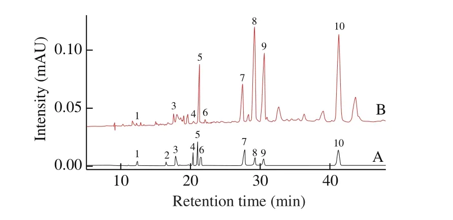
Based on the results mentioned above, the optimum buffer conditions for the separation of the 10 analytes were determined as follows: 500 mmol/L boric acid buffer with 150 mmol/L SDS at pH 9.0.A typical MEKC chromatogram of the 10 analytes under the described conditions was shown in Fig. 4A.

Fig. 4 Representative electrokinetic chromatograms of the 10 mixed standards (A) and Ganoderma sample (B).
3.2 Method validation
The optimized MKEC method has been validated by exploring the linearity, repeatability and recovery.
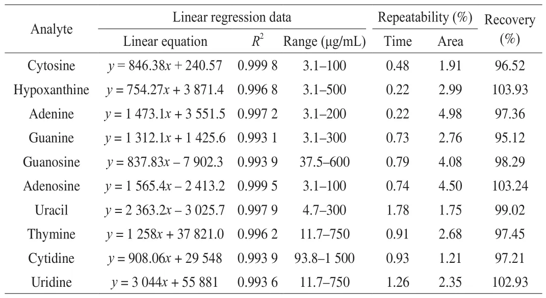
Standard stock solutions of the 10 analytes were individually diluted at specific percentage into a standards mixture with 20% MeOH. Each calibration curves were established by plotting 6 integrated peak areas and corresponding concentrations. As indicated in Table 1, correlation coefficients (R2) ranged from 0.993 1 to 0.999 8, indicating good linearity.

Table 1Linear regression data, repeatability and recovery of investigated analytes from Ganoderma.
Repeatability of the method was tested by running the standards mixture under optimized conditions for 5 replicates within one day.As shown in Table 1, the migration time of each analytes did not vary greatly, with relative standard deviation (RSD) no more than 1.78% .The RSD values of peak area of the 10 analytes were lower than 5% .The RSD was calculated using the following equation:

The recoveries were used to evaluate the accuracy of the method.Three different concentrations of standards were respectively spiked into the real samples whose contents of the investigated analytes were known. The mixture solutions were then analyzed in triplicate by the established MEKC method. As shown in Table 1, the values of recovery varied from 95.12% to 103.93% .
These results indicated that this method was repeatable and accurate for quantitative determination of nucleosides and bases inGanoderma.
3.3 Application of analysis of real samples
Under the optimized conditions, 23 batches ofGanodermacollected from different locations were subjected to analysis.Representative electrokinetic chromatogram of theGanodermasamples was illustrated in Fig. 4B. Each investigated analyte in real samples was identified by comparing migration time with those of corresponding standards. And standard spiking was also performed for accurate identifications in some samples with complex matrix. As shown in Fig. 4, the standards mixture andGanodermasamples could be well separated with high resolution. The contents of 10 target analytes in each sample were calculated and summarized in Table 2.

Table 2Contents (μg/g) of 10 investigated analytes in 23 batches of Ganoderma samples (n = 3).
According to the data, the contents of investigated analytes in different samples showed obvious variation. Five compounds,including guanosine, uracil, thymine, cytidine and uridine, were generally contained in differentGanodermaherbs, especially the contents of guanosine and cytidine were abundant. However, cytosine was hardly detected in the samples which originated in Yunnan and Tibet. Neither adenine nor adenosine contained inGanodermacollected from Tibet and northeast of China. In contrast, the contents of hypoxanthine and guanine in samples from northeast of China were generally higher than that in most other regions, such as Yunnan,Tibet, Jiangxi and Hubei provinces.
As multivariate analysis techniques, PCA and HCA are widely used in food and herb quality evaluation since they could visualize similarities or differences within multivariate data [22,24,30]. To further explore the relationship of the given samples collected from various regions, HCA was carried out based on PCA using SIMCA 13.0 software. Fig. 5A illustrated the distribution of 23 samples in 3D scatter plot based on the first three principle components with over 68.7% of the whole variance. It can be seen that all the loadings were divided into three groups: group 1 (sample 1–11), group 2(sample 12–17), and the group 3 (sample 18–23). Then the HCA tree (Fig. 5B) was consequent to visualize the result of hierarchical clustering calculation. It clearly demonstrated the three main branches and revealed the geographical relationships of 23 samples. Group 1(the green line) was formed byGanodermaherbs from Yunnan,Sichuan, and Tibet, which located in southwest of China. Group 2(the blue line) mainly consisted of samples grown in Jiangxi, Hubei,Hunan and Anhui provinces which mainly belong to middle part of China. Similar cluster was also found in group 3 (the red line), whose samples were mostly distributed in north part, Shandong province and northeast of China. These results revealed that the geographical origin might be the exclusive factor affecting nucleosides and bases accumulation inGanoderma.

Fig. 5 Classification analysis of Ganoderma samples from different regions.Scatter plot based on the first three principle components obtained by PCA (A);HCA tree illustration based on PCA (B).
4. Conclusion
In this study, a novel and simple MEKC method was proposed for the analysis of 10 nucleosides and bases inGanodermaherb for the first time. Under the optimized conditions, the established method demonstrated superiority in terms of good separation and sensitivity for quality analysis. The developed method was successfully used to determine 10 target compounds in 23 batches ofGanodermasamples.Furthermore, PCA and HCA were applied to quality classification of the given samples. The clustering results indicated that geographical origin is the significant factor affecting accumulation of nucleosides and bases inGanoderma. Therefore, these nucleosides and bases are promising features in terms of the distribution and quality assessment ofGanodermaherb. This strategy provides a practical tool for quality control and cultivation reference ofGanodermaherb.
Declaration of competing interest
The authors declare no conflicts of interests.
Acknowledgments
This work was financially supported by the Research Committee of the University of Macau (MYRG2018-00239-ICMS and MYRG2014-00089-ICMS-QRCM), the Macau Science and Technology Development Fund (162/2017/A3), and Guangzhou International Science and Technology Cooperation Project (File no.201807010044).
杂志排行
食品科学与人类健康(英文)的其它文章
- Potential application of proteolysis targeting chimera (PROTAC) modification technology in natural products for their targeted protein degradation
- Effects of vegetarian diet-associated nutrients on gut microbiota and intestinal physiology
- Recent progress in preventive effect of collagen peptides on photoaging skin and action mechanism
- Perspectives on diacylglycerol-induced improvement of insulin sensitivity in type 2 diabetes
- Effect of bacteriocin-producing Pediococcus acidilactici strains on the immune system and intestinal flora of normal mice
- A new Lactobacillus gasseri strain HMV18 inhibits the growth of pathogenic bacteria
