A new Lactobacillus gasseri strain HMV18 inhibits the growth of pathogenic bacteria
2022-11-28XingGoZixunWngXingLiXiolingZhngShengqingDuMiomioJibDilunHuXinxinJiBinCongYnZhngChunlingSongZhouJunZhng
Xing Go, Zixun Wng, Xing Li, Xioling Zhng, Shengqing Du,Miomio Jib,, Dilun Hu, Xinxin Ji,b,*, Bin Cong*, Yn Zhng*,Chunling M Song Zhou, Jun Zhng
a Department of Pathogen Biology, Institute of Basic Medicine, Hebei Medical University, Shijiazhuang 050017, China
b Research Unit of Digestive Tract Microecosystem Pharmacology and Toxicology, Chinese Academy of Medical Sciences, Shijiazhuang 050017, China
c Institute of Basic Medicine, Hebei Medical University, Shijiazhuang 050017, China
d Shijiazhuang Great Wall Integrated Traditional Chinese and Western Medicine Hospital, Shijiazhuang 050035, China
e Hebei Food Safety Key Laboratory, Hebei Food Inspection and Research Institute, Shijiazhuang 050227, China
f College of Forensic Medicine, Hebei Key Laboratory of Forensic Medicine, Collaborative Innovation Center of Forensic Medical Molecular Identification,Hebei Medical University, Shijiazhuang 050017, China
Keywords:
Lactobacillus gasseri
Antagonism
Bacteria
Safety evaluation
A B S T R A C T
To search for a new eco-friendly therapy for infectious disease caused by Escherichia coli, Staphylococcus aureus or Klebsiella oxytoca, we collected the vaginal swabs from healthy women, screened for Lactobacillus and found a strain repressing the growth of pathogenic bacteria. The new isolate was identified as L. gasseri by the colony morphology, Gram staining, biochemical reactions and confirmed by the 16S rDNA sequencing.The HMV18 strain inhibited the growth of food-borne pathogens such as E. coli, S. aureus and K. oxytoca. The HMV18 strain was sensitive to penicillin, ampicillin, erythromycin, tetracycline and chloramphenicol. The HMV18 strain produced α-hemolysis. Pathological histology of the mice ileum showed that the mucosa, villi,lamina propria and crypt depth remained intact and there was no inflammation or hyperemia in the L. gasseri HMV18 gavaged group. L. gasseri HMV18 could not up-regulate inflammatory cytokines level of plasma. All the results suggested L. gasseri HMV18 is a candidate probiotic to be an additive for food preservation or drug to prevent food-borne diseases.
1. Introduction
Probiotics are defined as “live microorganisms, which when administered in adequate amounts, confer a health benefit upon the host” by FAO/WHO in 2002. Probiotic microorganisms are closely related to human health [1], such as maintenance of mucosal homeostasis [2], proper activity of the immune system [3], part of normal flora [4], therapeutic applications for infectious disease [5],IBD [6], allergy [7,8]and improving metabolic diseases [9,10], and so on. Many of the well-defined probiotic strains areLactobacillus,andLactobacillus gasseriis one of this genus.L. gasserican be found at the oral cavity, gastrointestinal tract (GIT), vagina and breast milk.It was recognized as one of the first bacteria to colonize the GIT withBifidobacteriumand contribute to the formation of the anaerobic conditions in GIT [11-13].
L. gasserihas significant potential for probiotic application by fulfilling 11 criteria such as: human origin, nonpathogenic,nontoxigenic and noninvasive, devoid of transmissible antibiotic resistance genes, resistance to technological processes, demonstrated acid and bile tolerance, adhesion to epithelial tissue, transient persistence in the GIT, produce antimicrobial substances, antagonize pathogens and prevent infection, modulate immune responses and positively influence metabolic activity [14].
Escherichia coli,Staphylococcus aureusandKlebsiella oxytocaor their toxins always cause foodborne diseases by contaminating foods [15,16]. These bacteria can be antibiotic resistant, highly virulent,and high expense for the medication [17-19]. VariousLactobacillushave been found to be active in the antagonism of these pathogens and applied for the food preservation. Cano and Perdigon [20]reported thatL. caseiCRL 431 preventedSalmonellaandE. coliinfections in mice. Itoh et al. [21]reported thatL. gasseriLA 39 isolated from infant feces, had the widest inhibitory activity againstS. aureus,B. cereus, andListeria monocytogenesby the bacteriocin Gassericin A. Rybalchenko et al. [22]reported thatL. fermentum97 showed antimicrobial activity towards enterotoxin producingS. aureusandK. oxytoca.E. coli,K. oxytocaandS. aureusare not only food borne pathogens, but also normal flora which may be present at vagina. Since vaginal is a well-known reservoir ofLactobacillus, it would be more efficient to isolateLactobacillusfrom vagina.
In this article, we describe the characteristics of a new isolateL. gasseriHMV18 from the vagina of a healthy woman. We also clarify its inhibitory activity againstE. coli,S. aureusandK. oxytocafor its potential application for treatment of foodborne diseases caused by these pathogens.
2. Materials and methods
2.1 Materials
LB medium was used for culturing of indicator pathogens.E. coli,S. aureusandK. oxytocawere kept in this lab. Rogosa and Sharpe (MRS) medium was used for isolating or culturing ofLactobacillus. MRS broth was prepared according to ATCC Medium 416. Minor modification was made as follows: 10 g/L tryptone, 10 g/L beef extract, 5 g/L yeast extract, 20 g/L glucose, 1 mL Tween 80, 2 g/L K2HPO4, 5 g/L sodium acetate, 2 g/L ammonium citrate dibasic,0.2 g/L MgSO4·7H2O, 0.05 g/L MnSO4·4H2O, final pH 7.0, autoclave at 121 °C for 20 min. Blood agar plate was broth agar containing 5% defibred sheep blood. Tryptone and yeast extract were from Oxoid Ltd. (Basingstoke, Hampshire, UK). Beef extract, glucose and agarose were from Solarbio life science (Beijing, China). Biochemical reactions kits for test LAB were purchased from Hopebio (Qingdao,China). Antimicrobial Susceptibility Testing Kit for 30 kinds of antibiotics was purchased from Hangzhou Microbial Reagent Co.,Ltd. (Hangzhou, China).
2.2 Experimental methods
2.2.1 Isolation and screening of lactic acid bacteria (LAB)strains
Healthy women within age 22-39 with regular menstrual cycles were the volunteers. 100 female vaginal swabs were collected in Shijiazhuang Great Wall Integrated Traditional Chinese and Western Medicine Hospital in 2019. The exclusion criteria are the patients who had taken antibiotics in the past month or had diseases such as vaginitis, cancer, hepatitis B, hepatitis C, syphilis, AIDS, or diabetes.
The vaginal swab was inserted approximately 2 cm into the vagina. The swab was rolled on the wall below cervix for about 5 s.Taken out the swab and directly streaked on the MRS agar plates, and incubated for 72 h at 37 °C under anaerobic conditions.Lactobacillusstrains were separated by fractionated streaking techniques. The colonies were purified by at least three streaking. The grey smooth colonies were picked up and inoculated into 10 mL MRS broth in a screw-capped tube for anaerobic growth, and incubated for 24 h at 37 °C. Bacterial cells were concentrated by centrifugation, resuspended in 1 mL MRS broth containing 15% glycerol and stored at -80 °C.
2.2.2 Growth properties
The newL. gasseriHMV18 strain was inoculated in MRS medium and grown under anaerobic conditions as described in section 2.2.1. OD600nmwas recorded every hour from 0 to 30 h to plot the growth curve by Macy UV-1700PC (Shanghai, China). 105cells/mL was inoculated to start the growth at time point 0. Data plotted in the growth curve was the mean from three isolates.
The pH value at each time point was also recorded.
2.2.3 Identification of LAB
The identification of the new isolates was accomplished by morphology, biochemical and molecular methods. Firstly, the colony was made to a smear on a glass slide. Gram staining was conducted using the Rapid Gram Staining Kit (BASO, Zhuhai, China) according to the manufacturer’s instruction. The morphology was observed under 1 000 × microscope. The scanning electron microscopy (SEM)was also applied for the morphology observation. Specimens for SEM were prepared withL. gasseriHMV18 strain grown overnight in either MRS broth or MRS agar. The above procedure was done by the Electron Microscope Center of Hebei Medical University. Specimens were observed with an S-3500N scanning electron microscope(Hitachi, Tokyo, Japan).
Secondly, biochemical reactions were conducted by VITEK-2 System (bioMérieux, France). The biochemical identity for LAB was confirmed by the Hopebio LAB Biochemical Test Kit (Qingdao,China) according to the instruction manuals.
Thirdly, 16S rRNA analysis was also conducted for LAB strain identification. The genomic DNA was extracted from the 10 mL of LAB culture according to the instruction of DNAiso reagent (Takara,Dalian, China). The purification and concentration of the extracted genomic DNA were determined by Nanodrop2000C (Thermo Fisher Scientific Inc., USA). Genomic DNA was used as the template for polymerase chain reaction (PCR) (Multigene Optimax, Labnet International Inc., USA).
16S rDNA samples of the new isolates was amplified by PCR using universal bacterial 16S rDNA primer pair 27F(5’-AGAGTTTGATCCTGGCTCA-3’) and RT16U6 (5’-ATTGTAGCACGTGTGTAGC-3’) [23]. PCR amplification program comprised by predenaturation at 94 °C for 2 min, 30 cycles with denaturation at 94 °C for 30 s, annealing at 55 °C for 30 s and extension at 72 °C for 2 min, further extension at 72 °C for 10 min,and stored at 4 °C. The amplified product was tested by agarose electrophoresis and then sent to Sangon Co., Ltd. (Shanghai, China)for sequencing. The BLAST program (NCBI, USA) was used for sequence comparison. Phylogenetic and molecular evolutionary analyses were inferred using the Neighbor-Joining method [24].There were a total of 1 159 positions in the final dataset. Evolutionary analyses were conducted in MEGA7 [25].
2.2.4 Preparation of LAB cell free supernatant
L. gasseriHMV18 was cultured for 24 h to reach the stationary growth phase and harvested by centrifuged at 5 000 r/min for 10 min at 4 °C (Centrifuge 5804R, Beckman, USA). Cell pellets and the supernatant were separated. The supernatant was passed through a Millipore 0.22 μm filter (Cork, IRL).
2.2.5 Pathogen susceptibility assay
The Oxford cup method was conducted for the pathogen antagonizing test, with details as follows: The pathogenic bacteriaE. coli,S. aureusandK. oxytocawere grown in LB liquid medium,harvested in the exponential phase, washed with PBS and diluted to 106CFU/mL. 20 mL LB agar with 1.5% agarose was poured into each petri dish. After the agar was solidified, 100 μL of the diluted pathogen was spread on the surface of each plate. Then the Oxford cups were placed on the top of each plate. Three Oxford cups were placed on each plate with equal distance. 200 μL of the HMV18 cells free supernatantwas added into a cup. Then the plates were placed at 4 °C for 24 h for the absorbance of the tested supernatants, and then were transferred into the incubator at 37 °C for 24 h for the bacteria growth.
2.2.6 Genome sequencing, genome annotation and the transferable antibiotic risk analysis
The genome ofL. gasseristrain HMV18 wasde novosequenced by OE Biotech. Co., Ltd. (Shanghai, China) using the Pacbio Sequel platform. To annotate the protein functions, amino acid sequences were compared with NCBI GenBank (https://www.ncbi.nlm.nih.gov/) for NR analysis using Diamond software [26]. To analyze if theL. gasseristrain HMV18 contain the antibiotic resistance gene, the genes were submitted to the CARD database (https://card.mcmaster.ca/) for Antibiotic Resistance Ontology (ARO) analysis.
2.2.7 Hemolysis test
The recoveredL. gasseriHMV18 was streaked on a blood agar,and was cultured at 37 °C for 48 h anaerobically.
2.2.8 Antibiotic susceptibility test
The Kirby-Bauer Disk Diffusion Susceptibility Test was applied to determine the antibiotic sensitivity. The Test was carried according to the Hudzicki’s protocol with modifications, and the details were:L. gasseriHMV18 was smeared on an MRS agar. When the surface was dried, the antibiotic disks were placed on the agar surface with equal distance. There were 3 disks placed on each 90 mm diameter MRS agar. After incubated at 37 °C for 24 h, the diameter of the inhibition zone was determined for each kind of antibiotics.
2.2.9 Animals and treatments
All animal procedures were reviewed and approved by the Laboratory Animal Ethical and Welfare Committee of Hebei Medical University. Fifteen 8-weeks male Kunming mice were obtained from the Experimental Animal Center of Hebei Medical University. They were acclimatized for 3 days and randomly divided into 3 groups(n= 5). The mice were raised with constant temperature (25 °C)under standard conditions with 12 h light-12 h dark alteration, and were supplied with sufficient food and water. Mice in the tested group were gavaged with 109CFU in 200 μL per mouse. Mice in the inflammation (positive-control) group were gavaged with 1 mg/kg lipopolysaccharide (LPS), and that in the negative-control group were gavaged with 0.01 mol/L PBS. The oral administration was done twice with 2-day interval. 3 days after the second administration, the blood was collected by excising eyeball after anesthesia. Then the mice were sacrificed by cervical dislocation and ileums were obtained from all groups for the following studies.
2.2.10 Histologic examination
Mice ileums were perfused with saline and immediately fixed in 10% formaldehyde, dehydrated with a serial gradient of ethanol,embedded in paraffin, cut into 5 μm-thick sections, and stained with hematoxylin and eosin (H&E). The slides were examined with Olympus light microscope (Tokyo, Japan) at magnification of 200 × and the images were captured by a Cannon digital CCD camera(Tokyo, Japan).
2.2.11 Determination of inflammatory cytokines
The blood was collected and the serum was obtained by coagulation and separated by centrifugation at 3 000 r/min for 10 min at room temperature. The concentrations of inflammatory cytokines including IFN-γ, IL-4 and IL-10 were determined by the enzymelinked immunosorbent assay (ELISA) according to the manufacturers’instructions. The kits used were the Mouse IFN-γ ELISA Kit(ab100689, Abcam), IL-4 ELISA Kit (ab221833, Abcam), and IL-10 ELISA Kit (ab100697, Abcam).
2.3 Statistical analysis
Statistical analyses were performed using GraphPad Prism,version 8 (California, USA). Results are plotted with the means ± standard deviation (SD). Statistical comparisons were done by one-way ANOVA with studentt-test. Differences were considered significant atP< 0.001. All experiments were independent and repeated at least three times.
3. Results and discussions
3.1 Isolation and screening of the LAB strain
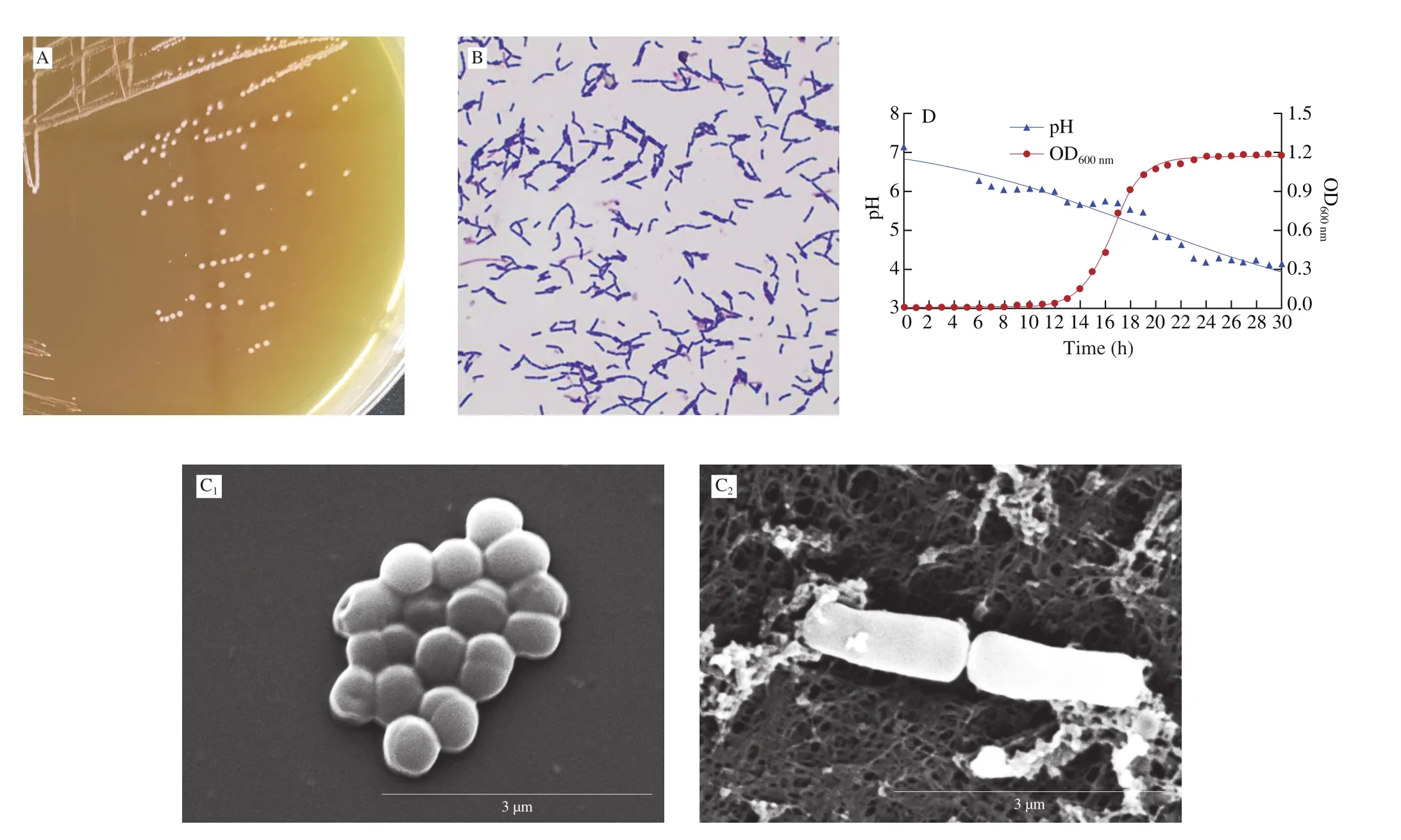
Fractionated streaking was performed for the colony purification,and the grey smooth colony (Fig. 1A) was picked, subjected to Gram staining, and observed under oil-immersion light-microscope. Grampositive bacilli were observed as shown in Fig. 1B.

Fig. 1 (A) The colony of L. gasseri HMV18 on MRS agar. (B) Gram staining and light microscopy of L. gasseri HMV18(magnification: 1 000 ×). (C) SEM of L. gasseri HMV18 grown in MRS broth (1) and on MRS agar (2) (magnification 12 000 ×).The scale bar represents 3 µm. (D) Growth properties of L. gasseri HMV18.
SEM was used to further examine the morphology ofL. gasseriHMV18. Bacterial cells grown in MRS broth appeared a spherical shape with diameter range from 0.5-0.7 µm (Fig. 1C1). Cells were fast dividing and always attached together to form a grapelike group.Bacterial cells grown on MRS agar were typical bacilli with width around 0.5-0.7 µm and length around 2-2.5 µm (Fig. 1C2). The phenotype exchange may be related to high glucose, acid production,and so on. The mechanism of the morphology variation needs to be further studied. The surface of the bacterial cell culturedin vitrowas smooth and no flagellum, fimbriae or pilus, capsule was observed.
3.2 Growth properties
The HMV18 cells were inoculated in MRS broth to the final concentration of 105CFU/mL. As shown in Fig. 1D, the latency time was approximately 10 h. The OD600nmincreased dramatically during the exponential phase from 14 h to 19 h. After that, the cells entered the stationary phase. During the cell growth, pH constantly decreased.There was a slightly decrease from time 6 h to 14 h, and a dramatic decrease from 16 h to 24 h. Taken together, the results suggested that acidic products were produced at the late exponential phase and the stationary phase. The production of acids may be useful in antagonistic against pathogens. The reduction in pH may also enhance the bacteriocin production during stationary phase [27].
3.3 Identification of L. gasseri HMV18
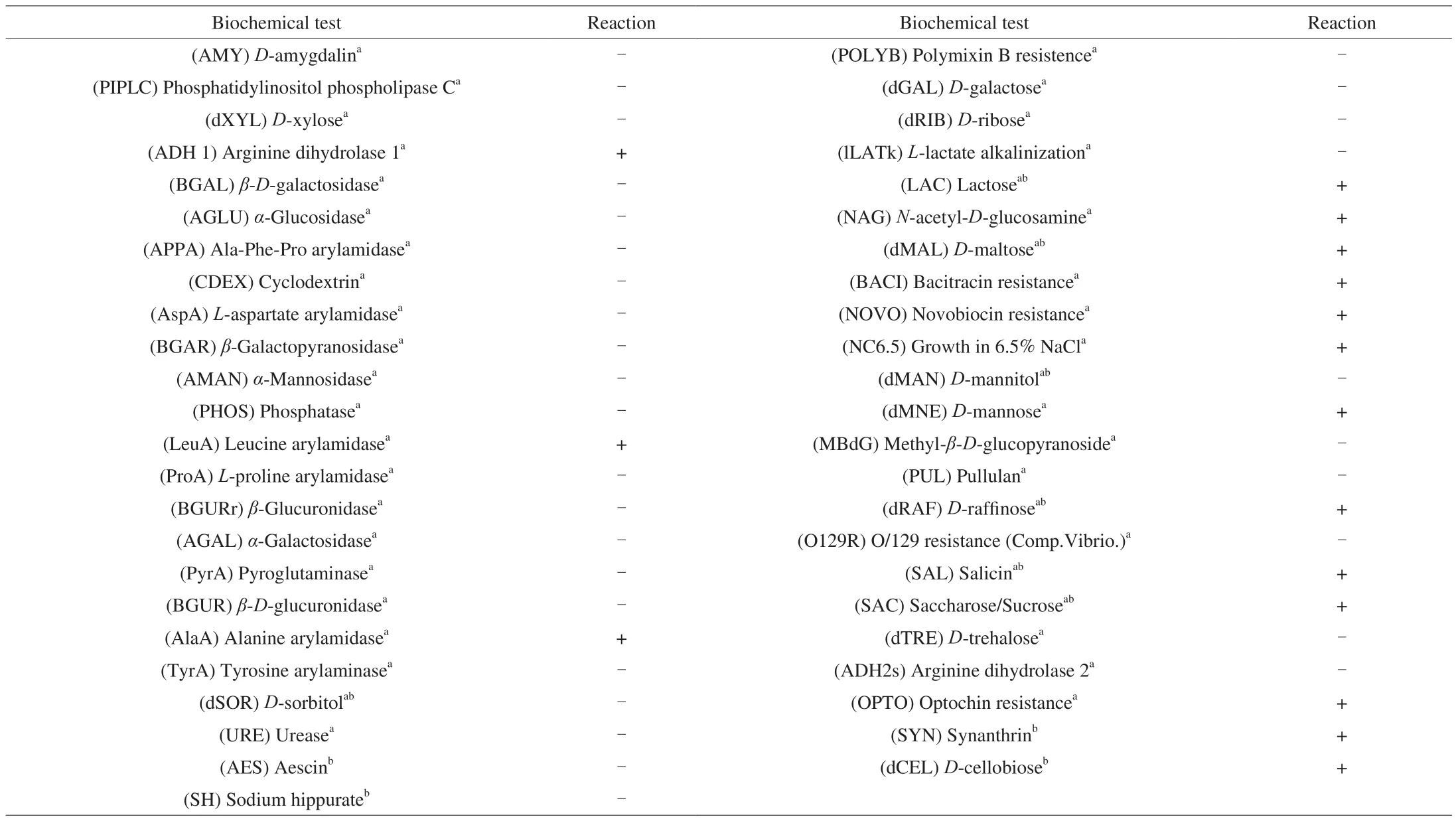
Single colonies were subjected for biochemical analysis and 16S rDNA identification. The biochemical test included the Grampositive identification automatic microorganism analysis and LAB Biochemical Test Kit. As shown in Table 1, there were 43 reactions in the automatic analysis system and 11 carbohydrates in the LAB Kit. Some of the carbohydrates were tested in both reactions and the results were consistent, such as the utilization of lactose,D-maltose,D-raffinose, salicin, sucrose, and non-utilization ofD-sorbitol orD-mannitol.

Table 1Biochemical test of L. gasseri.
16S rDNA of the HMV18 strain was obtained by PCR. The size of 16S rDNA amplification product was 1 253 bp (Fig. 2A), and the quantity was sufficient for sequencing. Sequencing peaks was shown in Fig. 2B. The sequence was aligned with the online bacterial 16S rDNA database using the NCBI Blast. The result showed a 99% identity withL. gasseri. Further phylogenetic and molecular evolutionary analyses suggested that was a newL. gasseristrain and was named asL. gasseristrain HMV18 (Fig. 3). The strain is stored in China Center for Type Culture Collection (Wuhan, China), and the ID No. is CCTCC M2019538.

Fig. 2 16S rDNA of the L. gasseri HMV18 strain. (A) PCR products of HMV18 isolates. Marker: 200–2 000 bp DNA marker; 1–5: L. gasseri HMV18 new isolates. (B) A representative of the sequencing peaks of 16S rDNA of the HMV18 strain.

Fig. 3 Phylogenetic analysis of L. gasseri HMV18. The tree was inferred from 16S rDNA of Lactobacillus downloaded from NCBI bacterial database. The scale bar represents a genetic distance of 0.005.
The whole genome ofL. gasseriHMV18 was obtained byde novosequencing, which showed that its genome was consisted of a 1.95 MB chromosome, a 4 626 bp small plasmid, a 32 KB large plasmid and 3.9 KB bacteriophage.
3.4 Pathogen susceptibility assay
The cell-free supernatant of theL. gasseriHMV18 was tested the antagonist effect againstE. coli,S. aureusandK. oxytocaby Oxford cup method. The results showed that the supernatant of theL. gasseriHMV18 inhibited all the three pathogens (Fig. 4). The diameter of the inhibition zone was 21, 16 and 18 mm, respectively, againstE. coli,S. auruesandK. oxytoca.

Fig. 4 The cell-free supernatant of the L. gasseri HMV18 inhibited (A) E. coli, (B) S. aurues, (C) K. oxytoca pathogenic bacteria. The cell-free supernatant tested were (3) L. fermentum, (4) L. gasseri HMV18, (8) L. reuteri.
3.5 Hemolysis test
Since the hemolysis characteristics always suggest the ability to invade the host of a pathogen, we streaked theL. gasseriHMV18 on blood agar to observe its hemolysis. It is also an important feature of the biosafety for the application of LAB in food. As shown in Fig. 5A,onlyα-hemolysis surround the colonies could be observed, which is characterized by a partial breakdown of the red blood cells (RBCs)in the medium, and is seen as a green zone surrounding the colony resulting from pigmentation of hemoglobin within RBCs. Although the termα-hemolysis is still used for this appearance, the property is due to oxidization of iron in hemoglobin molecules (reduction to methemoglobin), not lysis of RBCs [28].

Fig. 5 Safety evaluation of L. gasseri HMV18. (A) L. gasseri HMV18 was streaked on a blood agar plate. Representative image of H&E stained ileum section(magnification 200 ×). Control and HMV18 indicated the mice gavaged with (B) PBS and (C) L. gasseri HMV18. (D) Inflammatory cytokines in mice blood.Samples were collected from the LPS, L. gasseri HMV18 and PBS gavaged group respectively. The cytokines concentrations of IFN-γ, IL-4 and IL-10 were determined by ELISA Kits. The data were expressed in means ± SD. * P < 0.001, ns: no significant difference.
3.6 Antibiotic susceptibility test
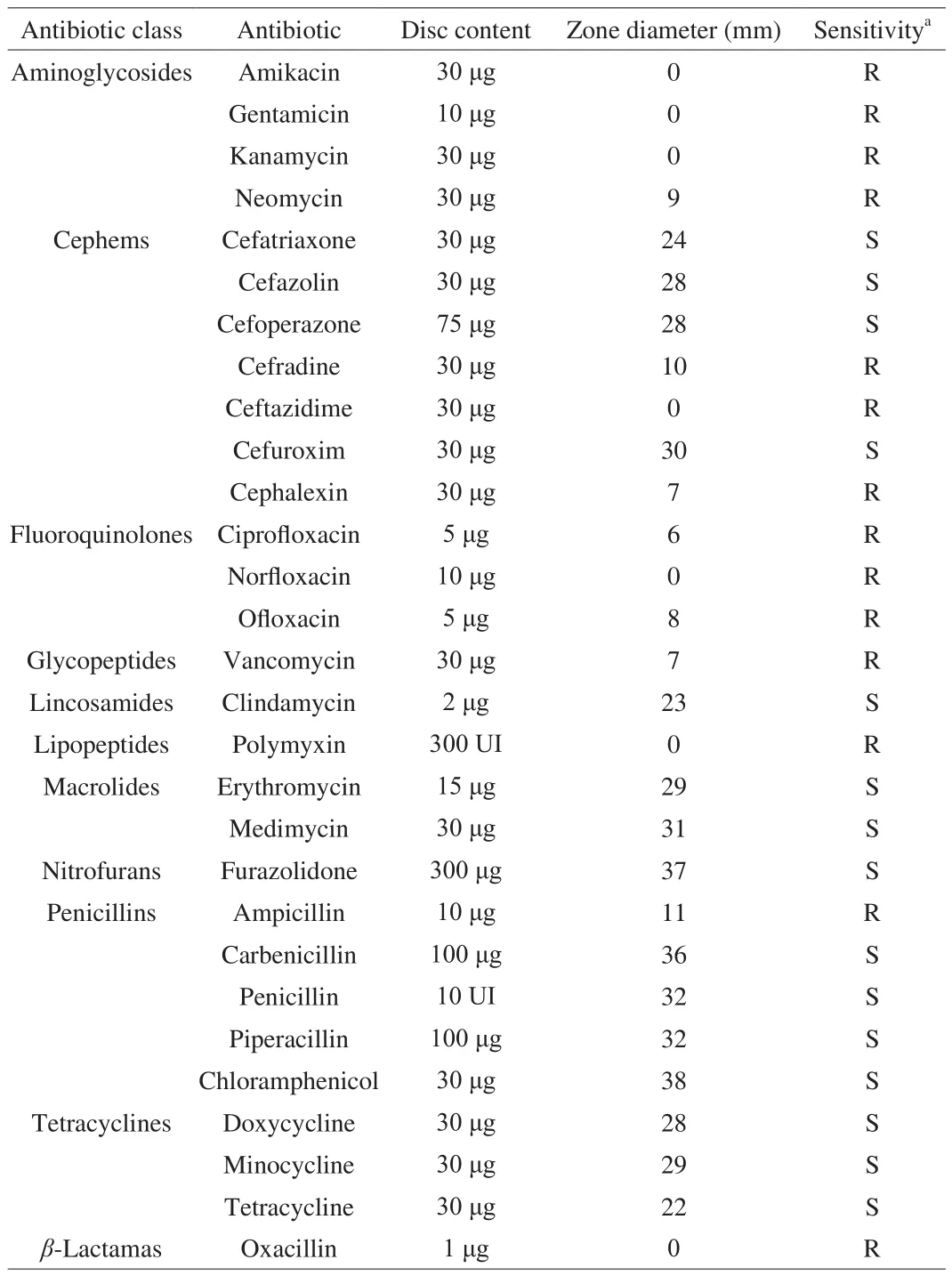
The automatic biochemical analysis showed that theL. gasseriHMV18 might be resistant to bacitracin and novobiocin (Table 1).The antibiotic resistance profile was analyzed by Disk Diffusion Testing Kit, and the results were shown in Table 2.L. gasseriHMV18 was sensitive to cefatriaxone, cefazolin, cefoperazone, cefuroxim,clindamycin, erythromycin, medimycin, furazolidone, carbenicillin,penicillin, piperacillin, chloramphenicol, and tetracyclines, while resistant to aminoglycosides and fluoroquinolones, vancomycin,polymyxin, ampicillin, and oxacillin. Antibiotic resistance genes were predicted using CARD Database. The results were shown in Table 3, and suggestedL. gasseriHMV18 might resistant to rifampin,lincosamides and elfamycins. The genes related to these antibiotic resistances were found in the chromosome but not in the plasmids or phage, which suggested that there was little possibility for the horizontal transfer of these antibiotic resistances and it can be easily eradicated by various antibiotics.

Table 2Results of antibiotic susceptibility tests on L. gasseri HMV18.

Table 3Antibiotic resistance annotation of L. gasseri HMV18.
3.7 Histologic examination
Intestinal micro flora adheres to the epithelial surface to maintain the homeostasis of the mucosa. In order to study whether the new isolateL. gasseriHMV18 induce the pathologic changes of ileum,L. gasseriHMV18 was oral administrated to mice and ileum histology was evaluated. As shown in Figs. 5B and 5C, the mucosa, villi, lamina propria and crypt depth remained intact and there was no significant inflammation, hyperemia, villi broken, or epithelial shedding in theL. gasseriHMV18 gavaged group, which was almost the same as the PBS group. The results suggested thatL. gasseriHMV18 could not induce inflammation and relative safe for further application in food and drug additions.
3.8 Determination of inflammatory cytokines
To further study whetherL. gasseriHMV18 could induce inflammation or not, the blood was collected and the concentrations of cytokines in serum were determined, including IFN-γ, IL-4 and IL-10. As shown in Fig. 5D,L. gasseriHMV18 could not induce the concentrations of IFN-γ and IL-4 compared with the PBS control group. However,L. gasseriHMV18 induced IL-10 significantly compared with the PBS control group. The induction of IL-10 byLactobacillushas been reported in Reyes-Díaz et al. [29]. Since IL-10 is considered as an anti-inflamatory cytokine, increasing its concentration may contribute to the immune-tolerance toL. gasseriHMV18 and other members in microflora. These results suggestedL. gasseriHMV18 could not induce inflammation or it can reduce inflammation.L. gasseriHMV18 is a candidate probiotic to be a food additive.
4. Conclusion
In this study, a newL. gasseriHMV18 strain was isolated from the vagina of a healthy woman. It was identified by colony morphology, Gram staining and bacterial cell morphology,biochemical characteristics and 16S rDNA alignment. The growth property and pH changing of the culture were monitored, which suggestedL. gasseriHMV18 grew fast in MRS medium. The production of acids may be useful in antagonistic against pathogens.Antibiotic susceptibility assay indicated the growth ofL. gasseriHMV18 is sensitive to various antibiotics, such as erythromycin,furazolidone, penicillin, chloramphenicol, retracycline, and so on.L. gasseriHMV18 showed ideal repression on the growth of foodborne pathogens, such asE. coli,S. aureusandK. oxytoca.L. gasseriHMV18 producedα-hemolysis, did not produceβ-hemolysis, did not induce inflammation in ileum as well. The mucosa, villi, lamina propria and crypt depth remained intact and there was no significant inflammation or hyperemia in theL. gasseriHMV18 gavaged group. There was little possibility for horizontal transfer of antibiotic resistance genes. The blood cytokines concentrations suggested thatL. gasseriHMV18 could not induce inflammation or it might reduce inflammation. To sum up,L. gasseriHMV18 is a candidate probiotic to be a food additive or drug to prevent food-borne diseases. The safety evaluation and further studies on the mechanism of inhibition of all the three food-borne pathogens are underway.
Acknowledgement
We would like to thank the Department of Electron Microscopy Center, Hebei Medical University, for our SEM work, and we are grateful to Wang Li and Cui Fang for their help in preparing EM samples, taking and analyzing EM images.
This work was supported by Natural Science Foundation of Hebei Province (H2020206579), S & T Program of Hebei (18277743D),CAMS Innovation Fund for Medical Sciences (2019-I2M-5-055), Key R & D projects in Hebei Province (20327125D), the Training Plan for Young Innovative Talents in Science and Technology (TJZR202008)and Spring rain project of Hebei Medical University (CYCZ201906).
Ethics statement
All subjects were fully informed and provided written consent for participation prior to enrollment. The protocol was reviewed and approved by the Medical Ethics Committee of Hebei Medical University No. 2017046.
Conflicts of interest
The authors declare that we have no conflict of interest with the contents of this article.
杂志排行
食品科学与人类健康(英文)的其它文章
- Potential application of proteolysis targeting chimera (PROTAC) modification technology in natural products for their targeted protein degradation
- Effects of vegetarian diet-associated nutrients on gut microbiota and intestinal physiology
- Recent progress in preventive effect of collagen peptides on photoaging skin and action mechanism
- Perspectives on diacylglycerol-induced improvement of insulin sensitivity in type 2 diabetes
- Effect of bacteriocin-producing Pediococcus acidilactici strains on the immune system and intestinal flora of normal mice
- Formation of advanced glycation end products in raw and subsequently boiled broiler muscle: biological variation and effects of postmortem ageing and storage
