Kaempferol attenuates knee osteoarthritis via inhibiting cartilage apoptosis in mice
2022-11-02MiYilinYiNanxingXuXiaotongYangLuojianLiJiabinChenShuyuanKuangGaoyanLuMin
Mi Yi-lin,Yi Nan-xing, Xu Xiao-tong, Yang Luo-jian,Li Jia-bin, Chen Shu-yuan, Kuang Gao-yan,Lu Min
1.The First Hospital of Hunan University of Chinese Medicine, Changsha 410007,China
Keywords:Kaempferol Knee osteoarthritis Apoptosis In vivo study Network pharmacology Molecular docking
ABSTRACT Objective: To study the mechanism of kaempferol in the intervention of knee osteoarthritis(KOA) in mice by inhibiting cartilage apoptosis. Methods: Firstly, the target genes of kaempferol were retrieved via TCMSP, and the genes related to KOA were obtained by GeneCards, OMIM, PharmGKB, TTD, and Drugbank databases. Then GO enrichment analysis and KEGG signaling pathway analysis were also performed. Subsequently,18 male C57 mice were randomly divided into the sham operation group, the model group, and the kaempferol group (50 mg/kg). Except for the sham operation group, the KOA mouse model was induced by destabilization of medial meniscus surgery. The sham operation group and model group were given the same amount of normal saline daily for 8 weeks while the kaempferol group was given 50 mg/kg kaempferol intragastrically. Results: A total of 63 targets of kaempferol were found that included 35 common target genes with KOA. GO and KEGG analyses showed that biological processes such as extrinsic apoptotic signaling pathway and response to oxidative stress, as well as signaling pathways such as cell apoptosis and regulation of TNF were closely related to common target genes. Molecular docking results also showed kaempferol has good binding properties with predicted targets Bcl-2, BAX, and CASP3.Compared with the model group, the pathological changes of cartilage in the kaempferol group were significantly reduced, OARSI scores were significantly decreased (P<0.001),and cartilage area was increased (P<0.01). In addition, Western blot analysis showed that kaempferol significantly decreased the protein expression of BAX and CASP3 (P<0.01,P<0.05), and increased the protein expression of BCL-2 (P<0.05). Conclusion: The treatment of KOA with kaempferol has the characteristics of multi-target and multi-pathway, and the mechanism may be related to the regulation of key genes such as Bcl-2, BAX, and CASP3 to inhibit cartilage apoptosis.
1. Introduction
Knee osteoarthritis(KOA) is a common chronic joint disease, and the main symptoms are articular cartilage degeneration, subchondral bone sclerosis, and synovial inflammation [1]. With the aging of population and the increase of obese population, the prevalence of KOA is rising, which has become an important social health problem facing China and even the world [2]. Non-steroidal antiinflammatory drugs (NSAIDs) are mainly used to relieve KOA symptoms in clinic. At present, there is still a lack of drugs that can effectively treat KOA., late joint replacement is the only treatment[3]. Therefore, it is urgent to study drugs for KOA.
Traditional Chinese Medicine is the Treasure House of Drug Research. This study intended to explore the effect of kaempferol on KOA based on the combination of network pharmacological analysis and experimental verification. Studies have shown that polyphenols may be beneficial to osteoarthritis, and flavonoids are the main members of polyphenol family [4]. Kaempferol is a bioflavonoid compound, which widely exists in Eucommia ulmoidesOliv., Achyranthes bidentata, Radix AngelicaePubescentis and other traditional Chinese medicines. Studies showed that kaempferol has many pharmacological activities, such as antiapoptosis, anti-inflammation and anti-oxidation [5]. It has been proved that kaempferol can alleviate IL-1β-induced chondrocyte inflammation by inhibiting NF-κB pathway [6]. It can also inhibit the ERK and p38 signaling pathways related to MAPK and the production of matrix metalloproteinases stimulated by IL-1β [7].
Network pharmacology is a new discipline based on bioinformatics and system biology, which provides a new method for revealing the complex mechanism of traditional Chinese medicine [8]. In this study,we explored the molecular mechanism of kaempferol in treating KOA through network pharmacology, and verified the combination of kaempferol and key target genes (BCL-2, BAX and CASP3)that interfere with KOA by molecular docking. Combined with in vivo experiments, we verified the influence of kaempferol on the histomorphology of knee joint and the expression of genes related to cartilage apoptosis signal pathway.
2. Materials and methods
2.1 Kaempferol-related targetscollection
The putative targets of kaempferol were obtained by TCM systematic pharmacology database and analysis platform TCMSP(https://tcmspw.com/tcmsp.php), and the related targets of kaempferol were uniformly transformed by protein standardized Uniprot database (https://www.uniprot.org/).
2.2 KOA-related targetscollection
OA related targets were obtained through searching database with the keyword “osteoarthritis”. The databases include: The Online Mendelian Inheritance in Man (OMIM) (http://omim.org/),the Therapeutic Targets Database (TTD) (http://bidd.nus.edu.sg/group/cjttd/), the PharmGKB (https://www.pharmgkb.org/), the DrugBank (https://www.drugbank.ca/), and the human gene database GeneCards (http://www.genecards.org/).Then we combined five disease database targets and deleted duplicate genes to get KOArelated target genes.
2.3 Kaempferol-KOA network construction
Based on the screened potential targets of kaempferol, a kaempferol-target-network -diagram was constructed by means of Cytoscape3.8.0. R 4.0.3 Venn Tool kit was used to draw Venn map of the related targets of kaempferol and KOA, and the common targets were obtained by taking the intersection of them, and then Cytoscape3.8.0 was used to construct the network map of the common targets of kaempferol and KOA.
2.4 Co-targets GO analysis and KEGG enrichment analysis
In order to further understand the specific role of the intersection network in gene function and related signal pathways, David 6.8 (https://david.ncifcrf.gov/) database was used to analyze the enrichment of GO function and KEGG pathway of the selected target proteins (the filtering conditions of P value and the corrected filtering conditions were less than 0.05), and the enrichment analysis results and pathways were visualized with the help of the clusterProfiler package in R 4.0.3 combined with enrichplot package.
2.5 Molecular docking
In order to further understand the binding pattern and affinity between kaempferol and the selected receptor targets in vivo, the 2D structure of kaempferol was downloaded from Pucheme data platform (https://pubchem.ncbi.nlm.nih.gov/), which was imported into Chem3D 14.0.0.117 software to optimize the structure and convert it into 3D structure. Download 3D PDB format file of target protein by RCSB data platform (https://www.rcsb.org/); PYMOL 4.6.0 was used to remove water molecules and small molecule ligands from protein, and then imported into Auto Dock Tools1.5.6 software to carry out hydrogenation and charge calculation on key targets. Adding polar hydrogen, loading charge, and constructing active pocket; Vina1.1.2 software was used to make molecular docking between the target and kaempferol, and the binding energy was used as docking evaluation index, which was imported into PYMOL 2.4.1 software for visualization.
2.6 Kaempferol intervention KOA confirmatory experiment
2.6.1 Experimental animals and groups18 C57BL male mice of SPF grade 8 weeks old were provided by the Animal Experimental Center of Hunan University of Traditional Chinese Medicine. Mice were randomly divided into sham operation group, model group and model+kaempferol group. Mice in each group drank water and ate freely under the same conditions, and were raised by the Experimental Animal Center of Hunan University of Traditional Chinese Medicine. This experimental scheme was reviewed by the Ethics Committee of Hunan University of Traditional Chinese Medicine (Ethics Number: LL2020121601).
2.6.2 Modeling and drug administration
The mice were anesthetized with 2% isoflurane, the right knee joint depilatory cream was used to depilate, the knee joint of mice was disinfected with iodophor, and the KOA model was induced by the operation of destabilization of the medial meniscus(DMM) [9]. Model group: A 0.5cm incision was made between the medial patellar ligament and medial collateral ligament of the right knee joint, and the medial meniscus tibial ligament was cut by microscopic shearing and the skin incision was sutured. Sham operation group: Sham operation model was made, 0.5 cm incision was made between medial patellar ligament and medial collateral ligament of right knee joint, and skin incision was sutured. Wipe the wound with iodophor, put it back into the cage, and wait for the mouse to wake up. Kaempferol group was given 50mg/kg kaempferol by gavage every day [10], and sham operation group and model group were given the same volume of normal saline by gavage every day. For eight weeks.
2.6.3 Drugs, reagents and instruments
Kaempferol (purity ≥ 98.0%, HPLC analysis, Jiangsu Yongjian Pharmaceutical Technology Co., Ltd.), depilatory cream (Lijieshi Company, India), isoflurane gas anesthetic (Rivard, Batch NO.217140901), paraformaldehyde (Sinopharm Reagent Co., Ltd.,80096618), absolute ethanol (Sinopharm Reagent Co., Ltd., 1000)Red O- fast green dye solution (Beijing Regan Biotechnology Co.,Ltd., production batch number DB0082), ammonia water (Sinopharm Reagent Co., Ltd., 10002118), xylene (Sinopharm Reagent Co., Ltd.,10023418), sodium hydroxide (General-Reagent, G19852H), Shitai tissue embedding box (Jiangsu Shitai Experimental Equipment Co.,Ltd. Total protein extraction kit (Nanjing Jiancheng), BCA protein concentration detection kit (Nanjing Jiancheng), rabbit anti-mouse CASP3, BAX and BCL-2 antibodies (Abcam, 1:500), goat antirabbit IgG labeled with HRP (CST, 1:1000), small animal anesthesia machine (Matrx VMR USA), dehydrator (Leica, TP), embedding machine (LEICA, EG1150C), etc.
2.6.4 Histological AnalysisAfter 8 weeks of continuous gastric administration, the mice were deeply anesthetized with small animal breathing anesthesia machine and 2% isoflurane, then the neck was removed and the knee joint was taken out and fixed with 4% paraformaldehyde. 10%EDTA decalcification for 4 weeks, tissue dehydration, paraffin embedding,5 micron sagittal continuous section, safranin O- fixation staining,the degree of cartilage degeneration was analyzed by the scoring system of Osteoarthritis Research Society International (OARSI) [11],and the area of tibia cartilage in mice was counted by Image-J v1.50i software.
2.6.5 Western blotThe levels of BCL-2, BAX and CASP3 protein in cartilage were detected by Western blot. The cartilage tissue was mixed with RIPA lysate, ground for 10-15 minutes, stirred on ice for 30 minutes,and the supernatant is collected. The protein concentration was determined by BCA method. After the gel electrophoresis was closed by membrane transfer, the corresponding primary antibody and secondary antibody were incubated, developed and images were collected.
2.7 Statistical Analysis
Statistical analysis was performed using the GraphPad Prism 7.0 software. All quantitative data were expressed as a mean ± standard deviation (±SD). One-way ANOVA followed by Dunnett’s t-test was used to determine the significance of the differences between groups.A value of P<0.05 was considered statistically significant.
3. Results
3.1 Network diagram of potential targets of kaempferol intervention in KOA
There were 63 targets of kaempferol found in TCMSP database,including BAX, PTGS2, MMP1, etc (Figure 1A). Searching five databases related to disease genes, such as Genecards, OMIM,PharmGKB, TTD and Drugbank, obtained 1637 target genes closely related to KOA diseases. Among them, there are 1579 genesin GeneCards, 6 in OMIM, 9 in PharmGKB, 2 in TTD and 71 in Drugbank, and the results of 5 databases were plotted into venn diagram (Figure 1B). The 63 targets of kaempferol obtained by the above screening were intersected with 1637 targets related to KOA diseases, and 35 common targets were obtained, including BCL2,BAX, CASP3, etc.(Figure 1C).
3.2 GO and KEGG Enrichment analysis of co-targets
With the help of DAVID 6.8 database, the signal pathway and biological process (BP), cell component (CC) and molecular functions (MF) enrichment analysis of the targets related to kaempferol treatment of KOA were carried out, and the bubble chart was made with R4.0.3 toolkit. The results show that kaempferol mainly participates in biological processes including exogenous apoptotic signaling pathway, negative regulation of apoptotic signaling pathway, response to oxidative stress, etc.(Figure 2A). The pathways involved mainly include apoptosis, tumor necrosis factor signaling pathway and so on (Figure 2B).

Figure 1 Network of potential targets for kaempferol treatment of KOA
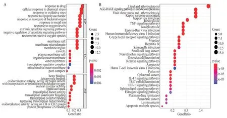
Figure 2 GO analysis and KEGG pathway enrichment analysis of kaempferol-KOA
3.3 kaempferol has good docking with BCL-2, BAX and CASP3
Molecular docking was used to evaluate the binding potential of protein and ligand to further understand the correlation between drugs and selected targets. Auto Dock1.5.6 was used to predict the binding energy of three targets (BCL-2, BAX and CASP3) of kaempferol in the treatment of KOA. According to the reference[12], the binding ability of ligand to receptor is -7.0kcal/mol with obvious binding activity, -5.0kcal/mol with good binding activity and -4.0kcal/mol with certain binding activity. The results showed that all the three targets had good binding capacity, and the binding capacity of kaempferol to BCL-2 receptor was -8.3 kcal/mol, that to BAX receptor was -6.6 kcal/mol, that to CASP3 receptor was -6.8 kcal/mol, and the binding activity was obvious. The docking results of kaempferol with BCL-2, BAX and CASP3 were introduced into PYMOL2.3.4 (Figure 3).

Figure 3 Visualization of kaempferol-targets (BCL-2, BAX, CASP3) docking
3.4 Kaempferol can reduce articular cartilage damage
Through safranine O- fast green staining, it was found that the articular cartilage of knee joint in sham operation group was intact, the cartilage surface was smooth, there was no roughness or defect, the meniscus was intact, and there was no obvious pathological manifestation. The thickness of tibial articular cartilage in model group and kaempferol group decreased significantly,and the cartilage surface was rough and defective. Statistically,compared with the sham operation group, the OARSI score of knee joint cartilage in the model group was significantly higher(5.2±0.57), while the area of tibial cartilage was significantly lower(1408.0±174.00). After kaempferol intervention, compared with the model group, the OARSI score of articular cartilage in kaempferol group was significantly improved (3.0±0.61), and the cartilage area was significantly increased (2522.4±439.10)(Figure 4).

Figure 4 Mice knee joints stained with safranin-O fast green F: Femur,T: Tibia,M: meniscus.***P<0.001,**P<0.01.
3.5 kaempferol can regulate the expression of BCL-2, BAX and CASP3 protein
Western blot shows that the expression of BAX and CASP3 protein in the knee cartilage of the model group was significantly higher (0.46±0.04,0.54±0.06)than that of the sham operation group (0.15±0.02,0.24±0.08), while the expression of BCL-2 was significantly lower (0.05±0.01). After kaempferol intervention, the expression of BAX and CASP3 protein in mouse cartilage decreased significantly (0.31±0.04,0.37±0.06), while the expression of BCL-2 protein increased significantly (0.17±0.02). It shows that kaempferol has significant inhibitory effect on chondrocyte apoptosis (Figure 5).
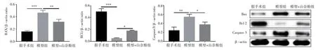
Figure 5 Western blots of BCL-2, BAX and CASP3 gene expression
4. Discussion
KOA is a common joint disease in middle-aged and elderly people,which can not only cause joint pain, deformity and dysfunction, but also significantly increase the risk of cardiovascular events, deep vein thrombosis of lower limbs, hip fracture and all-cause mortality[13]. The Osteology Branch of Chinese Medical Association [13] and the International Association for Osteoarthritis Research (OARSI)[14]all recommend basic treatment as the first choice. In this study, we are looking for potential drugs to treat KOA. Kaempferol, a flavonol widely found in natural medicines, has been proved to be one of the important natural anti-inflammatory compounds, which can improve the condition of arthritis, cancer, cardiovascular diseases and other diseases, which is closely related to reducing the risk of inflammation-related diseases [15-16]. Kaempferol widely exists in a variety of Chinese herbal medicines and compound preparations. It has been reported that flavonol compounds such as kaempferol are one of the effective chemical components of the classic prescription Duhuojisheng Decoction. Duhuojisheng Decoction has been widely used in clinical treatment of KOA and other orthopedic diseases for many years, and achieved good curative effect [17]. Our research group has confirmed that the serum containing Jiawei DuhuoJisheng Mixture can improve the pathological manifestations of chondrocytes induced by IL-1β by regulating Wnt signaling pathway [18], and this drug can improve joint function and pain by reducing the levels of IL-1, TNF-α and NO in joint fluid of OA patients [19]. In this study,the mechanism of kaempferol's intervention in KOA by regulating BAX, BCL-2 and CASP3 was systematically expounded for the first time.
As a chemical active ingredient of natural food and medicine,kaempferol has attracted more and more attention because of its small side effects and extensive effects. Many studies have confirmed that kaempferol has positive effects on KOA. Studies have found that kaempferol has strong anti-inflammatory and antiarthritis effects by regulating NF-κB and MAPK pathways [6-7];Jiang et al. found in 2019 that kaempferol may interfere with the apoptosis and inflammatory reaction of cartilage ATDC5 cells induced by lipopolysaccharide by down-regulating miR-146a and inhibiting the expression of Decorin [20]; Recent studies have found that kaempferol can inhibit inflammation and extracellular matrix degradation by regulating the XIST/miR-130a/STAT3 axis of chondrocytes [21], and clinical trials have proved that kaempferol is effective in relieving KOA pain and symptoms [22]. As a method of drug research, network pharmacology combines the multiple advantages of pharmacology and biology, and is characterized by the network model of drug-component-target-pathway, which provides a research method for the relationship between drugs and diseases,and can reflect the multi-target and multi-pathway interaction of drugs [8]. In this study, the mechanism of kaempferol in the treatment of KOA was expounded by network pharmacology. By digging out the target genes closely related to the occurrence and development of kaempferol and KOA diseases, and constructing the network relationship between kaempferol and KOA, we found that BCL2,BAX, CASP3, NOS2 and JUN are important targets for kaempferol to intervene KOA. Through GO and KEGG enrichment analysis,kaempferol is involved in exogenous apoptosis signal pathway,negative regulation of apoptosis signal pathway and oxidative stress reaction, and it is related to apoptosis, tumor necrosis factor signal pathway, etc. Apoptosis is a programmed cell death mode controlled by genes [23], which is very important for cartilage development and maintaining proper dynamic balance [24]. Cartilage degeneration is the key feature of KOA, and many experimental studies have shown that chondrocyte apoptosis plays a key role in the progress of KOA [25].During the course of KOA, chondrocytes apoptosis under the stimulation of inflammatory microenvironment caused by various reasons, resulting in the impairment of cartilage function[26]. Therefore, inhibiting chondrocyte apoptosis can be used as an important method to treat KOA. The mechanism of apoptosis is very complicated, which is related to a series of signal molecules,among which BCL-2, BAX and CASP3 are three key molecules in the apoptosis pathway [27]. The molecular docking in this experiment verified the binding ability of kaempferol to BCL-2, BAX and CASP3, and found that kaempferol not only had obvious binding ability, but also had strong binding activity.
Caspase family, as the core of apoptosis, was first found in 1985[28]. According to its structure and function, it can be divided into startup and execution types. Among them, startup-type Caspases can activate executive-type CASP3 and amplify the apoptotic signal pathway. The activated CASP3 can trigger the apoptotic cascade reaction and lead to rapid cell death [29]. BCL-2 protein family regulates mitochondrial membrane permeability, and can promote apoptosis and resist apoptosis. Among them, anti-apoptotic proteins include BCL-2, BCL-X, BAG, and pro-apoptotic proteins include BAX, BCL-10, and BID. These proteins have special significance because they can decide whether to apoptosis or to terminate apoptosis [30]. Therefore, inhibiting the expression of BAX and CASP3 and promoting the expression of BCL-2 are crucial to reducing the apoptosis of chondrocytes [27]. Studies have shown that oxidative stress is closely related to apoptosis and is a negative effect of free radicals in the body, which plays an important role in the development of KOA. The accumulation of reactive oxygen species will interfere with the related processes of chondrocyte anabolism,leading to chondrocyte dysfunction and degeneration, and further leading to apoptosis [31]. Extracellular stimulation activates exogenous apoptotic pathways through the TNF receptor gene family [32]. These biological processes and signaling pathways are consistent with our GO and KEGG enrichment results and, therefore,apoptosis is a potential molecular mechanism of kaempferol in the treatment of KOA.
This study also verified the efficacy of kaempferol in the treatment of KOA and its regulation of key apoptotic targets BCL-2, BAX and CASP3. The experimental results shows that DMM induced degeneration of cartilage in mice, while kaempferol significantly improve cartilage degeneration and regulate the protein levels of BCL-2, BAX and CASP3 in cartilage, which is consistent with the enrichment results of GO and KEGG. Our study shows that apoptosis level of cartilage in the kaempferol group is inhibited,and kaempferol plays a role in delaying the progression of KOA disease by reducing BAX and CASP3 in cartilage of KOA mice and increasing BCL-2 protein level.
In summary, the potential molecular mechanism of kaempferol in the treatment of KOA was investigated in this study based on network pharmacology and animal experiments. The results confirms that kaempferol is a potential effective drug for the treatment of KOA, and inhibition of apoptosis is possible mechanism.
Statement of conflict of interest: All authors have no conflict of interest.
Authors Contributions: Lu Min, Kuang Gaoyan designed the study. Mi Yilin, Xu Xiaotong retrived relevant literature. Mi Yilin,Yi Nanxing, Xu Xiaotong and Yang Laojian processed the data. Mi Yilin, Yi Nanxing, Li Jiabin, Chen Shuyuan produced the charts. Mi Yilin completes the first draft and Yi Nanxing checked.The research funds was obtained from Lu Min and KuangGaoyan.
杂志排行
Journal of Hainan Medical College的其它文章
- Advances in BRAF gene mutations in papillary thyroid carcinoma
- Research progress on the correlation between lncRNA and the pathogenesis of COPFD
- Analysis of laboratory and imaging examination results of patients with COVID-19 2 years after discharge in Chengdu
- Efficacy and prognosis of vacuum-assisted excision for benign intraductal papilloma of breast: A meta-analysis
- Clinical efficacy and perioperative safety of simultaneous or staged bilateral total hip arthroplasty:A Meta analysis
- Effect of Acacetin on flagellin induced NLRC4 inflammasome activation in mouse bone marrow-derived macrophages