Temporal retinal thinning might be an early diagnostic indicator in male pediatric X-linked Alport syndrome
2022-07-30RuiLinZhuLiangZhaoXiaoPengGuYaDiZhangFangWangYanQinZhangLiuYang
INTRODUCTION
株高与穗位高(0.45**)、株高与穗长(0.32**)、株高与穗行数(0.34**)、穗位高与空杆率(0.38**)、穗长与秃尖长(0.35**) 呈极显著正相关,活动积温与穗长(0.21*)、株高与出籽率(0.19*)、空杆率与秃尖长(0.21*) 呈显著正相关,空杆率与出籽率(-0.32**)、百粒重与秃尖长(-0.25**)、百粒重与穗行数(-0.43**) 呈极显著负相关,百粒重与空杆率(-0.22*)、秃尖长与出籽率(-0.19*)呈显著负相关。其余各项相关性均未达到0.05显著水平。
In male patients, the TTI of the total retina (
<0.0001), inner retina layers (
<0.0001), GCL (
<0.0001), IPL (
<0.0001),INL (
<0.0001), and ONL (
=0.0012) were significantly higher in the XLAS group. Twenty-seven male patients(77.14%) had severe pathological temporal retinal thinning.All the 27 patients had severe pathological temporal retinal thinning in inner retina layer, while 24 of the male patients(68.57%) had severe temporal thinning in the INL layer. The CRT of the XLAS group (233.40±31.11 μm) was significantly thinner than that of the control group (258.90±17.11 μm;
<0.0001). Figure 3 is a representative SD-OCT result of an XLAS patient.
SUBJECTS AND METHODS
Ethical Approval The study follows the tenets of the Declaration of Helsinki. The study was approved by the Institutional Review Board of Peking University First Hospital,Beijing, China (ID: 2017-1409). All patients’ parents gave informed consent for data collection and analysis.
Participants We retrospectively reviewed the SD-OCT data and medical records of XLAS patients aged under 18y who received ophthalmic examination from January 2017 to September 2019 at Peking University First Hospital. All patients received genetic testing with Sanger sequencing or targeted whole exome sequencing. All the patients were confirmed to have a pathogenic variant in the
gene,and the diagnosis of XLAS was made at the Department of Pediatrics of Peking University First Hospital. Data evaluated included patient demographics, ocular history, slit-lamp and dilated fundus examination records. SD-OCT studies were reviewed. We recruited healthy children as a control group who received SD-OCT and ocular examination. The refractive error of these participants was between -5.75 DS to +2.75 DS.No anterior segment or fundus changes were identified in the control group children. The systemic medical history was unremarkable in the control group.
Spectral Domain Optical Coherence Tomography SD-OCT(Spectralis; Heidelberg Engineering, Heidelberg, Germany)was performed in a high-resolution mode on all patients to assess macular thickness changes. The macular cube volumetric scans were obtained centered at the fovea, including 49 B-scans and 15 automated real-time repetitions. Retinal layer segmentation was executed automatically using Spectralis OCT built-in software (Heidelberg Eye Version 1.10.2.0).
进入21世纪以来,西藏印刷业不断进步,伴随着市场经济的发展,印刷业从官方主办逐步发展到市场主导,印刷,已走进西藏的千家万户。
The auto segmentation software determined 11 different retinal boundaries (Figure 1): the inner limiting membrane (ILM), the boundaries between the retinal nerve fiber layer (RNFL) and the ganglion cell layer (GCL), between the GCL and the inner plexiform layer (IPL), between the IPL and the inner nuclear layer (INL), between the INL and the outer plexiform layer(OPL), between the OPL and the outer nuclear layer (ONL),the external limiting membrane (ELM), two photoreceptor layers (PR1/2), the retinal pigment epithelium (RPE), and Bruch’s membrane (BM). Thickness of the following layers was automatically calculated: RNFL, GCL, IPL, INL, OPL,ONL, RPE, total retinal thickness (RT, comprising the ILM and BM), inner retinal layers (IRLs, comprising the ILM and the ELM), and outer retinal layers (ORLs, comprising the ELM and the BM).
All the images were reviewed by an ophthalmologist (Zhao L),who was masked to clinical information. Poor-quality images with a signal strength less than 20 dB, poor centration, or incorrect segmentation images were discarded.
当浆液扩散方位角90°
Temporal Thinning Index calculation According to Ahmed’s formula
, the TTI was calculated with the following formula:

N1 and N2 were the average thicknesses (μm) of the inner and outer nasal segments, respectively, and T1 and T2 were the average thicknesses (μm) of the inner and outer temporal segments, respectively (Figure 2). According to Ahmed’s report
, the TTI was subdivided into 3 categories based on the TTI mean value and SD value of the normal children in the control group. Normal physiological thinning was defined as a TTI<1 SD of the normal mean value, moderate pathological thinning was defined as a TTI 1-2 SDs above the normal mean value, and severe pathological thinning was defined as a TTI>2 SDs above the normal mean value.
据说这些智能产品用久了都会有灵性,说不定还会争风吃醋搞宫斗:倘若太过偏爱ipad,手机恐怕不高兴,会闹罢工发神经。这不是危言耸听,我的手机Siri就很皮,没少胡乱解读圣意,给不该联系的人错拨电话:比如前几天,我前脚给长官电话请假说今天上不了班了,头疼腿疼肚子疼哪儿哪儿都疼;后脚把手机塞包里,间谍Siri就发起了重拨,于是被听到刚才还奄奄一息的我正喜不自禁地哼着小调:“阳光彩虹小白马,你是最强哒最棒哒”!气得我呀,索性把语音控制设置成土耳其语,将暗害本宫的小贱人Siri打入冷宫。
As mentioned in Chen
’s study
, there was no difference in the TTI between the right eye and left eye, so the data of the right eyes were collected in our study. The TTI of the following layers was evaluated: RNFL, GCL, IPL, INL, OPL,ONL, RPE, RT, IRLs, and ORLs.
The acoustic impedance of the perforated plate is related to the acoustic resistance and mass reactance of the orifice on the assumption that the interaction between the orifices,if they are sparsely distributed,can be neglected.


Ocular abnormalities were found as below. Dot-and-fleck retinopathy was found in 8 patients (18.60%), and all of them were male. One male patient had pseudophakia due to lenticonus (2.33%). No corneal abnormality was found in our study. Visual acuity was recorded for the patients over 8 years old, and the best corrected visual acuity for all of them was 1.0. The total retinal thickness was shown in Table 2.Compared with the control group, the temporal inner sector retinal thickness (
<0.0001), the temporal outer sector retinal thickness (
<0.0001), and the nasal outer sector retinal thickness (
=0.0211) were significantly thinner in the XLAS male patients. There was no significant difference in retinal thickness between female XLAS patients and the control group. The TTI result was shown in Table 3. The TTI of the total retina (
<0.0001) was significantly higher in the XLAS group than in the control group. According to the criteria mentioned by Ahmed
, based on the TTI mean value and SD value of the normal children in the control group, there was temporal retinal thinning in 33 patients (76.74%), while 28 patients (65.11%) had severe pathological temporal retinal thinning and 5 patients (11.63%) had moderate thinning.
RESULTS
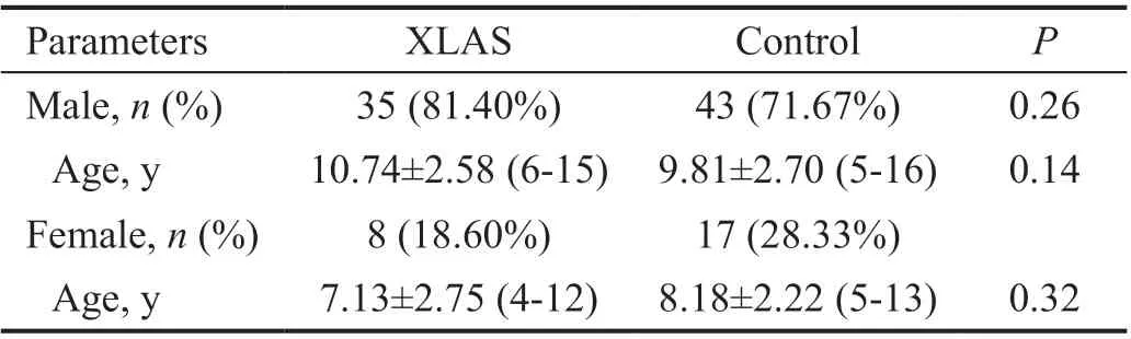
In this retrospective study, 43 patients diagnosed with XLAS were included. Thirty-five (81.40%) of the patients were male.The mean age of the patients was 10.07±2.95 (range 4-15)y when they received the SD-OCT examination. Sixty ametropia patients were included as control subjects. The mean age of thecontrol group was 9.28±2.65 (range 5-16)y. Demographic data of the patients are shown in Table 1. There were no significant differences in age or sex between the two groups.

Statistical Analysis GraphPad Prism 9 (GraphPad Software,San Diego, USA) was used for statistical analyses. Baseline descriptive characteristics (age, sex) were compared between the healthy and XLAS groups using an unpaired Student’s
-test for quantitative variables and Fisher’s exact test for categorical variables. The CRT and TTI of different layers were compared between the groups, and a
value <0.05 was considered statistically significant. The relationship between TTI and the age of XLAS patients was determined using Spearman’s
correlation coefficient.
根据常州市“十二五”河道长效管理评估[5],“十二五”期间,60条城市内河平均水质综合污染指数有所下降(RS=-0.800),污染等级由严重污染改善至重污染,水质呈现出一定改善趋势(见图1)。
With automated segmentation analysis, the TTI of the inner retina layers (
<0.0001), GCL (
<0.0001), IPL (
<0.0001),INL (
<0.0001), and ONL (
<0.0001) were significantly higher in the XLAS group. There were no significant differences in the TTI in other layers. The CRT of the XLAS patients was 234.40±28.69 μm (range 168-294 μm) and 257.30±16.62 μm (range 225-293 μm) in the control group.The CRT of the XLAS group was significantly thinner than that of the control group (
<0.0001).
Ocular abnormalities were generally more prominent in male XLAS patients. In our patient group, severe temporal retinal thinning was 27 (77.14%) in male patients
1 (12.50%) in female patients, and the retinal thickness measured by SDOCT was significantly different in the male group (Table 2).Thus, we further analyzed the TTI in different sexes. The results were shown in Tables 4 and 5.
Ocular abnormalities are common in AS patients, including corneal opacities, anterior lenticonus, and dot-and-fleck retinopathy
. In recent years, researchers from different groups have reported that temporal retinal thinning is a frequently detected ophthalmic feature in AS
. The occurrence of temporal retinal thinning is more common than other ocular abnormalities and is independent of other ocular features in AS patients. Ahmed
generated a temporal thinning index (TTI) to analyze temporal retinal thinning in X-linked Alport syndrome (XLAS) patients. Their reported data revealed that in XLAS patients, 70% of the patients had severe thinning, and 11% of them had moderate thinning
.Most studies measured total retinal thickness to calculate the extent of retinal thinning, and which retinal layer changed most was not addressed. Therefore, the characteristics of retinal thinning need to be elucidated. Previous studies on temporal retinal thinning in AS have been conducted almost all in adult cohorts, and little is known about pediatric AS patients. Recently, researchers have demonstrated that retinal temporal thinning is diagnostic for XLAS in men
. Since the frequency and severity of typical ocular anomalies increase with age
, temporal retinal thinning is more sensitive than typical ocular changes. Thus, in this study, we analyzed temporal retinal thinning with spectral domain optical coherence tomography (SD-OCT) and measured the retinal thickness of different retinal layers to investigate temporal retinal thinning changes in XLAS patients. We investigated temporal thinning in pediatric patients to study this feature at an early stage of the disease.
Temporal retinal thinning is more common than other AS ocular changes,
, anterior lenticonus, the lozenge sign, or dot-and-fleck retinopathy. Among the XLAS patients included in Ahmed
’s
study, 81% of the patients had moderate to severe thinning, while less than 20% eyes had other ocular findings
. Savige
reported that 89% male XLAS patients and 75% female XLAS patients had temporal retinal thinning. In our study, 33 (76.74%) patients had moderate to severe temporal retinal thinning, while dot-and-fleck retinopathy and lenticonus were found in only 18.60% and 2.33% patients,respectively. Since temporal retinal thinning is a sensitive and specific feature of AS, Chen
and Zhao
both have identified its diagnostic value in screening AS.
DISCUSSION
Retinal temporal thinning detected with SD-OCT is demonstrated as a common ocular change in AS. In 2004, Usui
reported an AS patient with symmetrical reduced thickness of the temporal macular area in both eyes, while the visual function of the patient was normal. During the following years,some other researchers reported AS patients with focal zones of inner retinal thinning in the temporal quadrant
. In 2013,Ahmed
suggested using the TTI parameter to assess the degree of retinal temporal thinning in AS patients. The TTI parameter was then adopted by other study groups and became an important index for evaluating temporal retinal thinning in AS patients. We compared the retinal thickness in nasal and temporal sectors between XLAS patients and the control group. Since the range of variation of the retinal thickness waslarge in different papers
, the TTI was useful to address the relative thinning of the temporal quadrant. Therefore, we use the TTI as our main result. It also made the results of our study comparable to previous reports by other investigators
.
Alport syndrome (AS) is a hereditary glomerular disease characterized by hematuria and progressive renal failure. The syndrome is usually associated with sensorineural hearing loss and distinct ocular abnormalities
, and it is estimated to affect 1 in 5000-10 000 individuals
. AS is caused by mutations in the
,
, and
genes, which encode the α3, α4, and α5 chains of collagen type IV
. Approximately 85% of affected patients show an X-linked dominant inheritance form caused by mutations in the
gene
.


In female patients, the TTI of the total retina (
=0.0248)was significantly higher in the XLAS group, while the TTI of the inner retina layers (
=0.0669) and outer retina layers (
=0.1070) were not significantly different between the two groups. The CRT of the female XLAS patients(238.10±17.33 μm) was significantly thinner than that of the control group (254.10±15.54 μm;
=0.0285). Age was not correlated with retinal temporal thinning in either male patients (
=-0.1356,
=0.4372) or female patients (
=0.1537,
=0.7163).
The foveal center was automatically identified by the SD-OCT software, and it was used as the center of the Early Treatment of Diabetic Retinopathy Study (ETDRS) grid. The ETDRS grid was used to analyze the average thickness of the different layers. As described by Ahmed
, the standard 6-mm macular OCT grid as defined by the ETDRS circle was labeled with 1-, 3-, and 6-mm-diameter circles centered at the fovea.The average of all measurements within the inner 1-mm circle was defined as central retinal thickness (CRT). The average retinal thickness in each of the nine macular sectors in the 6-mm diameter circle of each layer was evaluated automatically.
However, the pathogenesis of temporal retinal thinning is not fully understood. Previous studies suggested that thinning mainly affects the ILM and RNFL
. Some researchers assumed that the temporal thinning phenomenon in AS may be related to the postnatal development of the macula, tractional vitreoretinal forces, or aberrant Müller cell adhesion
. In our study, we used SD-OCT segmentation analysis to investigate the exact layer that changes most in AS. We included only XLAS pediatric patients to eliminate interference factors and observe the changes in the early stage of the disease.
民事审前程序的二元性价值改造并非要创设一个全新的审前程序,而是要将效率和公正这两个目标在审前程序的制度设计中进行恰当的安排。具体来说,目前的一元性价值的审前程序忽略了审前程序直接解决纠纷的功能,影响了司法效率,但改革决不能矫枉过正,例如正在修订的民事诉讼法欲将诉前先行调解纳入其中,但由于未设定严格的条件限制,虽然缓解了法院在结案率上的压力,但极有可能对民事审判确定社会行为的规范和标准这一基础性功能产生致命打击。同时,防止先定后审、避免单方接触、消除强制调解等也是我们在改造民事审前程序时必须考虑的。因此,对民事审前程序的二元性价值改造可以通过以下几个方面来进行.
We identified that temporal retinal thinning existed in XLAS patients at an early age. The youngest patient with temporal retinal thinning in our study was a 4-year-old girl whose TTI was 9.10. A 6-year-old boy in our patient group had severe temporal retinal thinning with a TTI of 11.27. Retinal thinning in male pediatric patients was more significant. With segmentation analysis, our results confirmed that the inner retinal structure change led to temporal retinal thinning, which was consistent with other researchers’ reports
. The GCL,IPL and INL thinning were significant.
Some investigators speculated that the ILM played an important role in retinal thinning. The key pathologic change in AS is the abnormality of type IV collagen, which is also the main component of ILM. Savige
reported the ILM thinning in AS patient. On the other hand, in some patients who underwent the ILM peeling procedure, similar retinal structure changes were also noted in non-AS patients.Fukukita
found that the retina of patients undergoing vitrectomy with ILM peeling had similar changes to AS, and their inner retina thinned predominantly in the temporal area.Hisatomi
found similar changes after ILM peeling.Imamura
observed retinal thinning occurring mainly in the temporal sector in macular hole patients after vitrectomy with ILM peeling. The retinal structure changes after ILM peeling indicated that the ILM was crucial in normal retinal structural maintenance, and temporal retinal thinning was caused by the loss of ILM
. Savige
indicated that since the ILM is a fusion of the foot processes of Müller cells,retinopathy may thus originate in Müller cells. However,the ultrastructural changes in these layers and cells still need further morphological studies for clarification.
2018年10月16日,农业农村部副部长于康震赴辽宁省锦州市北镇市现场指导非洲猪瘟疫情处置并组织召开东北三省非洲猪瘟防控工作现场会。于康震强调,要充分认识当前复杂严峻的疫情形势,把规模化养猪场和种猪场“两场”的疫病防控工作摆在更加突出的位置,统筹疫病防控和产业发展,采取有力措施,切实保护好生猪产业和市场供给的基础。
Our results showed that the central retinal thickness was significantly thinner in XLAS patients than in the control group. Stanojcic
also reported foveal thinning in an AS patient. Usui
reported a case with bilateral symmetrical temporal retinal thinning while preserving the foveal thickness within the normal range. Most studies on temporal retinal thinning in AS did not assess CRT changes. The normal range of CRT in children measured with Spectralis SD-OCT was reported to be 214-301 μm
. In our research, the CRT of XLAS children was significantly thinner than that of the control group.Although all female patients’ CRTs were within the normal range,some of the male patients’ CRTs were thinner than the lower limit of the normal range. Whether CRT thinning develops with disease progression needs long-term observation.
In our study, we did not observe a decrease in visual acuity in the XLAS patients. Whether retinal temporal thinning affects patients’ visual function is still debatable. Wong
tested visual function with microperimetry, and the results showed no definite decline in sensitivity corresponding to the areas of retinal thinning. Savige
demonstrated that retinal function was normal when there was thinning only. Borgman
reported an XLAS patient with temporal retinal thinning,and the mfERG test showed reduced signals of the temporal retina, which was correlated with the patient’s temporal retinal thinning. In our study, we only recorded visual acuity in patients over 8 years old, and no other tests were performed to assess the visual function of the patients. More investigations are required to identify the problem.
改革开放是中国共产党和中国人民的必然选择。经过40年改革开放,中国共产党团结带领全国各族人民开辟和拓展了中国特色社会主义道路,创立和发展了中国特色社会主义理论,健全和完善了中国特色社会主义制度,发展和繁荣了中国特色社会主义文化,中国特色社会主义进入了新时代。
The limitations of our research included that this study was a cross-sectional retrospective study and lacked long-term follow-up data. The number of patients included in this research was small, especially female patients. Although the results showed that the total retinal TTI of female patients was significantly higher than that of the control group, the TTI of IRLs did not show a significant difference compared with the control group, which was different from male patients. Therefore, more female cases are needed for further investigation. Moreover, we did not include AS patients who had other hereditary modes. The OCT measurement results were different with histologic status. Therefore, histological study is expected to determine the microstructure change of AS retina change. Although the frequency and severity of typical ocular anomalies increase with age
, we did not find a correlation between the TTI and patient age, and we did not analyze whether retinal thinning correlates with renal function. Whether temporal retinal thinning worsens as the disease progresses is unknown, and further study is needed.It should be noted the axis length and refractive error may affect the retinal thickness. To reduce the impact caused by axis length, we excluded the participants with high myopia or hyperpresbyopia.
In conclusion, our study analyzed the temporal retinal thinning characteristics with the automated segmentation algorithm of SD-OCT. We identified that temporal retinal thinning appeared early in XLAS patients and was mainly caused by structural abnormalities of the inner retina. This noninvasive examination could be helpful for the early diagnosis and follow-up of AS.
ACKNOWLEDGEMENTS
Conflicts of Interest: Zhu RL, None; Zhao L, None; Gu XP,None; Zhang YD, None; Wang F, None; Zhang YQ, None;Yang L, None.
1 Miner JH, Baigent C, Flinter F, Gross O, Judge P, Kashtan CE, Lagas S, Savige J, Blatt D, Ding J, Gale DP, Midgley JP, Povey S, Prunotto M, Renault D, Skelding J, Turner AN, Gear S. The 2014 International workshop on alport syndrome.
2014;86(4):679-684.
2 Kruegel J, Rubel D, Gross O. Alport syndrome—insights from basic and clinical research.
2013;9(3):170-178.
3 Warady BA, Agarwal R, Bangalore S, Chapman A, Levin A, Stenvinkel P, Toto RD, Chertow GM. Alport syndrome classification and management.
2020;2(5):639-649.
4 Hertz JM, Thomassen M, Storey H, Flinter F. Clinical utility gene card for: Alport syndrome - update 2014.
2015;23(9):2015 Sep;23(9).
5 Zhang HW, Ding J, Wang F, Yu LX. Attitudes toward genetic diagnosis and prenatal diagnosis of X-linked Alport syndrome in China.
(
) 2012;17(4):398-401.
6 Savige J, Sheth S, Leys A, Nicholson A, Mack HG, Colville D. Ocular features in Alport syndrome: pathogenesis and clinical significance.
2015;10(4):703-709.
7 Savige J, Colville D. Opinion: Ocular features aid the diagnosis of Alport syndrome.
2009;5(6):356-360.
8 Xu JM, Zhang SS, Zhang Q, Zhou YM, Zhu CH, Ge J, Wang L. Ocular manifestations of Alport syndrome.
2010;3(2):149-151.
9 Wong EN, Tay-Kearney ML, Chen FK. Structure-function correlation of focal and diffuse temporal perifoveolar thinning in Alport syndrome.
2014;42(7):699-702.
10 Igami TZ, Lavezzo MM, Ferraz DA, Takahashi WY, Nakashima Y.Unusual macular thickness in Alport syndrome: case report.
2012;75(4):283-285.
11 Gupta V, Jamil M, Luthra S, Puthalath AS. Alport syndrome with bilateral simultaneous anterior and posterior lenticonus with severe temporal macular thinning.
2019;12(8):e229554.
12 Ahmed F, Kamae KK, Jones DJ, Deangelis MM, Hageman GS,Gregory MC, Bernstein PS. Temporal macular thinning associated with X-linked Alport syndrome.
2013;131(6):777-782.
13 Chen Y, Colville D, Ierino F, Symons A, Savige J. Temporal retinal thinning and the diagnosis of Alport syndrome and Thin basement membrane nephropathy.
2018;39(2):208-214.
14 Borgman CJ, Duncan J, Martinez M. Temporal retinal thinning and increased foveal avascular zone blood vessel density in alport syndrome: a case report.
2021;104(8):874-875.
15 Usui T, Ichibe M, Hasegawa S, Miki A, Baba E, Tanimoto N, Abe H. Symmetrical reduced retinal thickness in a patient with Alport syndrome.
2004;24(6):977-979.
16 Navarro R, Casaroli-Marano R, Mateo C, Gris O, Adan A, Corcóstegui B. Optical coherence tomography findings in alport syndrome.
2008;2(1):47-49.
17 Fawzi AA, Lee NG, Eliott D, Song J, Stewart JM. Retinal findings in patients with Alport Syndrome: expanding the clinical spectrum.
2009;93(12):1606-1611.
18 Ghadiri NJ, Stanojcic N, Raja M, Burton BJ. A triad of retinal signs in Alport syndrome: the ‘stair-case’ fovea, choroidal thinning and peripheral schisis.
2019;29(1_suppl):10-14.
19 Adiyeke SK, Ture G, Mutlubas F, Aytogan H, Vural O, Uzakgider NK,Dayangaç GT, Talay E. Increased subfoveal choroidal thickness and retinal structure changes on optical coherence tomography in pediatric alport syndrome patients.
2019;2019:6741930.
20 Banc A, Ungureanu MI. Normative data for optical coherence tomography in children: a systematic review.
2021;35(3):714-738.
21 Krumova S, Sivkova N, Marinov V, Koleva-Georgieva D, Voynikova D. Normal reference ranges of optical coherence tomography parameters in children.
(
) 2020;62(2):338-344.
22 Motamedi S, Gawlik K, Ayadi N, Zimmermann HG, Asseyer S, Bereuter C, Mikolajczak J, Paul F, Kadas EM, Brandt AU.Normative data and minimally detectable change for inner retinal layer thicknesses using a semi-automated OCT image segmentation pipeline.
2019;10:1117.
23 Savige J, Liu J, DeBuc DC, Handa JT, Hageman GS, Wang YY,Parkin JD, Vote B, Fassett R, Sarks S, Colville D. Retinal basement membrane abnormalities and the retinopathy of Alport syndrome.
2010;51(3):1621-1627.
24 Zhao L, Zhu R, Yao XY, Xie J, Wang YQ, Wang F, Ding J, Yang L.Characteristics and diagnostic value of temporal retinal thinning in young patients with Alport syndrome.
2019;35:176-180.
25 Fukukita H, Ito Y, Iwase T, Kaneko H, Yasuda S, Kataoka K, Terasaki H. Inner macular changes after vitrectomy with internal limiting membrane peeling for rhegmatogenous retinal detachment: similarity with alport syndrome.
2019;39(12):2332-2340.
26 Hisatomi T, Tachibana T, Notomi S, Koyanagi Y, Murakami Y, Takeda A, Ikeda Y, Yoshida S, Enaida H, Murata T, Sakamoto T, Sonoda KH, Ishibashi T. Internal limiting membrane peeling-dependent retinal structural changes after vitrectomy in rhegmatogenous retinal detachment.
2018;38(3):471-479.
27 Imamura Y, Ishida M. Retinal thinning after internal limiting membrane peeling for idiopathic macular hole.
2018;62(2):158-162.
28 Stanojcic N, Raja MSA, Burton BJL. Choroidal thinning and “staircase” foveal sign in a patient with Alport syndrome.
2014;8(1):52-55.
29 Turk A, Ceylan OM, Arici C, Keskin S, Erdurman C, Durukan AH,Mutlu FM, Altinsoy HI. Evaluation of the nerve fiber layer and macula in the eyes of healthy children using spectral-domain optical coherence tomography.
2012;153(3):552-559.e1.
30 Yanni SE, Wang JY, Cheng CS, Locke KI, Wen YQ, Birch DG, Birch EE. Normative reference ranges for the retinal nerve fiber layer,macula, and retinal layer thicknesses in children.
2013;155(2):354-360.e1.
猜你喜欢
杂志排行

International Journal of Ophthalmology的其它文章
- Relative peripheral refraction and its role in myopia onset in teenage students
- Incidence and risk factors for vitreous loss in residents performing manual small-incision cataract surgery
- Comparison of minimally invasive glaucoma surgery with trabecular micro-bypass stent and microhook ab interno trabeculotomy performed in conjunction with cataract surgery
- A new bleb-independent surgery namely penetrating canaloplasty for corticosteroid-induced glaucoma: a prospective case series
- Dynamic changes of inducible nitric oxide synthase expression in rat’s retina and its role on blood-retinal barrier injury after acute high intraocular pressure
- Neuromyelitis optica spectrum disorders and anti-myelin oligodendrocyte glycoprotein positive optic neuropathies