Protective effects of tilapia (Oreochromis niloticus) skin gelatin hydrolysates on osteoporosis rats induced by retinoic acid
2022-07-11BingtongLiuLipingSunYongliangZhuang
Bingtong Liu, Liping Sun, Yongliang Zhuang*
Faculty of Food Science and Engineering, Kunming University of Science and Technology, Kunming 650500, China
ABSTRACT
Tilapia skin gelatin hydrolysates (TSGH) were obtained by complex protease hydrolysis. The amino acid sequences of 50 peptides in TSGH were identified, and most of these peptides were found to contain the-Gly-Pro-sequence. The osteoporosis (OP) rat model induced by retinoic acid was prepared, and the effects of different doses of TSGH on OP in vivo were evaluated. Serum calcium (Ca) and phosphate (P), alkaline phosphatase activity, and osteocalcin levels in OP rats were regulated by TSGH. The bone length, dry weight index, maximum load, and Ca content of OP rats significantly increased by treatment TSGH in a dosedependent manner. Micro-CT images of the femurs and tibias of the rats indicated that the bone mineral density, cortical bone thickness, and cortical/trabecular bone area ratios were recovered and that OP symptoms were improved. Tartrate-resistant acid phosphate and hematoxylin-eosin staining showed that osteoclast numbers and histomorphological changes in the femurs in OP rats could be recovered by TSGH.
Keywords:
Amino acid sequence
Bone growth
Micro-CT
Osteoporosis
Retinoic acid
Tilapia skin gelatin hydrolysates
1. Introduction
Osteoporosis (OP) refers to the microscopic structure of damaged bone tissue and is characterized by decreases in bone mineral composition, bone matrix, and trabecular bone; bone thinning;and increases in bone fragility and risk of fracture. OP is the main manifestation of systemic bone metabolic disorders. The incidence of OP has gradually increased as human life is prolonged and the population ages [1]. Therefore, methods to actively prevent and treat OP have attracted increased attention. Several substances, such as estrogens and bisphosphonates, have been developed to treat OP.However, these substances exhibit several side effects, including jaw necrosis and high risk of fracture [2]. Thus, new products must be prepared to prevent OP effectively and safely.
Seeking potential products with high OP inhibitory activity requires a suitable animal experimental model. Retinoic acid is a derivative of vitamin A acid, which is essential for body metabolism. However, excess intake of retinoic acid can cause OP in animals. The retinoic acid activates osteoclasts and promotes bone resorption but does not affect the activity of osteoblasts, thereby eventually causing bone resorption beyond bone formation [3]. Other models that can cause OP in rats include ovarian excision/removal [4],parathyroidectomy resulting in OP [5], and glucocorticoid-induced OP [6].Compared with these models, however, the OP rat model induced by retinoic acid is characterized by convenient operation and low time consumption and failure rates. Fahmy et al. [7] found reductions in bone mass and formation of OP in the morphological bone structure after treatment with retinoic acid for 1–3 weeks.
Evidence has consistently shown that gelatin hydrolysates play an active role in the treatment of OP, joint disease, and osteoarthritis [8,9].Gelatin hydrolysates could also stimulate the proliferation and differentiation of osteoblasts, inhibit the formation of osteoclasts, and improve calcium (Ca) absorption in vitro [10]. Intake of hydrolyzed gelatin in growing and mature rats could effectively increase bone mass and bone mineral density (BMD) [11]. Tilapia (Oreochromis niloticus) is a popular fresh water fish farmed worldwide and an important economic fish in China. Tilapia skin has high gelatin content. Tilapia skin gelatin hydrolysates (TSGH) have abundant biological activities due to the gelatin properties of their amino acid compositions and sequences. In our previous studies, TSGH showed antioxidant ability, antiphotoaging activity, angiotensin converting enzyme (ACE) activity inhibition, and high Ca-binding ability [12-15].
Reports on the regulation of TSGH on OP in vivo are limited.In the present study, TSGH were prepared by complex protease hydrolysis. The effects of TSGH on OP rats induced by retinoic acid,including serum Ca content, serum phosphate (P) content, alkaline phosphatase activity (ALP), and osteocalcin (OCN), were determined.The regulation of TSGH on bone formation and resorption were also evaluated by using bone biochemical parameters, BMD, bone biomechanical parameters, and bone histomorphology analysis.Furthermore, the amino acid sequences of the peptides in TSGH were determined. Thefindings provide an experimental basis for the use of TSGH to prevent and treat OP.
2. Materials and methods
2.1 Materials and chemicals
Fresh tilapia skins were provided by Kunming Ocean King Fishery Co., Ltd. (Kunming, China). Tilapia skin gelatin (TSG) was extracted by our pervious method [16], and TSG was hydrolyzed by the complex protease to obtain TSGH [17]. Female Wistar rats (12 weeks,220–230 g) were purchased from Chengdu Dashuo Experimental Animal Co., Ltd. (Chengdu, China). Retinoic acid (≥ 99% pure)and complex protease were purchased from Shanghai Yuanye Biological Technology Co., Ltd. (Shanghai, China). Acetonitrile and formic acid that (LC-MS grade) were purchased from Merck KGaA(Darmstadt, Germany). All other chemicals and reagents were of analytical purity.
2.2 Identification of amino acid sequences
The molecular weights and amino acid sequences of TSGH were determined by using a Q Exactive Hybrid Quadrupole-Orbitrap mass spectrometer (Thermo Scientific, USA) [14]. De NovoTMsoftware(Peak Studio 7.5, Bioinformatics Solutions, Inc., Waterloo, Canada)was employed to automatically select the peptide from the MS fragment information. The identified peptides were accepted when the molecular weights and amino acid sequences obtained had greater than 85% probability.
2.3 Animals and experimental design
Thirty-two female Wistar rats were kept under controlled temperature, humidity, and light ((22 ± 2) °C, (60 ± 5)% relative humidity, and 12 h/12 h light/dark cycle). All diets were prepared according to the AIN-93 formula [18]. After a week of adaptation, the rats were evenly divided into 4 groups according to body weight (bw).The normal control (NC) group was given 0.2 mL of 0.9% saline daily,and the 3 other groups received 75 mg/(kg·bw) retinoic acid daily for thefirst 2 weeks of treatment by intragastric administration [19].From the 3rdweek to the 5thweek, the different groups received different treatments by intragastric administration: (i) the NC group received 0.9% saline, (ii) the model control (MC) group received 0.9% saline, (iii) the TSGH-1 group received a low dose of TSGH(600 mg/(kg·bw)), and (iv) the TSGH-2 group received a high dose of TSGH (1 200 mg/(kg·bw)). All animal experiments were conducted in strict accordance with the animal experiment procedures approved by the Animal Care and Use Committee of Kunming University of Science and Technology.
All rats were weighed once a week, and the corresponding data were recorded. After the experiment, all rats were fasted for 12 h, and their body weight was measured. All rats were anaesthetized with chloral hydrate. Blood was taken from the abdominal cavity, and serum was obtained from the blood by centrifugation at 3 500 r/min for 15 min at 4 °C. The femurs and tibias of the rats were obtained and cleaned for subsequent study.
2.4 Serum parameters
Serum Ca and P contents were determined by the methylthymol blue and phosphomolybdic acid methods, respectively, using detection kits were purchased from Nanjing Jiancheng Bioengineering Institute(Nanjing, China). Serum ALP activity and OCN were determined by enzyme-linked immunosorbent assay using detection kits purchased from Shanghai Tongwei Biotechnology Co., Ltd. (Shanghai, China).All experimental procedures were operated strictly in accordance with the kit instructions.
2.5 Bone parameters
The lengths of the femurs and tibias of the rats were measured by using an electronic Verniercaliper (DL91150, Deli, China). Threepoint bending experiments were performed on left femurs and tibias using a universal testing machine (CMT5505, MTS Systems, China).A load was applied to the midpoint of the bone at increments of 10 kg,a loading speed of 2 mm/min and span of 20 mm until fracture occurred, and the maximum load of the bones were recorded.
Left femurs and tibias were dried at 110 °C for 2 h and weighed to calculate the bone dry weight (DW) index, as follows: DW =dry weight/body weight. After drying, the femurs and tibias were carbonized by using an electric furnace and ashed at 800 °C for 2 h in a muffle. Bone Ca contents were measured by flame atomic absorption spectrometry (NovAA®350, Analytikjena, Germany).
Right femurs and tibias were analyzed by using micro-CT (Latheta LCT-200, Hitachi Aloka Medical, Japan) to measure BMD, cortical bone thickness (CBT), cortical area ratio (CAR), and trabecular area ratio (TAR). Data processing and bone density imaging were performed by using micro-CT image processing software (Latheta V 3.61A Analysis). After micro-CT measurement, the adhesive tissues of the right femurs were cleaned off, and the bones were fixed in methanol, decalcified, embedded, and stained for morphological analysis. The transverse and longitudinal sections of the femurs were stained with hematoxylin-eosin (H&E), and their transverse sections were stained with tartrate-resistant acid phosphate (TRAP).Bone morphologies were then observed under an optical electron microscope (DP80, Olympus, Japan).
2.6 Statistical analysis
Data are expressed as mean ± SEM. Statistical differences of each index among all groups were assessed by Tukey’s test of one-way ANOVA using SPSS 18.0 software (SPSS Inc., Chicago, IL, USA).Differences were considered statistically significant whenP< 0.05.
3. Results
3.1 Amino acid sequences of TSGH
Table 1 shows the amino acid sequences of 50 peptides from TSGH with an average local confidence of ALC > 85%. Theidentified peptides had lengths in the range of 4–13 amino acid residues and molecular weights in the range of 390–1 125 Da. The amino acid sequences of these peptides revealed that TSGH contains large amounts of Gly, Pro, and Lys, which is in accordance with the characteristics of gelatin hydrolysates [12]. Most peptide sequences contained the amino acid sequence -Gly-Pro-, and Lys, Glu, and Arg often terminated these peptides.

Table 1Identification of the amino acid sequences of TSGH using UPLC-Q-Orbitrap-MS2.
3.2 Growth status of rats
During the experiment, rats in the NC group showed flexible activities, normal food intake, and normal hair color. However, rats injected with retinoic acid for 2 weeks had obviously slower reactions,reduced food intake, and body hair loss; back bending was also observed. Our results showed that TSGH could effectively regulate these abnormal changes compared with the MC group. As shown in Fig. 1, in thefirst 2 weeks of the experiment, the body weight of the NC group significantly increased (P < 0.05), but the body weight of the 3 other groups did not increase significantly (P > 0.05) after treatment with retinoic acid. During the next 3 weeks, the body weight of rats in the TSGH-1 and TSGH-2 groups increased significantly(P < 0.05) while that of rats in the MC group showed slow growth.No rats died during the experiment.

Fig. 1 The trend of body weight in rats. Different lowercase letters indicated significant differences in the NC group at different times (P < 0.05), and different uppercase letters indicated significant differences (P < 0.05).
3.3 Serum levels of Ca, P, ALP and OCN
The serum levels of Ca, P, ALP and OCN of the rats are indicated in Fig. 2. As shown in Figs. 2A and 2B, serum Ca and P contents in the MC group were significantly lower than those in the NC group(P < 0.05). Compared with those in the MC group, the serum Ca and P levels of rats in the TSGH groups increased significantly(P < 0.05). No significant difference between TSGH-1 and NC groups was observed (P > 0.05).

Fig. 2 Effects of TSGH on serum parameters. (A) Serum Ca content.(B) Serum P content. (C) Serum ALP. (D) Serum OCN. Different letters indicated significant differences among groups (P < 0.05).
Figs. 2C and 2D show the serum ALP and OCN levels of rats in the different groups. ALP and OCN levels in the MC group were significantly higher than those in the 3 other groups (P < 0.05). The serum ALP level of the TSGH-2 group was significantly lower than that of the TSGH-1 group (P < 0.05) and similar to that of the NC group (P > 0.05). Moreover, the serum OCN levels of the TSGH-1 and TSGH-2 groups were significantly lower than that of the MC group (P < 0.05) but not significantly different from that of the NC group (P > 0.05).
3.4 Biochemical and biomechanical parameters of bone
Figs. 3A and 3B respectively present the bone length and DW index of the rats. The femurs and tibias of the MC group were significantly shorter than those of all other groups (P < 0.05).Differences in the lengths of femurs and tibias between the NC and TSGH groups were not significant (P > 0.05). As shown in Fig. 3B,the DW indices of femurs and tibias in the MC group were significantly lower than those in the NC group (P < 0.05). The DW indices of femurs and tibias in the TSGH-1 and TSGH-2 groups were significantly greater than that in the MC group (P < 0.05).
Three-point bending tests of the femurs and tibias were performed, and Fig. 3C shows the maximum loads of the bones of rats in the different groups. The maximum load on the femurs and tibias of rats in the MC group was lower compared with that of rats in the NC group (P < 0.05). Treatment with TSGH increased the maximum load in a dose-dependent manner. Fig. 3D shows that the bone Ca content of the MC group was significantly lower than that of the NC group(P< 0.05). The bone Ca content of rats in the TSGH-1 group was not significantly different from that of rats in the NC group (P> 0.05).The bone Ca content of rats in the TSGH-2 group was significantly higher than that of rats in the NC group (P< 0.05).
Fig. 3E shows the BMD of the rats. The BMDs of the femurs and tibias of rats in the MC group were significantly lower than those of rats in the three other groups (P< 0.05). The responses of the TSGH-1 and TSGH-2 groups were dose dependent, and the BMD of femurs in the TSGH-2 group was higher than that in the TSGH-1 group(P< 0.05). Figs. 3F-3H revealed the CBT, CAR, and TAR of all rat groups respectively. These indicators were significantly lower in the MC group than in the NC and TSGH groups (P< 0.05).No significant difference between the TSGH and NC groups was observed (P> 0.05).
The bone density electronic images of the rats were evaluated.As shown in Fig. 4A, gaps between the trabecular bone increased and the inside was slacker in the MC group after treatment with retinoic acid. After treatment with TSGH, the trabecular bone was recovered.The bone density of the TSGH-2 group did not differ considerably compared with that of the NC group. As shown in Fig. 4B, the CBT of the MC group decreased. No significant difference in the CBTs of the TSGH-2 and NC groups was found.

Fig. 3 Effects of TSGH on bone parameters. (A) Bone length. (B) DW index. (C) Bone maximum load. (D) Bone calcium content. (E) BMD. (F) CBT.(G) CAR. (H) TAR. Different lower case letters indicated significant differences among femur groups and different capital letters indicated significant differences among tibia groups (P < 0.05).
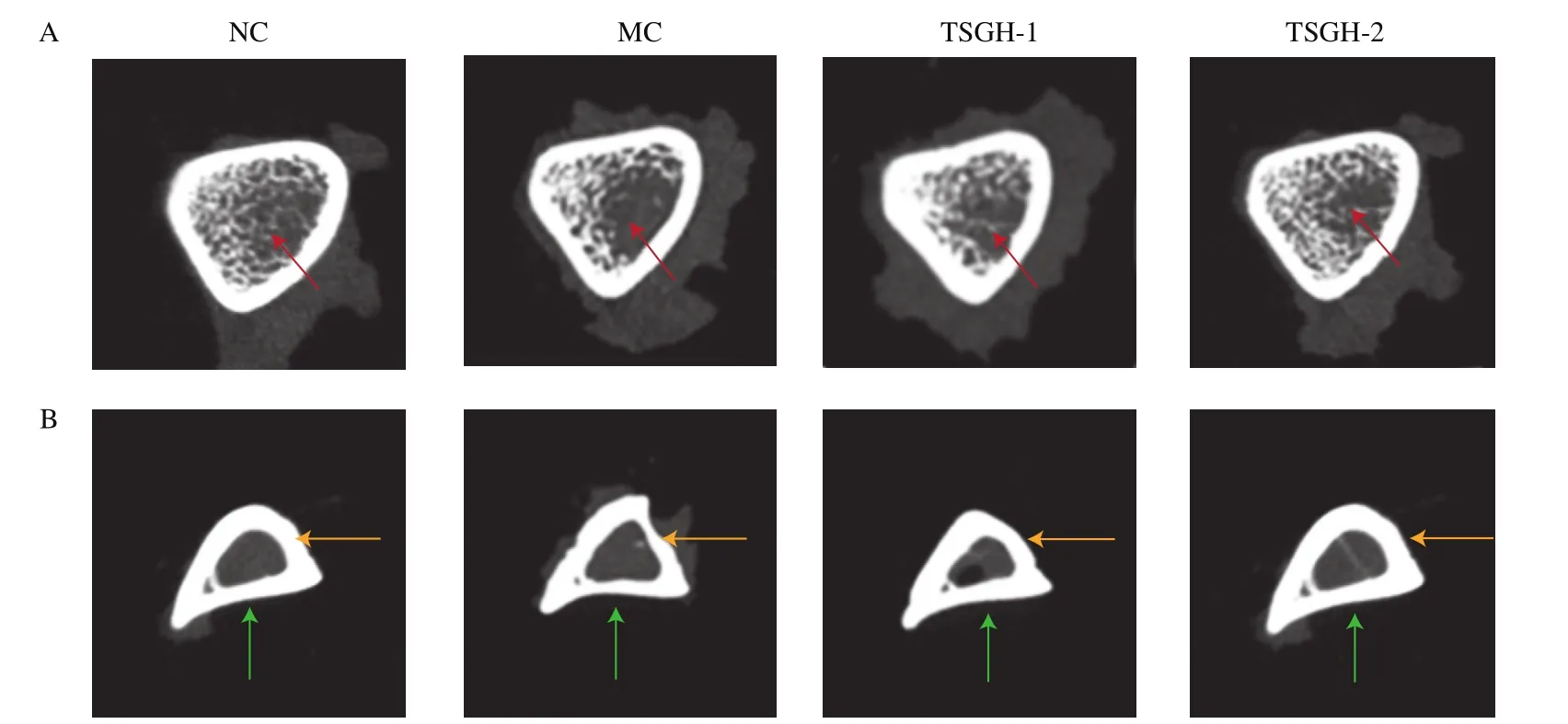
Fig. 4 Bone density electron images obtained by micro-CT detection. (A) The femurs of rats; (B) the tibias of rats. The red arrows indicated the density of trabecular bone and the yellow and green arrows indicated cortical bone thickness in different parts.
3.5 Analysis of bone histomorphology
Histomorphological changes in transverse sections of the femurs were studied by H&E staining and observation under an electron microscope. As shown in Fig. 5A, the femurs of the NC group revealed a good trabecular bone structure, compact gaps, and normal density. Compared with this group, the MC group showed a trabecular bone structure that was remarkably thinned and separated,indicating a severe degree of OP. However, OP was restored in the TSGH groups, and the bone histomorphology of the TSGH-2 group was clearly better than that of the TSGH-1 group. Fig. 5B shows histomorphological changes in the longitudinal section of the femurs as observed under an electron microscope at a magnification of 40×.The CBTs of the left and right sides of the femurs of the MC group were significantly thinner than those of the three other groups. No significant difference in CBT between the TSGH and NC groups was observed.
The osteoclast activity of the femurs was determined by TRAP staining. After TRAP staining, the cytoplasm of the osteoclasts became red. As shown in Fig. 5C, color intensity in the MC group was significantly deeper than that in the other groups, there by indicating that the osteoclasts of this MC group have high activity. The activities of osteoclasts in the femurs could be reduced after treatment with TSGH. Inhibition of osteoclast activity by TSGH appeared to occur in a dose-dependent manner.
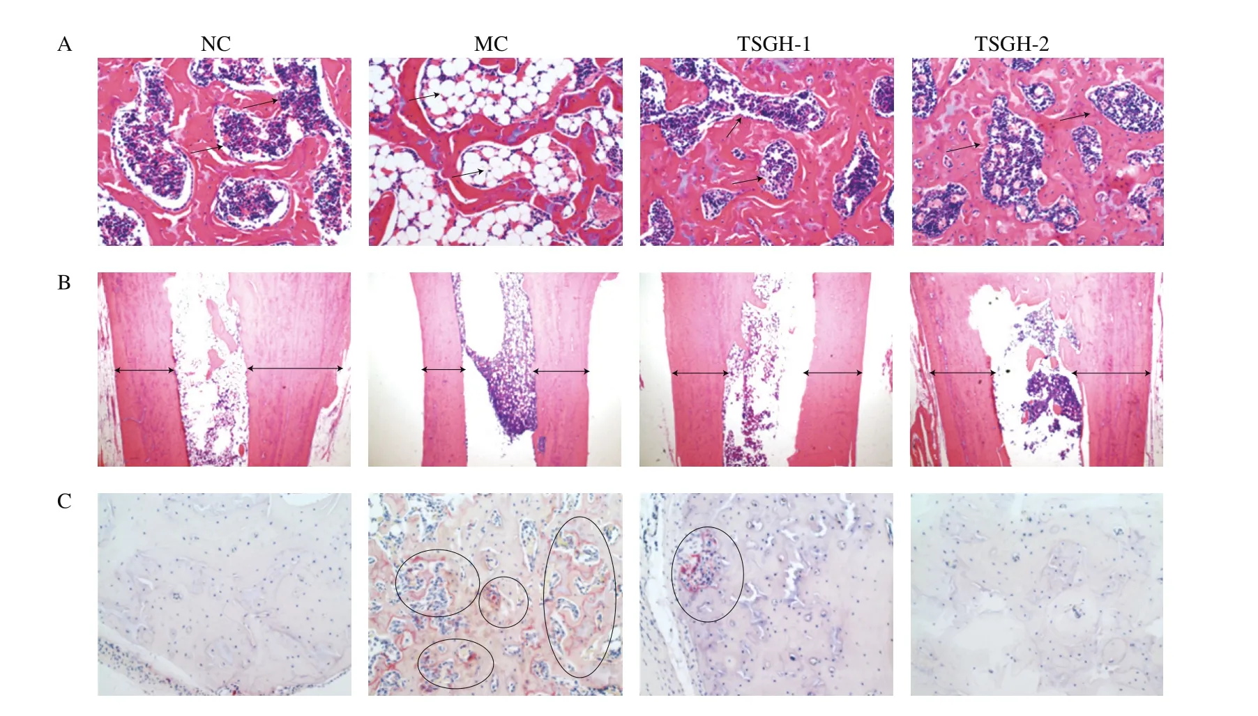
Fig. 5 Effect of TSGH on the histomorphology of bone (right femur). (A) H&E staining microscope images of the femurs transverse section at a magnification of 200×; the arrows indicated that tight and loose of trabecular bone structure. (B) H&E staining microscope images of the femurs longitudinal section at a magnification of 40×; the arrows indicated cortical bone thickness. (C) The microscope images of TRAP staining at a magnification of 200×; the circles indicated the red areas with high osteoclast activity.
4. Discussion
In this study, retinoic acid successfully induced OP in rats within 2 weeks. The model of OP rats induced by retinoic acid is a practical,meaningful, and widely used preclinical model because of its convenient operation, short preparation time, and low failure rate [3,20]. The mechanism of this model is that retinoic acid activates osteoclasts and promotes bone resorption but does not inhibit osteoblast activity,eventually causing bone resorption to exceed bone formation and leading to OP in rats.
The effects of TSGH on Ca absorption and bone growth in OP rats induced by retinoic acid were studied. Previous studies indicated that the OP and bone loss could be ameliorated by restoring intestinal Ca absorption [21,22]. The Ca absorption and bone growth activity of TSGH depend on its amino acid composition and sequence [23].In this study, UPLC-Q-Orbitrap-MS2was applied to identify the amino acid sequences of the peptides in TSGH. Fifty peptides were identified, and most peptides contained -Gly-Pro- sequences, such as GPAGPK, GPLGPR, GPSGPRGPA, and GPAGPRGPSGE, which is accordance to the characteristics of gelatin hydrolysates. TSGH also contained large amounts of Pro and Lys and lacked of Trp and Cys.It has been reported that the high ratio of Gly to Pro in crucian skin collagen peptides (CPs) helps bind Ca ions. Gly, Pro, Glu, and Lys in CPs are the most important amino acids promoting Ca absorption in vivo [18]. A previous study indicated that gelatin hydrolysates have many advantages for bone growth. For example, gelatin hydrolysates could promote the growth and differentiation of osteoblasts, reduce the differentiation of osteoclasts, and regulate bone formation and mineralization [24]. Our previous study showed that TSGH has high Ca-binding capacity in vitro [17]. Therefore, TSGH may be a suitable candidate to improve Ca absorption and regulate bone growth in vivo.
During the experiment, the body weight of the rats, which directly re flects their pathological status, was monitored. Retinoic acid could reduce the appetite of rats, and minimum increases in rat body weight may be expected. During treatment with TSGH, the body weight of the rats significantly increased, and no rats died. The increase rates of the body weight of rats in the TSGH groups were similar to those of rats in the NC group from the third week onward. These results demonstrate that TSGH could essentially regulate the survival state and health conditions of OP rats.
Any disturbance during bone formation and resorption could lead to bone diseases [3]. OP is characterized by changes in bone turnover markers. Serum Ca, P, ALP, and OCN contents are the most important bone turnover markers for assessing bone formation and resorption [25,26]. The results of serum Ca and P assays showed that TSGH could promote Ca absorption and increase the bioavailability of the mineral in rats. Serum ALP is a marker of osteoblast activity or functional status. Previous studies showed that ALP plays a major role in bone calcification, and its expression could be increased during abnormal calcification [26,27]. High ALP levels may inactivate Ca absorption [28]. In this study, TSGH could restore the bone calcification of OP rats to normal levels and improve Ca absorption and calcification. A high dose of TSGH exerts better effects than a low dose. OCN is often found during bone synthesis after bone injury and considered an accurate indicator of osteoblast differentiation [26].Indeed, high OCN levels indicate high Ca deficiency in vivo. When serum Ca levels fall below the normal level after treatment with retinoic acid, OCN is brought into the blood to maintain the necessary Ca level [29]. Our results showed that the serum OCN level in the MC group significantly increased compared with the NC group, which was similar to previous study [19,21]. After treatment with TSGH,the rates of bone synthesis in OP rats were regulated effectively. A previous study indicated that Antarctic krill-derived peptides could recover the levels of bone turnover markers in OP rats [30]. Thisfinding is consistent with the results of the present study.
Osteometry is a key aspect of evaluations of the effectiveness of the new products for OP [31]. Thus, the bone length and DW index of the rats were measured in this study. After treatment with TSGH,the length and DW index of the femurs and tibias of OP rats returned to the normal range, which confirms the positive effects of TSGH on bone synthesis. Bone biomechanical property is a comprehensive index that could directly re flect the anti-fracture ability and strength of bone [32]. Thus, the femurs and tibias of the rats were subjected to three-point bending tests. The results showed that TSGH could effectively increase bone maximum loads and enhance bone strength.A previous study indicated that egg white peptides could regulate the bone length, DW index, and tensile strength of OP rats induced by retinoic acid, similar to ourfindings [33].
Ca is the main component of bone minerals and an important determinant of the degree of OP risk. BMD is thefirst indicator used to judge bone loss and OP [34]. In this study, the bone Ca content and BMD of OP rats could be recovered to a certain extent by TSGH, and a high dose of TSGH exerts better effects than a low dose. The results of bone mineral analysis indicated that TSGH promotes bone growth.The bone density electronic images also showed that TSGH could recover trabecular bone effectively, thereby revealing that TSGH could promote bone formation and prevent resorption [35]. TSGH also regulated the bone parameters, such as CBT, CAR, and TAR,thus further confirming its positive effect on OP rats.
Histomorphological studies, including H&E and TRAP staining,were conducted to illustrate the effect of TSGH on the femurs of OP rats and verify the preventive effect of the hydrolysate on bone damage. H&E staining was applied to the transverse section of the femurs. The trabecular bone structure of the MC group was significantly separated compared with that of the NC group. By contrast, an increase in trabecular bone was observed in the TSGH groups. A similar phenomenon was observed in a previous study, Cabinding peptide prepared from Pacific cod bone nearly completely restored the trabecular bone structure and bone density of OP rats when applied at a high dose [21]. The CBT between groups was compared by H&E staining of the longitudinal sections of the femurs to assess the effects of TSGH on cortical bone further. After treatment with TSGH, the thickness of the left and right sides of the cortical bone significantly increased. The microscope images of H&E staining were consistent with the micro-CT results of CBT, CAR, and TAR(Fig. 3). A previous study indicated that the CBT of rats treated with a new anti-OP agent significantly thickened, similar to our results [36].Therefore, H&E staining of the transverse and longitudinal sections of bone confirms the protective effects of TSGH on trabecular bone loss and CBT. One mechanism of OP induced by retinoic acid is reduction of estrogen level with concurrent increases in osteoclast number.Thus, TRAP staining, a useful method for measuring osteoclast numbers, was carried out to evaluate the effect of TSGH on osteoclast formation. When osteoclasts are stained with TRAP, their cytoplasm becomes red and their nucleus turnsblue [37]. As shown in Fig. 5C,red coloration of the MC group revealed great intensity, but the number of osteoclasts in the TSGH groups effectively decreased in a dose-dependent manner. A previous study revealed that the number of osteoclasts in OP rats induced by retinoic acid could be significantly reduced by treatment with desalted duck egg peptides, which was consistent with our results [33].
5. Conclusions
In this study, TSGH were obtained by complex protease hydrolysis. Fifty peptides were identified, and most of these peptides contained the amino acid sequence -Gly-Pro-. The protective effects of TSGH on Ca absorption and bone growth were evaluated by using rats with retinoic acid-induced OP. Serum parameters showed that TSGH can improve Ca absorption, and bone parameters indicated that the hydrolysate can promote bone formation and prevent resorption.Bone histomorphological studies showed that the bone trabecular structure and CBT were restored by TSGH. These results suggest that TSGH is a potential novel alternative for OP treatment.
Con flict of interest
The authors declare no con flicts of interest.
Acknowledgments
We gratefully thank the National Natural Science Foundation of China (Grant No. 31360381) for thefinancial support on this research.
杂志排行

食品科学与人类健康(英文)的其它文章
- Production of antihypertensive and antidiabetic peptide fractions from quinoa (Chenopodium quinoa Willd.) by electrodialysis with ultrafiltration membranes
- Identification and characterization of a novel tetrapeptide from enzymatic hydrolysates of Baijiu byproduct
- Effects of phosvitin phosphopeptide-Ca complex prepared by efficient enzymatic hydrolysis on calcium absorption and bone deposition of mice
- Structural requirements and interaction mechanisms of ACE inhibitory peptides: molecular simulation and thermodynamics studies on LAPYK and its modified peptides
- Anti-diabetic and anti-hyperlipidemic effects of sea cucumber(Cucumaria frondosa) gonad hydrolysates in type II diabetic rats
- Antibacterial and antibiofilm activity of peptide PvGBP2 against pathogenic bacteria that contaminate Auricularia auricular culture bags