Gypenoside ameliorates insulin resistance and hyperglycemia via the AMPK-mediated signaling pathways in the liver of type 2 diabetes mellitus mice
2022-06-23MengxueSongDehongTnBinLiYnqunWngLinShi
Mengxue Song, Dehong Tn,b, Bin Li,b, Ynqun Wng,b, Lin Shi,b,*
a College of Food Science, Shenyang Agricultural University, Shenyang 110866, China
b Key Laboratory of Healthy Food Nutrition and Innovative Manufacturing of Liaoning Province, Shenyang Agricultural University, Shenyang 110866, China
Keywords:
Gypenosides
Gynostemma pentaphyllum
Diabetes
Insulin resistance
Gluconeogenesis
Adenosine monophosphate-activated protein
kinase (AMPK)
A B S T R A C T
Gynostemma pentaphyllum, also called “Southern Ginseng” in China, is a traditional Asian folk medicinal plant. Gypenosides (Gps) are the biologically active constituents of G. pentaphyllum, which have been reported with hypoglycemic activity. However, the underlying mechanisms are unclear. The effects of two Gps (Gp-I and Gp-II) on type 2 diabetic mellitus (T2DM) mice, induced by high-fat and high-sugar diet and streptozotocin, were evaluated to explore the mechanism of their hypoglycemic actions. Gps reduced fasting blood glucose and serum lipids, as well as significantly improved T2DM mice glucose tolerance and insulin resistance (IR). After Gps treatment, the severity of liver injury was reduced and liver glycogen content increased. In addition, Gps promoted the phosphorylation of adenosine monophosphate-activated protein kinase (AMPK), and downregulated the key proteins phosphoenolpyruvate carboxy kinase and glucose-6 phosphatase, in the AMPK signaling pathway. Thus, our study suggests that Gps mediate hepatic gluconeogenesis and improve IR via activating AMPK signaling pathway in T2DM mice.
1. Introduction
Gynostemma pentaphyllum(Thunb.) Makino, mainly distributing in China, Japan, North Korea and Southeast Asia, is an edible and medicinal plant [1]. As the main active constituents ofG. pentaphyllum,a group of triterpene saponins-gypenosides (Gps) are found to pose very similar action to ginsenosides [2]. It was reported that Gps exhibited various biological activities, such as anti-cancer [3,4],anti-inflammation [5-8], prevention of atherosclerosis [9], inhibition of apoptosis [10], protection of nerve [11,12], and anti-hypolipidemic [13,14]and hypoglycemic effects, etc. [15,16]. The hypoglycemic activity of Gps has been demonstrated in previous researches [17], however,there are still few reports about the mechanisms by which Gp improves insulin resistance (IR) and inhibits hepatic gluconeogenesis.
It is known, the primary type 2 diabetic mellitus (T2DM) is mainly caused by IR, which is the leading cause of nonalcoholic fatty liver disease, while hepatocellular injury and liver fatty change would aggravate IR [18]. Besides, the greatest source of whole-body IR is the defection of transduction pathways of the insulin signal in liver [19].The liver is the main target organs of insulin action and plays a significant role in glucose regulation and lipid metabolism [20].T2DM causes the increase of free fatty acid and steatolysis,accumulated in the liver, and then causes lipotoxicity damage [21].Therefore, improving the steatosis pathological changes in the liver is essential for intervening T2DM IR.
Adenosine monophosphate-activated protein kinase (AMPK)is a kinase and an energy sensor that is widely involved in various metabolic regulation and plays a key role in regulating glucose homeostasis, insulin sensitivity and all aspects of cell function [22,23].In diabetic patients, increased activity or synthesis of key enzymes of hepatic gluconeogenesis leads to increased hepatic glucose output, causing the occurrence of IR, which is the primary cause of fasting blood glucose (FBG) abnormal increase [24,25].Furthermore, activated phosphorylation of AMPK (p-AMPK)plays an important role in the regulation of lipid synthesis via inhibiting related transcription factors [26]. It also restrains the level of gluconeogenesis-related key enzymes, such as PC (pyruvate carboxylase), PEPCK (phosphoenolpyruvate carboxykinase),G6Pase (glucose-6-phosphatase), thereby reducing blood glucose concentration to control blood sugar stability [27,28]. Accordingly,it is of great significance and value to investigate whether natural products regulate glycolipid metabolism and improve IR through the AMPK-mediated signaling pathway.
In our serious of studies on Gps, we have found some active compounds. Gp-I and Gp-II are the ones more abundant among them, about 0.8% and 1.0%, respectively. As a continuation of our work for discovering the functional factors [29,30], in the present study, we evaluated the effects of two groups (Gp-I and Gp-II) on the hypoglycemic mechanism and liver pathology in T2DM mice induced by high-fat and high-sugar diet (HFSD) and streptozotocin (STZ).
2. Materials and methods
2.1 Plant material and reagents
The crude saponins ofG. pentaphyllum(> 80%) were obtained from Tianyi Bio-Tech Inc. (Shaanxi, China). The voucher specimen (No. 2014010) was deposited at our laboratory.
Dimethyl sulfoxide (DMSO), 3-(4,5-dimethylthiazol-2-yl)-2,5-diphenyltetrazolium bromide (MTT), MeOH and cell lysis solution were purchased from Sigma-Aldrich Chemical Co. (St. Louis, MO,USA). Penicillin streptomycin solution, trypsin, phosphate-buffered saline (PBS, pH 7.4) were from Solarbio Science and Technology Co.,Ltd. (Beijing, China). Biochemical kits were obtained from Jiancheng Bioengineering Institute (Jiangsu, China). Blood glucose meter and test strips were obtained from Yuyue Medical Equipment Co.,Ltd. (Jiangsu, China). Antibodies, secondary antibodies, STZ, RIPA Lysis Buffer, polyvinylidene fluoride (PVDF) membranes, AMPK,p-AMPK, PEPCK, G6Pase and PC were gathered from Solarbio Science and Technology Co., Ltd. (Beijing, China). Additionally, the other chemicals used in this study are of analytical grade.
2.2 Sample preparation
The totalG. pentaphyllumsaponins (100 g) were chromatographied repeatedly over silica gel with CH2Cl2-MeOHH2O (7:2:1, 7:3:1, 7:4:1). Six fractions, A-F were obtained. Fr. E was separated into five fractions, Ea-Ee, by ODS column (85% MeOH). Fr. Ec was purified by preparative HPLC using MeOH-H2O(75:25) as eluent to afford Gp-I (810 mg,tR= 33 min). Fr. D was purified on preparative HPLC (82% MeOH) to yield Gp-II (1 020 mg,tR= 17 min).
2.3 Cell culture
The HepG2 cell line was obtained from Chinese Academy of Sciences (Beijing, China). They were grown as previously described [31].Cells in the mid-log phase were used for experiments.
2.4 Cytotoxicity evaluation
According to the previous method [32], the cytotoxicity toward HepG2cells was determined. The CC50(median cytotoxic concentration) of Gp-I and Gp-II were calculated. The absorbance value was measured at 570 nm using a Microplate Reader.Experiments were performed in quintuplicate and repeated three times. The viability was calculated by the equation indicated below.

2.5 Animal experimental design
Four-week-old healthy male Kunming mice, clean-grade, male,weighing (18 ± 2) g were purchased from Changsheng Biotechnology Co., Ltd. (license number SCXK (Liao)-2015-0004, Liaoning,China). All animals were subjected to adaptive feeding at (23 ± 3) °C,(55 ± 5)% relative humidity and 12 h light/dark cycle for 1 week prior to the beginning of the animal experiment. The animal experiment was compliant with the World Medical Association Declaration of Helsinki, and approved by the Animal Ethics Committee of Shenyang Agricultural University.
Afterwards, they were randomly divided into the normal control group (NC) and the experiment groups, which were fed with normal pellet diet and high-fat and high-sugar diet (58.8% basic feed, 15% saccharose, 15% lard oil, 10% protein, 2% cholesterol,1% vitamins, 0.2% cholate, Maohua Biotechnology Co., Ltd.(Shenyang, China)), respectively. Four weeks later, the experiment groups were intraperitoneal injected with STZ (60 mg/kg bw),dissolved in citric acid-sodium citrate buffer, pH 4.4) after being fasted for 12 h to induce type 2 diabetes [33]. Meanwhile, the NC group was treated with equal amounts of citric acid-sodium citrate buffer. After 1 week, the mice with fasting blood glucose (FBG)levels ≥ 16.7 mmol/L were considered successfully induced to T2DM and selected for further treatment. Diabetic mice were randomly divided into 4 groups (n= 8): diabetic model group (DM), positive control group (MH, administered with metformin hydrochloride tablet, 50 mg/kg bw), Gp-I group and Gp-II group. The sample dose being intragastric administered was 50 mg/kg bw. The normal control group and diabetic model group were given with an equal volume of normal saline for 4 weeks. All animals were sacrificed 24 h after the last Gps treatment with phenobarbital sodium (i.p., 100 mg/kg)anesthesia, blood and liver samples were taken for assays.
2.6 Oral glucose tolerance test (OGTT)
After 4 weeks, the mice, which underwent 12 h fasting, were treated separately with metformin hydrochloride tablet, Gp-I and Gp- II. The method of OGTT as the same as Veerapur et al. [34].Glucose contents were calculated as the area under the curve (AUC).

Where,G1andG2are indicate of blood glucose at different time points,T1andT2are different tested time points.
2.7 Determination of metabolic parameters
Body weight, food intake and water consumption (weighted the water bottle every 24 h and calculated the difference) of mice were recorded once daily. FBG concentration was measured once a week using a blood glucose meter (Yuyue Medical Equipment Co., Ltd., Jiangsu, China). After the last injection and fasting for 12 h, mice eyeballs were removed and blood was collected from the orbit, centrifuged to take serum. The liver was obtained after the mice were sacrificed by cervical dislocation. Liver glycogen content and serum parameters, e.g. fasting serum insulin (FINS),non-esterified fatty acid (NEFA), triglyceride (TG), total cholesterol (TC),high-density lipoprotein cholesterol (HDL-C), low-density lipoprotein cholesterol (LDL-C), aspartame aminotransferase (AST) and alanine aminotransferase (ALT), were measured according to the instructions in biochemistry reagent kits (Nanjing Jiancheng Bioengineering Institute, Jiangsu, China). The parameters for evaluation were calculated as follow:

Where HOMA-IR indicates the homeostasis model assessment of insulin resistance, HOMA-β is the homeostasis model assessment of β-cell function, QUICKI is Quantitative insulin sensitivity index and ISI refers to the insulin sensitivity index.
2.8 Histopathological examinations
Histopathological observation of the liver was conducted according to the reported experimental method. In short, liver tissues were fixed in 10% formaldehyde solution (pH 7.4). After rinsing, the tissue was cut into small pieces, dehydrated in gradient ethanol solution, and embedded in paraffin. The 4 µm thick sections were prepared using a Leica Microsystem microtome (Model RM 2265, Germany), and stained with hematoxylin and eosin (H&E).The slides were observed and photographed under an Olympus microscope (4X-1, Japan).
2.9 Western blot analysis
Western blot was used to evaluate target protein levels. Brie fly,the mice liver was homogenized in RIPA buffer and then centrifuged at 14 000 r/min for 15 min at 4 °C. BCA protein assay was used to detect the total protein concentration. SDS-PAGE (sodium dodecyl sulfate-polyacrylamide gel electrophoresis) was used to isolate protein from equal amounts of samples and the protein was transferred to a PVDF membrane. Then, a blocking reagent,consisting of Tris-buffered saline with Tween-20 (TBST) and 5% skimmed milk powder, was prepared to incubate the membrane at room temperature for 2 h, followed by primary antibodies probing overnight at 4 °C. The primary antibody was diluted in TBST and included anti-GAPDH (1:10 000), anti-PC (1:1 000), anti-G6Pase(1:1 000) anti-PEPCK (1:1 000), anti-AMPK (1:1 000) and anti-p-AMPK (1:1 000). After 3 times TBST washing, the membrane was diluted with blocking solution containing the secondary antibody(1:5 000), incubated at room temperature for 2 h. The protein bands were visualized with ECL reagent and observed by autoradiography.The result was quantitatively analyzed by ImageJ software.
2.10 Statistical analysis
Data were expressed as mean ± standard deviation. Data were subjected to one-way ANOVA, and significant differences were analyzed using SPSS 22.0 software (SPSS, Chicago, IL, U.S.).P< 0.05 andP< 0.01 indicate significant differences between groups.
3. Results
3.1 Structural identification of compounds Gp-I and Gp-II
In this research, we isolated and identified two gypenosides (Table 1)via Preparative HPLC (CXTH Corp., Beijing, China) and NMR Spectra (Bruker AV-600 and ARX-400 spectrometers). The two known compounds were (20S, 23S)-3β,20-dihydroxydammar-24-en-21-oic acid 21,23-lactone 3-O-[α-L-rhamnopyranosyl(1→2)][β-D-xylopyranosyl(1→3)]-6-O-acetyl-β-D-glucopyranoside (Gp-I) and gypenoside XLIX (Gp-II). And they were identified by comparing the1H and13C NMR data with that in the literatures [35,36].

Table 1Cytotoxicity assays of two compounds on the growth of HepG2 cells.
3.2 Cytotoxicity assays on liver cells
Using HepG2 cell line, the antitumor activities of 2 Gps were evaluatedin vitroand the results were listed in Table 1. The CC50values of them were (125.76 ± 2.47), (169.62 ± 3.09) μg/mL,respectively. It was clear that the cell viabilities were 109.13% and 113.27% at 50 μg/mL.
3.3 Mice body weight, liver index, food intake and water consumption
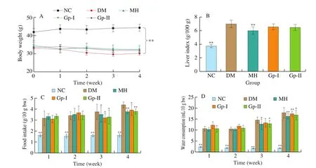
As shown in Fig. 1, compared with NC mice, diabetic mice showed gradually decreasing body weight, increasing food intake and water consumption (P< 0.01). The liver indices of mice in the Gp-I and Gp-II groups were lower than that of the DM group, but with no significant difference (P> 0.05). These characteristics may be related to weight loss, polydipsia and polyphagia in diabetes with impaired energy metabolism [37,38]. The initial body weight (0 week) of diabetic mice was less than the NC group, which was attributed to STZ injection. The weight of mice in the NC group showed an increasing trend with the increase of feeding time, which was significantly different from the DM group (P< 0.01), while the other groups were not. After 4 weeks feeding, the body weight of mice in the DM, MH,Gp-I and Gp-II groups were reduced by 10.09%, 6.05%, 5.03% and 5.27%, respectively. The liver index of diabetic mice was significantly higher than that in the NC group, and no clear difference was observed in experimental groups except the MH group (Fig. 1B). The food intake of the DM group mice increased from 3.19 g/10 g bw to 4.41 g/10 g bw (Fig. 1C), and the water consumption increased from 10.81 mL/10 g bw to 13.17 mL/10 g bw (Fig. 1D). From the data, the treatment of Gp-I and Gp-II alleviated the thirst of diabetic mice and improved their consumption of food.

Fig. 1 The effect of Gps on body weight (A), liver index (B), food intake (C) and water consumption (D) in diabetic mice. *indicates significant difference compared with the DM group, *P < 0.05, **P < 0.01.
3.4 Effects of Gp on FBG and OGTT in T2DM mice
The FBG data of mice in each group were measured weekly.As shown in Fig. 2A, at 0 week (the initial blood glucose value of administration), the fasting blood glucose of the model group (24.07 mmol/L) was significantly higher than that of the NC group (P< 0.01), which suggested that the T2DM model was successfully constructed. It can be seen that the levels of FBG mice in MH, Gp-I and Gp-II groups were significantly lower than those in the DM group at the end of the experiment. Meanwhile, the OGTT curves and the AUC for each group were presented in Fig. 2. It was found that the highest blood glucose level occurred at 0.5 h after giving 2 g/kg glucose and it began to decline at 1 h. Following this, at 2 h,the blood glucose of Gp-I and Gp-II groups returned to the initial blood glucose level of OGTT, as same as that of the MH and NC groups. However, for the DM group, the 2 h blood glucose value increased by 20% compared with the 0 h blood glucose value. In addition, it can be seen from the AUC data (Fig. 2C) that the AUC of the Gp-I and Gp-II groups was significantly lower than that of the DM group, and both were 30% lower than the DM group (P< 0.05).These results suggested that the hypoglycemic effects of Gp-I and Gp-II on T2DM mice are similar to those of metformin hydrochloride tablets.

Fig. 2 (A) Changes of FBG in different groups, (B) blood glucose curves of different groups during OGTT and (C) the AUC of blood glucose curves. *indicates significant difference compared with DM group, *P < 0.05, **P < 0.01.
3.5 Analysis of insulin level and insulin sensitivity
It can be observed that the glucose tolerance of T2DM mice was lower than normal mice, while insulin levels were in contrast. The analysis of IR and higher insulin levels in T2DM serum are essential for coping with glucose tolerance. As shown in Table 2, compared with the NC group, the T2DM mice FINS and HOMA-IR increased significantly, indicating that pancreatic islet cells secreted too much insulin, resulting in IR. However, under the effect of metformin hydrochloride, Gp-I and Gp-II, the HOMA-IR of T2DM mice decreased significantly (P< 0.05 orP< 0.01). On the other hand,the data of QUICKI, ISI, and HOMA-β of experimental groups were distinctly lower than normal mice (P< 0.01). In terms of HOMA-β,the DM group was 16.09 ± 2.08, significantly lower compared to the NC group with 107.86 ± 9.90, which indicated that the β-cell function of T2DM mice was impaired. It was noted that the values of HOMA-β in Gp-I group and Gp-II group were higher than that in the DM group. In addition, there was no significant difference in the level of Fins between the 3 groups (Gp-I, the Gp-II and DM). The results suggested that the intervention of Gp-I and Gp-II reduced islet β-cell damage and improved insulin sensitivity, and significantly improved IR caused by diabetes, but there are no obvious effects on FINS.

Table 2Fasting insulin levels and insulin sensitivity of mice in different groups.
3.6 Effects of Gps on serum lipids
Data analysis of mice serum indices after the 4 weeks administration is shown in Table 3. It can be seen from the data that the TC, TG, and LDL-C levels of diabetic mice were obviously higher than ones of the NC group (P< 0.05 orP< 0.01), indicating their lipid metabolism was disordered. Moreover, Gp-I had a positive effect on TG and LDL-C with a significant decrease (P< 0.05 orP< 0.01), while Gp-II reduced TG only. The ratio of TC and HDL-C were (2.21 ± 0.35), (3.50 ± 0.67), (2.65 ± 0.48), (3.01 ± 0.59) and(3.09 ± 1.12) mmol/L, related to NC, DM, MH, Gp-I and Gp-II groups, respectively. Accordingly, the value of ratio for Gp-I and Gp-II groups were lower than that of the DM group, indicating their advancements in improving hepatic injury. Besides, the NEFA of the DM group was 1.9 times higher than the NC group, indicating the damnification of diabetic mice β-cells. Compared with the DM group, the NEFA concentration of Gp-I and Gp-II groups showed an approximate decrease of 0.39 and 0.25 mmol/L, respectively. Oral administration of Gps ameliorated serum lipid metabolism in T2DM mice effectively.

Table 3Lipids (mmol/L) in serum of mice in different groups.
3.7 Effects of Gp on liver glycogen content and liver tissue histopathological
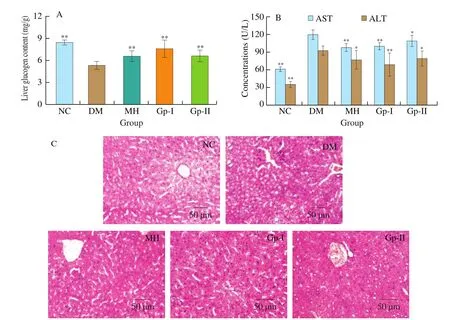
The levels of AST and ALT in serum, which reflect the liver damage severity, were analyzed and shown in Fig. 3B. It has shown that Gps significantly reduced the concentration of AST and ALT in the serum of T2DM mice. Compared with the DM group, the liver glycogen content of Gp-I and Gp-II groups increased significantly(increased by 42% and 24%, respectively,P<0.01). Consistent with the results obtained by H&E staining, it is suggested that Gps regulates blood glucose levels by protecting T2DM mice from liver damage and promoting the production of liver glycogen content.Pathological evaluations of liver sections display representative photomicrographs from mice after 4 weeks of 4 groups (Fig. 3C).There were no histological changes in the NC group. The hepatocytes were neatly arranged around the central vein radially, and the structure was clear. In contrast, the DM group mice had an extensive liver injury, there were disordered arrangement and obvious cellular swelling of the hepatocytes. After treatment with Gp-I and Gp-II,there was less edema, the structure basically returned to normal, and the treatment effects were similar to that of the MH group.

Fig. 3 (A) Liver glycogen content. (B) AST, ALT concentrations in serum. (C) Histopathological change of the liver in different groups, hypertrophy assessed by H&E. *indicates significant difference compared with DM group, *P < 0.05, **P < 0.01.
3.8 Effects of Gp on AMPK protein signaling pathway in T2DM mice
As shown in Fig. 4, compared with the NC group, the expression of total AMPK protein in mice of each group had no significant difference (P> 0.05). Whereas, the lower level of AMPK phosphorylation in diabetic mice was observed to be restored in various degrees in Gp-I and Gp-II groups (P< 0.05). Compared with the blank group, the PC, PEPCK and G6Pase of the DM group were significantly increased, and the intervention of two Gps reduced the expression of the PEPCK and G6Pase in the liver (P< 0.05 orP< 0.01). Therefore, our data revealed that Gps treatment was associated with enhanced AMPK phosphorylation levels along with decreased key enzyme levels of gluconeogenesis, suggesting that reduced hepatic gluconeogenesis is responsible for the hypoglycemic effects of Gps.

Fig. 4 Relative expression of key proteins of mice with type 2 diabetes, as tested by (A) Western blot analysis, (B) AMPK, (C) p-AMPK, (D) PC, (E) G6Pase and(F) PEPCK. *indicates significant difference compared with DM group, *P < 0.05, **P < 0.01. #indicates significant difference compared with NC group, #P < 0.05, ##P < 0.01.
4. Discussion
T2DM is a metabolic disease accompanied by a series of symptoms, such as hyperglycemia, hyperlipidemia, and hepatic steatosis. The main characteristics are insufficient insulin secretion and insulin resistance. Long-term hyperglycemia can lead to the damage of the function of islet β-cells and insulin receptors, resulting in IR and lacking of insulin. Thus, it is essential to control the hyperglycemia tightly to protect the patients from suffering serious symptoms. Previous studies have shown that Gps have hypoglycemic effects. We purified and obtained Gp-I and Gp-II from the total saponins ofG. pentaphyllumand con firmed the hypoglycemic effect of these compounds. In present study, T2DM mice had worse glucose tolerance as well as the ability to recover blood glucose in the DM group. However, Gp-I and Gp-II markedly reduced the levels of FBG and alleviated the IR in T2DM mice induced by HFSD and STZ after 4 weeks of treatment. It demonstrated that Gp-I and Gp-II both have hypoglycemic effect similar to metformin hydrochloride.
In addition to insulin resistance, in obese T2DM patients,the excessive lipid in their diet can lead to various complications of diabetes. On the one hand, high levels of harmful blood fats are the primary cause of dyslipidemia, which increases the risk of cardiovascular disease. On the other hand, T2DM causes the disorder of lipid metabolism boosting concentrations of the free fatty acid (e.g. TC, TG and LDL-C) in blood and accumulating fat in tissues, such as the liver and pancreas, which result in hepatocellular injury and liver fatty change. It was reported that the glucose intake in the muscle will decrease with the increase of NEFA concentration,while promoting the synthesis of triglycerides. Meanwhile, it will also induce gluconeogenesis in the liver and result in β cells failure [39].Besides, the ratio of TC/HDL-C is an important indicator related to non-alcoholic fatty liver disease and cardiovascular disease [40].In the study, Gp-I and Gp-II significantly decreased the levels of TG, LDL-C and NEFA and increased TC/HLD-C levels in serum of T2DM mice, which was attributed to the positive effects of Gps on blood glucose levels and insulin secretion probably. Liver is both the main target organs of insulin action and the main organ of gluconeogenesis, which plays a key role in regulating lipid and glucose metabolism and maintains the dynamic balance of blood glucose. Meanwhile, insulin activity and glucose metabolism can be indirectly reflected by the severity of liver damage and liver glycogen synthesis content [41]. Accordingly, it is an effective way for intervention of T2DM by improving the steatosis pathological histological changes in liver. Furthermore, the content of ALT and AST in serum and edema in liver were relieved via the intervention of Gps, which also increased the liver glycogen content of diabetic mice.These results indicated that Gp-I and Gp-II have a direct effect on the protecting liver and recovering the liver damage by improving T2DM mice hyperlipidemia.
Gluconeogenesis plays an important role in maintaining blood sugar balance and fasting hyperglycemia in diabetic patients is also related to the excess glucose from the pathway [42]. It is well known that AMPK is a pharmacological target for the regulation of gluconeogenesis and glycogen synthesis. Meanwhile, the expression and activity of enzymes in gluconeogenesis, including PC, PEPCK and G6Pase, is an important factor for IR [43]. In fact, increased gluconeogenesis is considered to be one of the early pathological changes in diagnosed T2DM patients. Western blot results showed,key proteins involved in AMPK-mediated gluconeogenesis, such as PC, PEPCK and G6Pase, were down-regulated and p-AMPK was up-regulated in Gps-treated T2DM mice. Thus, it can be concluded that Gp-I and Gp-II have the potential to treat hyperglycemia by improving IR and inhibiting gluconeogenesis through AMPK-mediated signaling pathway. Gp-I was a little more effective for than the other one, suggesting that the number of sugar moieties, and the kind of side chain may affect their biological activity. However, the underlying mechanisms and whether it modulates signaling pathways in other organs need further in-depth researches to explore.
5. Conclusions
In this work, we found Gp-I and Gp-II ameliorated insulin resistance and hyperglycemia of STZ-induced T2DM mice, which may be achieved through AMPK-mediated signaling pathway. Taken together, our studies may make an opportunity to develop Gps as potential anti-diabetic functional factors.
conflict of interest
The authors declare that there are no conflicts of interest.
Acknowledgements
This work was supported by the National Natural Science Foundation of China (81602983).
杂志排行
食品科学与人类健康(英文)的其它文章
- Hypoglycemic natural products with in vivo activities and their mechanisms: a review
- Bacteroides utilization for dietary polysaccharides and their beneficial effects on gut health
- Capsular polysaccarides of probiotics and their immunomodulatory roles
- Natural compounds may contribute in preventing SARS-CoV-2 infection: a narrative review
- A comprehensive review on the effects of green tea and its components on the immune function
- A review on current and future advancements for commercialized microalgae species
