Enigmatic rapid organization of subdural hematoma in a patient with epilepsy:A case report
2022-06-23HongTaoLvLinYunZhangXiaoTongWang
lNTRODUCTlON
Subdural hematomas(SDHs)following traumatic head injury constitute an essential proportion of the cases seen in daily practice in the field of neurosurgery.An SDH is considered chronic(CSDH)when it is discovered > 3 wk after the initiating trauma[1].Organized chronic SDH(OCSDH)is a rare category of CSDH characterized by the formation of areas of solid consistency encapsulated by thick membranes[2,3],which has been extensively reported regarding the diagnosis,treatment options and outcomes.Only a minority of CSDHs develop into OCSDHs,and OCSDHs have various clinical manifestations.Although the mechanisms underlying the development of OCSDHs in these cases remain unclear,the process of hematoma organization is undoubtedly slow(probably lasting at least 6 mo)[2,4,5].It is conceivable that OCSDH develops only when the hematoma is chronic.By reviewing the literature,we found no previous reports of the rapid formation of membranes around a hematoma,causing it to appear organized,which is an interesting presentation.In this article,we report a rare case of organized acute SDH(ASDH)with an unusual appearance found in a deceptively simple craniotomy performed to evacuate a hematoma,and we believe this is the first report of this phenomenon.
CASE PRESENTATlON
Chief complaints
A 56-year-old female farmer had a seizure 13 h prior to presentation along with a slight fever.
History of present illness
The patient developed a cold approximate 5 d prior to presentation after being exposed to rain.After a careful investigation of her history,we learned that the patient had two separate tonic-clonic seizures 2-3 h after the primary seizure,characterized by oral and linguistic automatism in sleeping.
History of past illness
Her major medical history included hypertension and diabetes for > 10 years;both of which were marginally controlled by medication,a hysteromyomectomy 20 years before,cerebral infarction 12 years before,and renal dysfunction incidentally detected 3 years before of unknown current extent.
Personal and family history
Three of her family members,including the patient’s mother and two brothers,died of bronchitis.The brothers passed away at young ages.
Physical examination
The patient exhibited clear consciousness and normal motor function[Glasgow Coma Score(GCS)15 points]with occasional delirious speech and a positive Babinski sign on the right side.Both pupils were 3 mm in diameter(d)and sensitive to light.No other specific physical signs were found.
Laboratory examinations
This hematoma did not have characteristics consistent with those of ASDH,CSDH or even SSDH with acute rebleeding.We cautiously present a theory that it may have been a subarachnoid hematoma in which the outer membrane of the hematoma was actually the arachnoid that was thickened for some reason,and what was left on the brain surface was just pia mater(it looked dry,unlike arachnoid covered with CSF).Regardless of the true nature of this hematoma,a large craniotomy was the only correct choice for the patient.
Imaging examinations
Computed tomography(CT)on admission showed multiple malacia sites,mainly in the right lobe,without any signs of hemorrhage(Figure 1A).Magnetic resonance imaging(MRI)on day 3 after admission showed no signs of intracranial hemorrhage(Figure 2).On the morning of day 10 after admission,sudden deterioration of the patient prompted us to perform emergency CT,which showed a left supratentorial SDH with considerable mass effect(Figure 1B).
FlNAL DlAGNOSlS
The main diagnosis was symptomatic epilepsy and ASDH.
TREATMENT
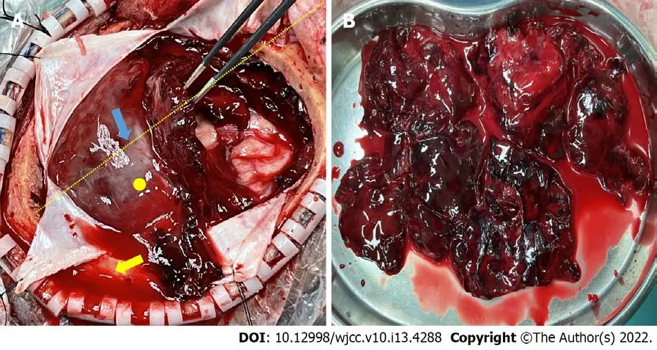
The process of treatment was rather dramatic.Immediate antiepileptic treatment as well as treatment for the comorbidities were administered on admission,and the patient seemed to remain stable over the course of the following week while necessary examinations were conducted.Just when we thought everything was going well,surprisingly,the patient experienced projectile vomiting and a severe headache on the morning of day 10.However,only 20 min later,she began to show arresting weakness on her right side but she could still response to calling and her pupils were good.We ran an emergency CT scan(Figure 1B).By the time the patient was returned to her room,she was in complete coma[GCS 5 points,E(eye opening)1 points;V(verbal response)1 points;M(motor response)3 points],and her left pupil was dilated to 4 mm and fixed.After quickly debriefing her family members on the lifethreatening situation,we operated as soon as possible with large craniotomy.The intraoperative findings were surprising.The hematoma strongly resembled an organized hematoma and assumed a form we had not previously observed.We inclined to think the hematoma was acute according to the clinical course and CT scan despite some low-density components inside it.Therefore,the observation of the absence of a liquefied hematoma with only a small amount of clear CSF when we started to open the dura mater was an anomaly.The hematoma was better exposed after we cut the dura completely open in a cruciform fashion,and we found it under a solid covering of a thick membrane(Figure 3).This finding made us even more confused because we were confident that it was definitely not a CSDH based on strong evidence of recent CT and MRI scans.The flap range was believed to be sufficient for a typical ASDH but not necessarily for an organized hematoma,so a tentative tumorectomy-like technique was used to remove the hematoma in pieces.Fortunately,the attempt proved successful,which enabled us to evacuate the hematoma thoroughly without any unwanted damage.However,the subarachnoid space macroscopically appeared dry,and a thinnish inner membrane of the hematoma was detected that did not adhere significantly over the brain surface(Figure 3).We washed the cavity with plenty of saline until it cleared,during which minor frontal lobe superficial arterial bleeding was easily controlled by bipolar electrocoagulation hemostasis.The procedure was successful.The brain tissue remained collapsed without emerging pulsations before closure,and the patient was then transferred to the intensive care unit(ICU)for further observation.


We performed an electroencephalogram on day 2 after admission,but re-examination was required for conclusive findings.However,we were unable to perform the re-examination due to the unexpected craniotomy.On day 3,MRI revealed nothing to alert us to the hemorrhage.Another day later(day 4 of hospitalization),we tested her CSF by a regular lumbar puncture.The pressure was approximately 190 mmH
O,and approximate 7 mL of clear CSF was obtained and sent for evaluation.On the morning of day 9,the patient suffered a gelastic seizure without warning that was later disassembled by injection of diazepam and dilantin
representing the only recurrence since admission.Then on day 10,it happened as above.However,the husband revealed that the patient hit her head heavily against the wall in a bathroom several minutes prior to deterioration.Carelessness was to blame for her injury,according to her husband,who was confident that it was not another seizure.
OUTCOME AND FOLLOW-UP
During the initial treatment,oral aspirin was given to the patient who was not a routine user of the drug in the past for ischemic considerations in the nine consecutive days before this unexpected operation.We are not sure whether this treatment played a role in the hemorrhagic event.However,some authors suggest that an elevated risk of SDH(SSDH/CSDH)is related to the use of antiplatelet and/or anticoagulation agents[10,11].
I slouched down in the passenger seat of our old Pontiac ’cause it was the cool way to sit when one is in the fourth grade. My dad was driving downtown to shop and I was going along for the ride. At least that’s what I had told him -- actually I had an important question to ask that had been on my mind for a couple of weeks and this was the first time I had been able to maneuver1 myself into his presence without being overt2 about it.
He thought, in fact, that the pile of wood rather grew bigger than smaller, in spite of what he took off it; so he let his hands fall by his side, and dried the sweat from his forehead, and was ill at ease, for he knew that it would be bad for him if he was not finished with the work before the witch came home
DlSCUSSlON
When the moon appeared they got up, but they found no crumbs, for the thousands of birds30 that fly about the woods and fields had picked them all up
When the question becomes who s catching9 who, little Georgie knew what to do. It s silly to fish when fishing s no fun, so he dropped his pole and started to run.
The proportion of cases of CSDH and SSDH that are classified as organized SDHs is low,whereas no cases of ASDH with organizational characteristics have been reported.There is still no feasible explanation of the complete formation of encapsulating membranes around this hematoma in such a short time,and even an educated guess cannot be made.The general principles of neurosurgical practice are not compromised even under peculiar circumstances.More discreetness and more individualized treatment should be administered by neurosurgeons when dealing with SDHs,especially acute or subacute SDHs,as they can be very diverse with regard to their features.We should reconsider and further explore the natural history of SDHs.
Upon arrival at the ICU,the patient relied on a ventilator with GCS 5 points(E1 V1 M3),and the left pupil was still dilated(d = 4.5 mm)and fixed.The family members decided to give up further treatment right on day 2 after surgery due to financial problems.So,the patient was released from the hospital,and follow-up was not performed,as it would have been unavailable and pointless.She died shortly thereafter because she had still been on a ventilator when the decision to terminate treatment was made.
We would like to acknowledge professor Xu YH,Chairman,Health Commission of Liaoning Province,and professor Liu RY,Director of Neurosurgery at The First Affiliated Hospital of Dalian Medical University,for their general support in preparing this manuscript.
First,the diagnosis is of particular interest given that the most recent scan with the exception of that performed on the day of surgical intervention was an MRI performed exactly 7 d before.Even if the head injury did not immediately cause hemorrhage,it was a subacute SDH(SSDH)at most with no chance of being a CSDH.Despite the husband’s statement,did the patient have another seizure when she hit the wall? We will never know.Currently,we believe that the essence of CSDH is a fibrous capsule enclosing bloody fluids that requires 3-4 wk to complete encapsulation[2,6].No previous study has reported such a rapid development of an organized SDH,as noted in our case,and we regret that a pathological examination of the specimen,which could have revealed the structural features of the membranes,was not accomplished in the end.Even if it was subacute,which it was likely not,we cannot explain the almost complete absence of a liquefied hematoma,which conflicts with both our experience and historical reports.Cai
[7]and Bosma
[8]reported surgical treatment for several SSDH cases with a transcranial neuroendoscopic approach and traditional burr holes,respectively.These studies reported major liquefaction of the hematomas(subacute),as the liquid was easily drained by regular suction.Making the boldest assumption that CT findings on the day of surgery were due to fresh bleeding secondary to an SSDH,fresh clotted blood derived from recent bleeding within a CSDH or an SSDH usually undergoes rapid liquefaction[2].However,the hematoma was as solid as organized but with marked differences from fresh clotted blood.Thus,we were put in a predicament because we had to evacuate the hematoma like resecting a convexity meningioma.Youn
[9]reported a tumorlike presentation of an organized SDH,but the hematoma was chronic.Moreover,the capsular membranes of SSDHs are usually yellowish,whereas those in our case were light in color.
Laboratory assessments showed a high level of C-reactive protein(9.47 mg/L,reference range 0-8.0 mg/L);compromised renal function,with high serum creatinine(164 μmol/L,reference range 46-92 μmol/L);high blood urea(13.53 mmol/L,reference range 2.90-8.20 mmol/L);and increased serum myoglobin(268.35 ng/mL,reference range < 110 ng/mL).The test results for cerebrospinal fluid(CSF)obtained through a lumbar puncture were uneventful.
CONCLUSlON
All along he had found his girl. That was why he did not bother to look further when he realized she was not coming back. It was not any specific girl he was seeking! It was perfection that he wanted, and yes... perfection!!
ACKNOWLEDGEMENTS
CSDH organization involves the formation of a solid hematoma to replace the primary liquefied bloody contents when the inner and outer membranes completely fuse due to a slowly increased volume of fibrous material by unknown mechanisms[12].Killeffer
[13]noted that the liquid characteristic of CSDHs is linked to prevalent local hyperfibrinolytic activities within them,so fibrinolytic abnormalities may be related to the strange organized appearance of the hematoma in our case.However,solid evidence is lacking.
FOOTNOTES
Lv HT contributed to manuscript drafting and literature research;Zhang LY provided important intellectual content to the manuscript and processed the figures;Wang XT contributed to manuscript drafting and reviewed the literature;all authors issued final approval for the version to be submitted and participated in the surgical procedure as neurosurgeons.
Informed written consent was obtained from the patient’ husband for publication of this report and any accompanying images.
The authors declare that they have no conflict of interest.
The authors have read the CARE Checklist(2016),and the manuscript was prepared and revised according to the CARE Checklist(2016).
But though she was very pretty, with a skin as white as a gull s breast, for which her neighbours gave her the name of the Seagull, he did not think about her at all, for he was dreaming of the green eyes of the Princess
This article is an open-access article that was selected by an in-house editor and fully peer-reviewed by external reviewers.It is distributed in accordance with the Creative Commons Attribution NonCommercial(CC BYNC 4.0)license,which permits others to distribute,remix,adapt,build upon this work non-commercially,and license their derivative works on different terms,provided the original work is properly cited and the use is noncommercial.See: https://creativecommons.org/Licenses/by-nc/4.0/
China
Hong-Tao Lv 0000-0002-5226-2577;Lin-Yun Zhang 0000-0002-6665-0634;Xiao-Tong Wang 0000-0001-
7190 -6836.
Guo XR
On Friday morning, Susan took the bus to work as usual. As she was exiting the bus, the driver said, Miss, I sure envy you. Curious, Susan asked the driver why.
Kerr C
That s the very thing for a man like me, thought the little tailor; one doesn t get the offer of a beautiful princess and half a kingdom every day
Guo XR
1 Zacko JC,Harris L,Bullock MR.Youmans neurological surgery.6th ed.In: Surgical management of traumatic brain injury.Philadelphia: Elsevier,2011: 3424-3452
2 Prieto R,Pascual JM,Subhi-Issa I,Yus M.Acute epidural-like appearance of an encapsulated solid non-organized chronic subdural hematoma.
2010;50: 990-994[PMID: 21123983 DOI: 10.2176/nmc.50.990]
3 Tanikawa M,Mase M,Yamada K,Yamashita N,Matsumoto T,Banno T,Miyati T.Surgical treatment of chronic subdural hematoma based on intrahematomal membrane structure on MRI.
2001;143: 613-618;discussion 618[PMID: 11534679 DOI: 10.1007/s007010170067]
4 Kuwahara S,Miyake H,Fukuoka M,Koan Y,Ono Y,Moriki A,Mori K,Mokudai T,Uchida Y.Diffusion-weighted magnetic resonance imaging of organized subdural hematoma--case report.
2004;44: 376-379[PMID: 15347216 DOI: 10.2176/nmc.44.376]
5 Rocchi G,Caroli E,Salvati M,Delfini R.Membranectomy in organized chronic subdural hematomas: indications and technical notes.
2007;67: 374-80;discussion 380[PMID: 17350406 DOI: 10.1016/j.surneu.2006.08.066]
6 Stoodley M,Weir B.Contents of chronic subdural hematoma.
2000;11: 425-434[PMID: 10918011 DOI: 10.1016/S1042-3680(18)30104-9]
7 Cai Q,Guo Q,Zhang F,Sun D,Zhang W,Ji B,Chen Z,Mao S.Evacuation of chronic and subacute subdural hematoma
transcranial neuroendoscopic approach.
2019;15: 385-390[PMID: 30787612 DOI:10.2147/NDT.S193548]
8 Bosma JJ,Miles JB,Shaw MD.Spontaneous chronic and subacute subdural haematoma in young adults.
2000;142: 1307-1310[PMID: 11201648 DOI: 10.1007/s007010070030]
9 Youn DK,Sohn YK,Park J.Tumor-like presentation of organized chronic subdural hematoma.
2006;40: 199-201
10 Kolias AG,Chari A,Santarius T,Hutchinson PJ.Chronic subdural haematoma: modern management and emerging therapies.
2014;10: 570-578[PMID: 25224156 DOI: 10.1038/nrneurol.2014.163]
11 Rust T,Kiemer N,Erasmus A.Chronic subdural haematomas and anticoagulation or anti-thrombotic therapy.
2006;13: 823-827[PMID: 16997707 DOI: 10.1016/j.jocn.2004.12.013]
12 Baek HG,Park SH.Craniotomy and Membranectomy for Treatment of Organized Chronic Subdural Hematoma.
2018;14: 134-137[PMID: 30402432 DOI: 10.13004/kjnt.2018.14.2.134]
13 Killeffer JA,Killeffer FA,Schochet SS.The outer neomembrane of chronic subdural hematoma.
2000;11: 407-412[PMID: 10918009 DOI: 10.1016/S1042-3680(18)30102-5]
杂志排行

World Journal of Clinical Cases的其它文章
- Capillary leak syndrome:A rare cause of acute respiratory distress syndrome
- lmproving outcomes in geriatric surgery:ls there more to the equation?
- Mass brain tissue lost after decompressive craniectomy:A case report
- Primary intracranial extraskeletal myxoid chondrosarcoma:A case report and review of literature
- Spinal canal decompression for hypertrophic neuropathy of the cauda equina with chronic inflammatory demyelinating polyradiculoneuropathy:A case report
- Multiple stress fractures of unilateral femur:A case report