Primary pulmonary meningioma:A case report and review of the literature
2022-06-23DanBinZhangTaoChen
lNTRODUCTlON
Primary ectopic meningiomas are rare tumors that occur in the head,neck,skin,peripheral nerves,bone,retroperitoneum,and lungs.They account for approximately 2% of meningiomas[1,2].Primary pulmonary meningiomas(PPMs)are rare.Since the first case report in 1982 by Kemnitz
[3],only 67 cases of PPMs have been reported domestically in the medical literature.Among these cases,only five were malignant meningiomas,and PPMs were more likely to be benign.
PPMs usually appear as isolated pulmonary nodules that are accidentally detected on chest radiographs or computed tomography(CT).Despite advancements in radiological examination such as enhanced CT and positron emission tomography(PET),it remains difficult to assess indeterminate isolated pulmonary nodules or masses,and many benign PPMs are misdiagnosed.The present paper reports a rare case of PPM.We also summarized the clinical imaging characteristics of PPMs in the literature to provide a reference for PPM diagnosis.
I was out of breath by the time I knocked on the teacher s door and peered through the glass. With one finger, she motioned for me to wait. She said something to Jonathan and handed him and two other children crayons and a sheet of paper.
CASE PRESENTATlON
Chief complaints
A 47-year-old woman had a pulmonary mass on physical examination 1 mo ago.
Hand in hand, we strolled by the stores. People smiled and nodded. Lots of smiling and nodding, in fact. I never realized there were so many friendly people as we have seen this evening, dear, I observed.
History of present illness
The patient was hospitalized due to chest CT findings of a pulmonary mass in the left lower lobe of the lung upon physical examination 1 mo prior.
History of past illness
The patient had a free previous medical history.
Personal and family history
The patient had no personal and family history.
Physical examination
In conclusion,the accurate diagnosis of PPM is challenging because the tumors are rare and show variable radiological manifestations.A single
F FDG PET or contrast-enhanced CT examination may not be sufficient to evaluate patients with PPM.Surgical resection is the main treatment strategy,and no relapse has been reported in benign cases after complete resection.In clinical practice,attention shouldbe paid to common isolated pulmonary nodule or mass,especially in asymptomatic patients.PPM should be considered in the differential diagnosis of lung diseases.
Laboratory examinations
All tumor marker results were within the normal range.
Imaging examinations
Contrast-enhanced chest CT revealed a 6.9 cm diameter mass with a well-circumscribed margin in the left lower lobe of the lung.The adjacent left lower lobar bronchus and lingual segment of the left upper lobar bronchus were compressed by the mass.The lesion was confined to the lung parenchyma and showed striated calcification.After contrast enhancement,the mass showed mild homogeneous enhancement,from a pre-contrast attenuation of 40 HU to a postcontrast attenuation of 60 HU(Figure 1).
Pathological identification is necessary to allow PPM diagnosis;however,diagnosis can sometimes be difficult using needle biopsy alone[32].False positives are sometimes reported,in addition to negative reports.For instance,in the case reported by Žulpaitė
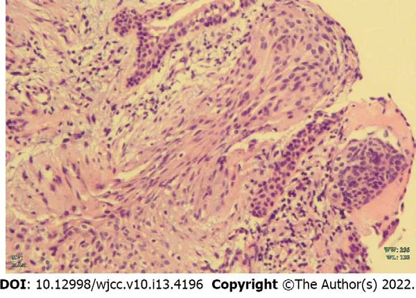
[38],a false positive diagnosis of paraganglioma was given based on preoperative transthoracic needle biopsy.The present patient was misdiagnosed as having low-grade neuroendocrine tumor based on preoperative bronchoscopic biopsy.

FlNAL DlAGNOSlS
The final diagnosis of the presented case was PPM.
TREATMENT
Zhang DB was responsible for collecting the medical history of the patient and drafting the paper;Chen T reviewed the literature and revised the manuscript;all authors read and approved the final manuscript.
OUTCOME AND FOLLOW-UP
The patient was disease-free after 3 mo of follow-up.
And it is well known that if the yard-cock belonging to this family happened to crow at midnight, they would declare it was morning, although the watchman and all the clocks in the town were proclaiming the hour of twelve at night
DlSCUSSlON
But it was not dark on the green lawn, for instantly the eyes of all the skulls on the wall were lighted up and shone till the place was as bright as day. When she saw this Vasilissa trembled so with fear that she could not run away.
Patient characteristics
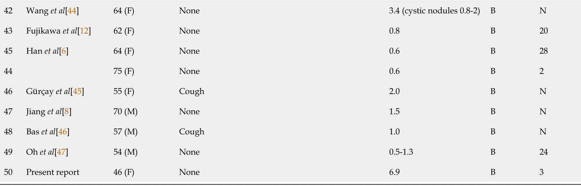
The study group comprised 50 patients: 19 men and 31 women.The age range was 18-108 years(median age: 58.0 years).Thirty-five patients were asymptomatic and only occasionally showed pulmonary nodules or masses on chest CT or X-ray.Thirteen patients had respiratory symptoms,including chest pain,chest tightness,hemoptysis,cough,and sputum).In addition,two patients had non-specific symptoms[3,4].There were nine patients with a history of malignancy: two had suffered lung adenocarcinoma[5,6],two colorectal cancer[7,8],two breast cancer[9,10],one buccal cancer[11],one papillary thyroid carcinoma[12],and one thymoma and kidney cancer[13](Table 1)[14-47].

Radiological characteristics
Primary ectopic pulmonary meningiomas are very rare,and only 67 cases(including our report)of PPM have been reported in the English language medical literature.The present study reported a case with very complete clinical procedure and imaging data,including preoperative enhanced CT examination,PET-CT examination,bronchoscopy biopsy,and postoperative pathological results.There were rare signs of calcification on CT,false positives on PET-CT and errors in our biopsy results.Thissuggests that we need to be cautious when excluding PPM only through auxiliary examination or even needle biopsy in clinical work.
I want to tell her that the physical wounds of child bearing will heal, but becoming a mother will leave her with an emotional wound so raw that she will be vulnerable forever
The CT enhancement patterns were described in 11 patients: six cases showed homogeneous enhancement,one showed heterogeneous enhancement[39],two showed mild enhancement[37,44],one showed mild concentric enhancement[8],and one showed no significant enhancement[13].
F-fluorodeoxyglucose-PET was performed in 12 patients,including our reported case.The PET scans of four patients showed no accumulation of 18F-FDG in lung lesions[8,10,45,46].Seven patients showed metabolically active lesions suspicious for malignancy,with a reported SUV range from 2.46 to 12.9 in seven cases.No other extra-pulmonary sites with increased FDG uptake were detected in any of the patients.
Maggie turned away, her thoughts racing1, her peace shattered. She thought about retreating to the safety of her car. But having come so far she was determined2 not to turn back. She ambled3 along the path, picking the occasional berry, enthusiasm gone.
And then, for the next few minutes, he read to me with more expression, clarity, and ease than I’d ever thought possible from him. The pages were already dog-eared, like the book had been read thousands of times already.
The prognosis of benign PPM resection is good,with almost no recurrence or metastasis.Follow-up was reported in 35 benign cases,ranging from 2 to 96 mo(median: 24 mo).However,two malignant PPMs relapsed[23,30].The above summary is presented in Table 3.
A total of 68 patients diagnosed with PPM were reported in the English literature from 1982 to 2021.All of these patients received histological assessment confirming PPM.Eighteen cases were excluded because(1)They underwent no radiological examination;or(2)They received no radiological evaluation of the CNS negative for meningioma.Ultimately,50 patients(including the case reported above)were included in the analysis.


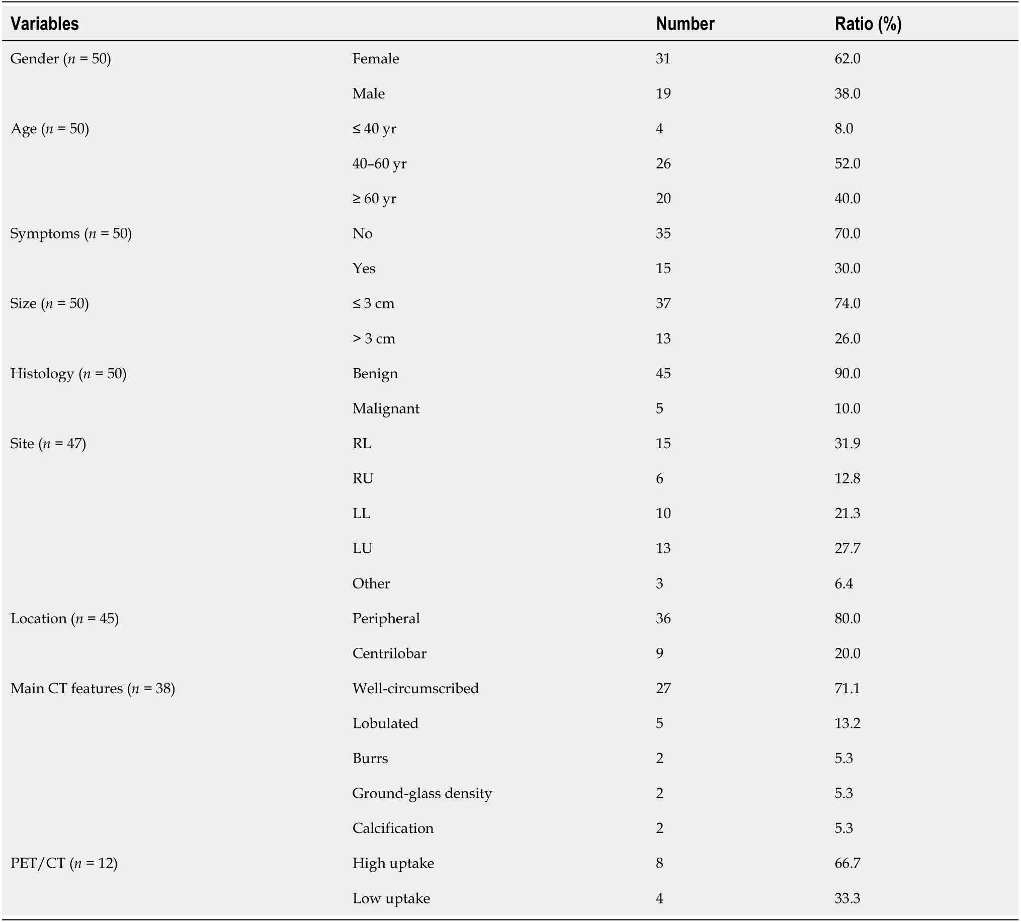
Most PPMs were benign,and only five cases were malignant[4,13,23,30,38].Benign PPMs were generally well-circumscribed on radiological studies,with diameters ranging from 0.4 to 6 cm(median:2 cm).The five malignant PPMs ranged in diameter from 1.5 to 15 cm(median: 6.4 cm).On chest CT scan,benign PPMs usually appear as isolated,rounded,solid,well-defined nodules or masses,with or without lobulation.Five cases were lobulated[24,31,32,37,40],two manifested as ground glass density[11,45],and two showed burrs on the edges[6].In addition,one recent study reported that the PPM showed multiple cystic lesions with a solid component[44].The CT features of the lesions were not described in the remaining eight cases(Table 2)[14-47].
The pathogenesis of PPMs remains unclear.One hypothesis is that the tumors develop from multipotent mesenchymal cells.Another states that PPMs originate from minute pulmonary meningothelial nodules that are occasionally found in approximately 1% of autopsies and excised lung specimens[48].However,the incidence of meningiomas is much lower than that of meningeal epithelial nodules.Moreover,previous genotypic comparisons have failed to demonstrate pulmonary meningeal epithelial nodules or intracranial meningiomas,further supporting the hypothesis[49].
To date,approximately 90% of PPMs reported in the literature have been benign,while five have been malignant[4,13,23,30,38].Most patients with PPM have no obvious symptoms,while some have respiratory or non-specific symptoms.Clinical symptoms may be related to the lesion location.As previously reported,benign PPMs are usually located in the peripheral pulmonary region,with no involvement of the bronchi,blood vessels,or pleura.Some PPM patients have a known history of malignancy[8],so a comprehensive and careful evaluation of pulmonary lesions must be carried out to avoid the misdiagnosis of metastasis.
Radiologically,PPMs usually appear as isolated,solid,and well-defined parenchymal coin-like lesions,ranging in size from 0.4 to 6.5 cm.Approximately 74.0% of PPMs are less than 3 cm in diameter.The lesions may present with burrs,lobulation,ground-glass density,or calcification,but these features are uncommon.Furthermore,one study reported a PPM presenting as multiple cystic lesions[44].PPMs have diverse enhancement CT manifestations.They usually show different degrees of enhancement,or even no significant enhancement.Hence,the pattern of lesion enhancement may not help to determine whether the lesion is benign or malignant.On
F-FDG PET,most PPMs exhibit high or mildly high metabolic activity,as in in our reported case.Only four PPMs showed low uptake of
F-FDG[8,10,45,46].However,one recent study reported a patient with both benign and malignant PPMs,both characterized by increased glucose uptake[13].This suggests that the malignancy of PPMs may not be related to
F-FDG uptake.
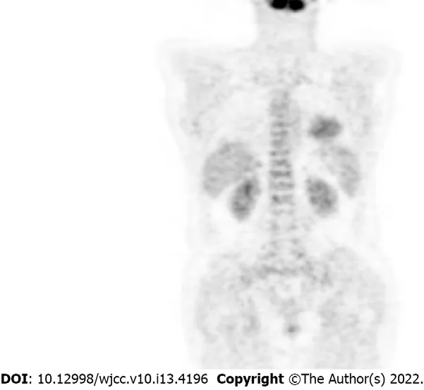
On 18-fluoro-2-deoxy-D-glucose(FDG)PET imaging,the standardized uptake value(SUV)of the mass increased unevenly,with a maximum value of 4.4,which suggested malignant lesion(Figure 2).No other lesions were detected on PET/CT.Moreover,enhanced magnetic resonance imaging(MRI)of the brain showed no evidence of intracranial tumors or metastases.Bronchoscopy revealed partial obstruction of the lower left lobe by the mass and narrowing of the lingual opening in the upper left lobe.A subsequent transbronchial biopsy result suggested a low-grade neuroendocrine tumor(Figure 3).
CONCLUSlON
Physical examination revealed no obvious positive signs.
FOOTNOTES
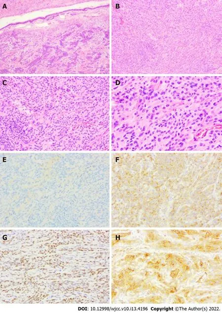
Considering the CT and PET features of the mass and the results of transbronchial biopsy,an open cuff resection of the left lower lobe and wedge resection of the lingual segment were performed.Gross examination revealed a 6.5 cm,off-white,tenacious texture mass.Microscopic examination revealed a tumor with focal bronchial cartilage involvement,no pleural involvement,and fusiform nests of cells arranged in fascicles or whorls.Immunohistochemistry showed positivity for epithelial membrane antigen(EMA),progesterone receptor(PR),somatostatin receptor 2(SSTR2),D2-40,and CD34,and negativity for S-100,cytokeratin(CK),glial fibrillary acidic protein,CgA,SOX10,and SMA;the Ki-67 index was about 5%-10% positive(Figure 4).These morphological and immunohistochemical features were suggestive of a PPM.Preoperative contrast-enhanced chest CT,contrast-enhanced brain MRI,and PET-CT did not reveal evidence of intracranial or spinal meningioma.
When he saw her well-disposed towards him, he exclaimed: Madam, I have a most important secret to confide49 to you, and I beg you not to be alarmed by what I am about to say
Written informed consent was obtained from the patient for the publication of this case report and accompanying images.
All authors declare no conflict of interest related to this study.
The authors have read the CARE Checklist(2016),and the manuscript was prepared and revised according to the CARE Checklist(2016).
This article is an open-access article that was selected by an in-house editor and fully peer-reviewed by external reviewers.It is distributed in accordance with the Creative Commons Attribution NonCommercial(CC BYNC 4.0)license,which permits others to distribute,remix,adapt,build upon this work non-commercially,and license their derivative works on different terms,provided the original work is properly cited and the use is noncommercial.See: https://creativecommons.org/Licenses/by-nc/4.0/
China
Dan-Bin Zhang 0000-0002-4897-6358;Tao Chen 0000-0001-5013-323X.
He promised faithfully to come back to her as soon as he could and begged her to await his return under the lime-tree where they had spent so many happy hours
Ma YJ
A
Ma YJ
1 Kershisnik M,Callender DL,Batsakis JG.Extracranial,extraspinal meningiomas of the head and neck.
1993;102: 967-970[PMID: 8285520 DOI: 10.1177/000348949310201211]
2 Muzumdar DP,Vengsarkar US,Bhatjiwale MG,Goel A.Diffuse calvarial meningioma:A case report.
2001;47: 116-118[PMID: 11832603]
3 Kemnitz P,Spormann H,Heinrich P.Meningioma of lung: first report with light and electron microscopic findings.
1982;3: 359-365[PMID: 7157498 DOI: 10.3109/01913128209018558]
4 Weber C,Pautex S,Zulian GB,Pusztaszeri M,Lobrinus JA.Primary pulmonary malignant meningioma with lymph node and liver metastasis in a centenary woman,an autopsy case.
2013;462: 481-485[PMID: 23443940 DOI:10.1007/s00428-013-1383-7]
5 de Perrot M,Kurt AM,Robert J,Spiliopoulos A.Primary pulmonary meningioma presenting as lung metastasis.
1999;33: 121-123[PMID: 10225315 DOI: 10.1080/14017439950141948]
6 Han D,Deng H,Liu Y.Primary pulmonary meningiomas: report of two cases and review of the literature.
2020;216: 153232[PMID: 33045659 DOI: 10.1016/j.prp.2020.153232]
7 Falleni M,Roz E,Dessy E,Del Curto B,Braidotti P,Gianelli U,Pietra GG.Primary intrathoracic meningioma:histopathological,immunohistochemical and ultrastructural study of two cases.
2001;439: 196-200[PMID:11561761 DOI: 10.1007/s004280000387]
8 Jiang M,Chen P,Huang R,Zhang J,Zheng J.A case report of primary pulmonary meningioma masquerading as lung metastasis in a patient with rectal carcinoma: role of
F-FDG PET/CT.
2021;16: 153[PMID:34051819 DOI: 10.1186/s13019-021-01546-3]
9 Picquet J,Valo I,Jousset Y,Enon B.Primary pulmonary meningioma first suspected of being a lung metastasis.
2005;79: 1407-1409[PMID: 15797095 DOI: 10.1016/j.athoracsur.2003.10.071]
10 Lepanto D,Maffini F,Petrella F,Colandrea M,Putzu C,Barberis M,Paganelli G,Viale G.Atypical primary pulmonary meningioma: a report of a case suspected of being a lung metastasis.
2014;8: 414[PMID:24761155 DOI: 10.3332/ecancer.2014.414]
11 Juan CM,Chen ML,Ho SY,Huang YC.Primary Pulmonary Meningioma Simulating a Pulmonary Metastasis.
2016;2016: 8248749[PMID: 27974986 DOI: 10.1155/2016/8248749]
12 Fujikawa R,Arai Y,Otsuki Y,Nakamura T.A case of a primary pulmonary meningioma mimicking a metastasis from a papillary thyroid carcinoma due to a size reduction after radioactive iodine therapy.
2020;6: 57[PMID:32221747 DOI: 10.1186/s40792-020-00823-y]
13 Cimini A,Ricci F,Pugliese L,Chiaravalloti A,Schillaci O,Floris R.A Patient with a Benign and a Malignant Primary Pulmonary Meningioma: An Evaluation with 18F Fluorodeoxyglucose Positron Emission Tomography/Computed Tomography and Computed Tomography with Iodinated Contrast.
2019;34: 45-47[PMID: 30713380 DOI: 10.4103/ijnm.IJNM_101_18]
14 Chumas JC,Lorelle CA.Pulmonary meningioma.A light- and electron-microscopic study.
1982;6:795-801[PMID: 7168461 DOI: 10.1097/00000478-198212000-00011]
15 Zhang FL,Cheng XR,Zhang YS,Ding JA.Lung ectopic meningioma.A case report.
1983;96: 309-311[PMID: 6413150]
16 Kodama K,Doi O,Higashiyama M,Horai T,Tateishi R,Nakagawa H.Primary and metastatic pulmonary meningioma.
1991;67: 1412-1417[PMID: 1991305 DOI:10.1002/1097-0142(19910301)67:5<1412::Aid-cncr2820670523>3.0.Co;2-v]
17 Drlicek M,Grisold W,Lorber J,Hackl H,Wuketich S,Jellinger K.Pulmonary meningioma.Immunohistochemical and ultrastructural features.
1991;15: 455-459[PMID: 2035740 DOI: 10.1097/00000478-199105000-00005]
18 Flynn SD,Yousem SA.Pulmonary meningiomas: a report of two cases.
1991;22: 469-474[PMID: 1709609 DOI: 10.1016/0046-8177(91)90133-a]
19 Maiorana A,Ficarra G,Fano RA,Spagna G.Primary solitary meningioma of the lung.
1996;88: 457-462[PMID: 8988660]
20 Kaleem Z,Fitzpatrick MM,Ritter JH.Primary pulmonary meningioma.Report of a case and review of the literature.
1997;121: 631-636[PMID: 9199633]
21 Lockett L,Chiang V,Scully N.Primary pulmonary meningioma: report of a case and review of the literature.
1997;21: 453-460[PMID: 9130993 DOI: 10.1097/00000478-199704000-00012]
22 Ueno M,Fujiyama J,Yamazaki I,Uchiyama T,Ishikawa Y,Satoh Y.Cytology of primary pulmonary meningioma.Report of the first multiple case.
1998;42: 1424-1430[PMID: 9850654 DOI: 10.1159/000332179]
23 Prayson RA,Farver CF.Primary pulmonary malignant meningioma.
1999;23: 722-726[PMID:10366156 DOI: 10.1097/00000478-199906000-00013]
24 Spinelli M,Claren R,Colombi R,Sironi M.Primary pulmonary meningioma may arise from meningothelial-like nodules.
2000;4: 35-39[PMID: 10936897]
25 Cesario A,Galetta D,Margaritora S,Granone P.Unsuspected primary pulmonary meningioma.
2002;21: 553-555[PMID: 11888783 DOI: 10.1016/s1010-7940(01)01174-5]
26 Cura M,Smoak W,Dala R.Pulmonary meningioma: false-positive positron emission tomography for malignant pulmonary nodules.
2002;27: 701-704[PMID: 12352110 DOI: 10.1097/00003072-200210000-00003]
27 Comin CE,Caldarella A,Novelli L,Janni A.Primary pulmonary meningioma: report of a case and review of the literature.
2003;89: 102-105[PMID: 12729374 DOI: 10.1177/030089160308900123]
28 Rowsell C,Sirbovan J,Rosenblum MK,Perez-Ordoñez B.Primary chordoid meningioma of lung.
2005;446: 333-337[PMID: 15714337 DOI: 10.1007/s00428-004-1192-0]
29 Kaneda Y,Miyoshi T,Hiratsuka M,Yamamoto S,Kato F,Maki K,Hayashi H,Shiraishi T,Iwasaki A,Iwasaki H,Nabeshima K,Shirakusa T.[Primary pulmonary meningioma;report of a case].
2005;58: 512-515[PMID:15957430]
30 van der Meij JJ,Boomars KA,van den Bosch JM,van Boven WJ,de Bruin PC,Seldenrijk CA.Primary pulmonary malignant meningioma.
2005;80: 1523-1525[PMID: 16181912 DOI: 10.1016/j.athoracsur.2004.04.015]
31 Meirelles GS,Ravizzini G,Moreira AL,Akhurst T.Primary pulmonary meningioma manifesting as a solitary pulmonary nodule with a false-positive PET scan.
2006;21: 225-227[PMID: 16915069 DOI:10.1097/01.rti.0000203639.66629.68]
32 Incarbone M,Ceresoli GL,Di Tommaso L,Cappuzzo F,Inzirillo F,Infante M,Alloisio M.Primary pulmonary meningioma: report of a case and review of the literature.
2008;62: 401-407[PMID: 18486986 DOI:10.1016/j.lungcan.2008.03.031]
33 Izumi N,Nishiyama N,Iwata T,Nagano K,Tsukioka T,Hanada S,Suehiro S.Primary pulmonary meningioma presenting with hemoptysis on exertion.
2009;88: 647-648[PMID: 19632430 DOI:10.1016/j.athoracsur.2008.12.058]
34 Kim YY,Hong YK,Kie JH,Ryu SJ.Primary pulmonary meningioma: an unusual cause of a nodule with strong and homogeneous enhancement.
2016;40: 170-173[PMID: 26452726 DOI: 10.1016/j.clinimag.2015.08.004]
35 Jiang GY,Zhang Y,Yu JH,Lin XY,Fan CF,Sun CL,Xu HT,Wang EH.Primary pulmonary meningioma:A case report and a review of the literature.
2016;9: 4467-4472
36 Oide T,Hiroshima K,Shibuya K,Nakatani Y.Primary Pulmonary Meningioma Presenting as a Coin Lesion.
2017;56: 2073-2074[PMID: 28768984 DOI: 10.2169/internalmedicine.56.8481]
37 Huang S,Chen L,Mao Y,Tong H.Primary pulmonary meningioma:A case report.
2017;96: e6474[PMID: 28489736 DOI: 10.1097/MD.0000000000006474]
38 Žulpaitė R,Jagelavičius Ž,Mickys U,Janilionis R.Primary Pulmonary Meningioma With Rhabdoid Features.
2019;27: 457-463[PMID: 30563401 DOI: 10.1177/1066896918819257]
39 Hong S,Jiang J,Zhou F,Liu J.Computed tomography findings of primary pulmonary meningioma:A case report.
2018;97: e9651[PMID: 29480880 DOI: 10.1097/MD.0000000000009651]
40 Luo JZ,Zhan C,Ni X,Shi Y,Wang Q.Primary pulmonary meningioma mimicking lung metastatic tumor:A case report.
2018;13: 99[PMID: 30285886 DOI: 10.1186/s13019-018-0787-5]
41 Xu KK,Tian F,Cui Y.Primary pulmonary meningioma presenting as a micro solid nodule: A rare case report.
2018;9: 874-876[PMID: 29718593 DOI: 10.1111/1759-7714.12639]
42 Ohashi-Nakatani K,Shibuki Y,Fujima M,Watanabe R,Yoshida A,Yoshida H,Matsumoto Y,Tsuchida T,Watanabe SI,Motoi N.Primary pulmonary meningioma: A rare case report of aspiration cytological features and immunohistochemical assessment.
2019;47: 330-333[PMID: 30548187 DOI: 10.1002/dc.24126]
43 Bae SY,Kim HS,Jang HJ,Chung WS,Kim H,Kim YH,Lee JH,Bang SS.Primary Pulmonary Chordoid Meningioma.
2018;51: 410-414[PMID: 30588452 DOI: 10.5090/kjtcs.2018.51.6.410]
44 Wang X,Li P,Zhou P,Fu Y,Lai Y,Che G.Intrapulmonary metastasis from primary pulmonary meningioma presenting as multiple cystic lesions: a case report.
2019;19: 8[PMID: 30621651 DOI: 10.1186/s12890-018-0773-7]
45 Gürçay N,Öztürk A,Demirağ F,İncekara F.Primary pulmonary meningioma mimicking pulmonary metastasis:A rare case report.
2020;28: 699-701[PMID: 33403148 DOI:10.5606/tgkdc.dergisi.2020.19370]
46 Bas A,Valiyev E,Ozkan ND,Tombul I,Yonat S,Sayan M,Kurul IC.A Rare Entity: Primary Pulmonary Meningioma.
2021[PMID: 34514565 DOI: 10.5146/tjpath.2021.01535]
47 Oh JH,Cho HS,Hwang HS,Ji W.Primary pulmonary meningioma presenting as multiple lung nodules: A case report.
2022;13: 141-143[PMID: 34878222 DOI: 10.1111/1759-7714.14270]
48 Gaffey MJ,Mills SE,Askin FB.Minute pulmonary meningothelial-like nodules.A clinicopathologic study of so-called minute pulmonary chemodectoma.
1988;12: 167-175[PMID: 2830799 DOI:10.1097/00000478-198803000-00001]
49 Ionescu DN,Sasatomi E,Aldeeb D,Omalu BI,Finkelstein SD,Swalsky PA,Yousem SA.Pulmonary meningothelial-like nodules: a genotypic comparison with meningiomas.
2004;28: 207-214[PMID: 15043310 DOI:10.1097/00000478-200402000-00008]
杂志排行

World Journal of Clinical Cases的其它文章
- Capillary leak syndrome:A rare cause of acute respiratory distress syndrome
- lmproving outcomes in geriatric surgery:ls there more to the equation?
- Mass brain tissue lost after decompressive craniectomy:A case report
- Primary intracranial extraskeletal myxoid chondrosarcoma:A case report and review of literature
- Spinal canal decompression for hypertrophic neuropathy of the cauda equina with chronic inflammatory demyelinating polyradiculoneuropathy:A case report
- Enigmatic rapid organization of subdural hematoma in a patient with epilepsy:A case report