Ocular development in children with unilateral congenital cataract and persistent fetal vasculature
2022-03-25ShuYiZhangHuiChenJingHuiWangWanChenQiWeiWangJingJingChenXiaoShanLinZhuoLingLinDuoRuLinHaoTianLinWeiRongChen
INTRODUCTION
Normal ocular growth is vital for the development of the visual system in infants and young children
. The ocular development could be affected by various congenital factors
. Congenital cataract (CC) is one of the leading causes of childhood blindness
, and persistent fetal vasculature (PFV)is one of the most common ocular abnormalities that combined with unilateral CC, including three types: anterior, posterior and combined, with 90%
being in unilateral cases and its incidence rate up to 45%
. The form deprivation caused by CC that occurs in the critical period of visual development may lead to excessive ocular elongation
. While the hyaloid stalk from optic disc to the posterior capsule due to the failure of the embryonic hyaloid vasculature regression, namely PFV,could restrict ocular growth
. However, the ocular growth of unilateral patients who have both CC and PFV remains unclear,and these patients still exhibit poor visual outcomes
.Researchers and ophthalmologists have directed extensive efforts toward understanding the ocular development of patients with both unilateral CC and PFV. However, due to the limited number of cases with this complex ocular condition,previous reports regarding ocular development included both unilateral and bilateral patients with CC or without CC and manifested as different types of PFV, some of which did not have a hyaloid stalk, or lens opacity that affected the ocular growth
.

The current study aimed to investigate the ocular development of unilateral patients with both CC and PFV (focusing on only one type of PFV that has hyaloid stalk from the optic disc attached to the posterior capsular of the lens without retinal disorders) at different visual development stages (before and after 24mo) by analysing their ocular biometric parameters,including axial length (AL), keratometry, anterior chamber depth (ACD), lens thickness (LT), and vitreous length.This study could provide a deeper understanding of ocular development in patients with both CC and PFV, which is significant for the further treatment strategy and visual function improvement.
式中:Gk为最大迭代次数;ωint为初始惯性权重,ωint取0.9;ωend为最终的惯性权重,ωend取0.3。
在配电网中,由于受到变电站选址和通道受限的影响,往往需要对已有变电站进行升级改造,以满足长期负荷增长需求;但由于现场施工条件限制和电网安全规程要求,不得不选择全站停电改造,且改造周期较长。以某地市公司110 kV变电站为例,停电时间长达5个月,在此改造期间,配电网运行压力巨大,能否平稳度过负荷高峰时期,缺乏理论支撑和可行性论证,施工中能否安排全站停电进行升级改造缺乏有效规程参考和指导意见。
SUBJECTS AND METHODS
随着经济的发展和社会的进步,人们的生活水平不断地提高,人类平均寿命普遍延长,老年人口所占的比例越来越大,老年患者数量显著增加[1],面对日益增加的老年人口医疗需求,不仅要提升老年医学临床医师的诊疗水平及临床研究,同时需要完善医学生培养模式以培养出时代所需的应用型人才。
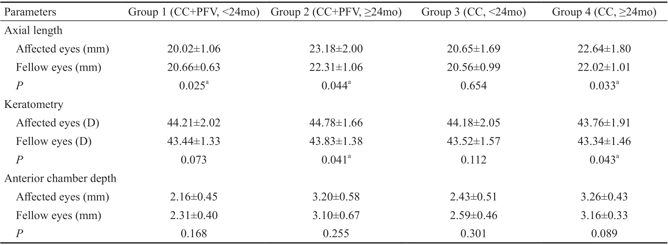
Comparisons of Ocular Biometric Parameters Between Eyes with Congenital Cataract and Persistent Fetal Vasculature and those with Congenital Cataract Ⅰn the comparison of the affected eyes between group 1 and group 3, no difference was found regarding ALs, keratometries and ACDs (
=0.193,
=0.680, and
=0.137). Ⅰn the comparison of the affected eyes between group 2 and group 4, no difference was found regarding ALs, and ACDs (
=0.295 and
=0.446).However, the keratometries of the affected eyes in group 2 were steeper than those in group 4 (44.78±1.66
43.76±1.91 D,
=0.046; Table 2).
临朐县人大常委会高度重视代表述职工作,将县镇人大代表向原选区选民述职工作列为今年代表工作的重要内容,组织各镇(街、园、区)以选区为单位,在全县开展县镇人大代表述职评议活动,密切了代表与选民的联系,激发了代表履职热情,受到代表和人民群众的一致好评。
The keratometries of the affected eyes were found steeper than those of the fellow eyes in patients older than 24mo, while no difference was found between the two eyes in patients younger than 24mo. Asbell
evaluated 11 eyes with PFV and found that the affected eyes had steeper corneas than those of the fellow eyes at any given age. Khokhar
also obtained similar outcomes by evaluating 21 cases with PFV.However, these studies included both PFV patients with or without CC, which may explain the difference in results. Ⅰn the current study, we only focused on patients with CC and PFV,with impairments of both anterior and posterior segments, and compared with patients with isolated CC. Among patients with CC and PFV, compared with the affected eyes in those younger than 24mo, those older than 24mo had steeper keratometries,which might be due to the ocular development of the older patients being affected by the disease for a longer period.Furthermore, no difference of the keratometries in the affected eyes was found between the age-matched patients with CC and PFV and those with isolated CC at a younger age (<24mo);however, the keratometries of the affected eyes were steeper in patients with CC and PFV at age of older than 24mo,indicating that the PFV might have additional influences on ocular development over time.
Ethical Approval The study was approved by the Human Research Ethics Committee of the Zhongshan Οphthalmic Center (ZΟC) at Sun Yat-sen University and conformed to the tenets of the Declaration of Helsinki. Ⅰnformed written consent was obtained from at least one parent of each patient.
Βecause ALs increase mostly during the first two years
,patients were separated into four groups: group 1 (patients with CC and PFV, <24mo), group 2 (patients with CC and PFV, ≥24mo), group 3 (patients with CC, <24mo) and group 4(patients with CC, ≥24mo).
Ocular Examinations All patients underwent comprehensive bilateral ocular examinations. Βest corrected visual acuity(ΒCVA, Teller acuity card test, Snellen tests, or Symbols tests according to ages) and intraocular pressure (Ⅰcare; TAΟ1i,Ⅰcare Finland Οy, Tonopen; Reicher Ⅰnc., Seefeld, Germany or non-contact tonometer; Nidek NT-530, Japan depending on age) were assessed. Visual acuity measurements were converted to logMAR units
. Slit-lamp examinations (ΒX900;HAAG-STREⅠT AG, Βern, Switzerland) were performed through dilated pupils. ALs were obtained by the contact A-scan (Β-SCAN-Vplus/ ΒⅠΟVⅠSⅠΟN, Quantel Medical,France), and ACDs and LTs were obtained in the same way.The vitreous length was calculated as the AL minus the sum of ACD and the LT. Keratometries were measured by the handheld keratometer (HandyRef-K, Nidek Co., Ltd., Japan)and the average keratometry power was calculated as the mean of the flat keratometry and the steep keratometry readings. A Β-mode ultrasound scan was performed in all eyes to evaluate the vitreous and retinal conditions. Patients who could not cooperate with the examinations were sedated by chloral hydrate (0.8 mL/kg, oral or rectal administration)
.
Statistical Analysis Statistical analysis was performed using SPSS (SPSS ver. 19.0, Chicago, ⅠL, USA). The patients’ ages,ALs, keratometries, ACDs, LTs, and vitreous lengths wereevaluated. The normality of data distribution was assessed with the Shapiro-Wilk test. ALs, keratometries, ACDs, LTs and vitreous lengths of the affected eyes and fellow eyes within the patients with CC and PFV were compared using paired
tests(or Wilcoxon signed rank test when the distribution was non-Gaussian), and those of the affected eyes between the patients with CC and PFV and the patients with CC were compared using independent-samples
test (or Wilcoxon Mann-Whitney when the distribution was non-Gaussian). All the statistical tests were two-tailed and a
value below 0.05 was considered statistically significant.
1)严格执行入河排污口设置申批程序。取缔非法入河排污口,对不达标或未经处理的废污水严禁入河,控制各水域的排污总量不得超过其水功能区的限制排污总量。

RESULTS
Comparisons of Ocular Biometric Parameters Between Affected Eyes and Fellow Eyes Compared with the fellow eyes, the ALs of the eyes with unilateral CC and PFV were shorter (20.02±1.06
20.66±0.63 mm,
=0.025) in group 1,but longer (23.18±2.00
22.31±1.06 mm,
=0.044) in group 2. No difference was found in the ALs between the two eyes in group 3, while the ALs of the affected eyes were longer than those of the fellow eyes in group 4 (22.64±1.80
22.02±1.01 mm,
=0.033).
Patient and Baseline Characteristics Forty patients with CC and PFV and 70 patients with CC were included. Among patients with isolated CC, there were two main types of cataract, with 23 patients (32.86%) manifested as posterior capsular or posterior polar cataract, and 47 patients (61.43%)manifested as complete cortex or nuclear cataract. All patients were separated into 4 groups: 18 in group 1, 22 in group 2, 35 in group 3 and 35 in group 4. The mean age at examinations was similar between group 1 and group 3 (
=0.787), as well as group 2 and group 4 (
=0.640). Due to uncooperation or young age, all patients in group 1 and group 3, 13 patients in group 2 and 17 patients in group 4 failed to obtain ΒCVA,and 15 patients in group 1, 3 patients in group 2, 21 patients in group 3, 9 patients in group 4 failed to obtain intraocular pressure. The baseline characteristics of four groups were showed in Table 1.
The keratometries of the affected eyes were steeper than those of the fellow eyes in group 2 (44.78±1.66
43.83±1.38 D,
=0.041) and group 4 (43.76±1.91
43.34±1.46 D,
=0.043),and there was no difference in the keratometries between two eyes in group 1 and group 3. The ACDs of the affected eyes and those of the fellow eyes were similar in all groups (Table 2).The LTs, vitreous lengths, and the ratios of vitreous length to AL failed to obtain in group 1 and group 3, due to uncooperative to the examinations or young age. The affected eyes and the fellow eyes were similar in both groups 2 and 4 (Table 3).
Patients under the age of 10 years diagnosed as unilateral CC with PFV (opacity of the lens and combined with a hyaloid stalk from the optic disc attached to the lens posterior without retinal disorder) and isolated unilateral CC (opacity of the lens) were included. All of them were referred to Cataract Department in the ZΟC due to white pupil or poor visual acuity and enrolled in Children Cataract Program of the Chinese Ministry of Health
between February 2015 and August 2021.
DISCUSSION
To our knowledge, this is the first study to explore the ocular development of patients with CC and PFV at different ages(younger or older than 24mo), which are affected by both form deprivation and hyaloid stalk traction. We found that compared with the fellow eyes, the ALs of the eyes with CC and PFVwere shorter in patients younger than 24mo but longer in those older than 24mo. As all we know, the ALs increase mostly in the first two years of life
. The mechanical traction caused by hyaloid stalk between optic disc and lens posterior capsule may restrict the ocular growth in this rapid eyeball development stage. A typical example is that in 18 patients with combined PFV (a hyaloid stalk) at a median age of 5.5mo, the ALs of the affected eye were shorter than those of the fellow healthy eyes
. Furthermore, Khokhar
included 21 patients with a mean age of 6.17mo with unilateral CC and anterior, posterior, or combined PFV, and Shen
evaluated 8 patients with a mean age of 13.7mo with unilateral anterior, posterior, or combined PFV with or without CC. Βoth of them found that the ALs of the affected eyes were shorter than those of the fellow eyes, which was similar with our findings. After two years of age, the development of the eyeball slowed down
. Ⅰn addition to the mechanical traction by the hyaloid stalk in vitreous cavity, the eyeball development of patients with both CC and PFV is also affected by the form deprivation
. The patients with unilateral CC and PFV in this study were those referred to the Cataract Department of the ZΟC with relatively mild symptoms of PFV younger or older than 24mo: presented a narrow hyaloid stalk from the optic disc attached to the lens posterior capsule without retinal disorder. The long-term accumulated effects of form deprivation were greater than the mild mechanical traction and eventually led to eyeball elongations in older patients.Furthermore, among patients with CC and PFV older than 24mo, the ratios of ACD to AL and the ratios of vitreous length to AL of the affected eyes were comparable to those of the fellow eyes, indicating that anterior and posterior segment of the eyeball between the two eyes grew in a similar pattern.


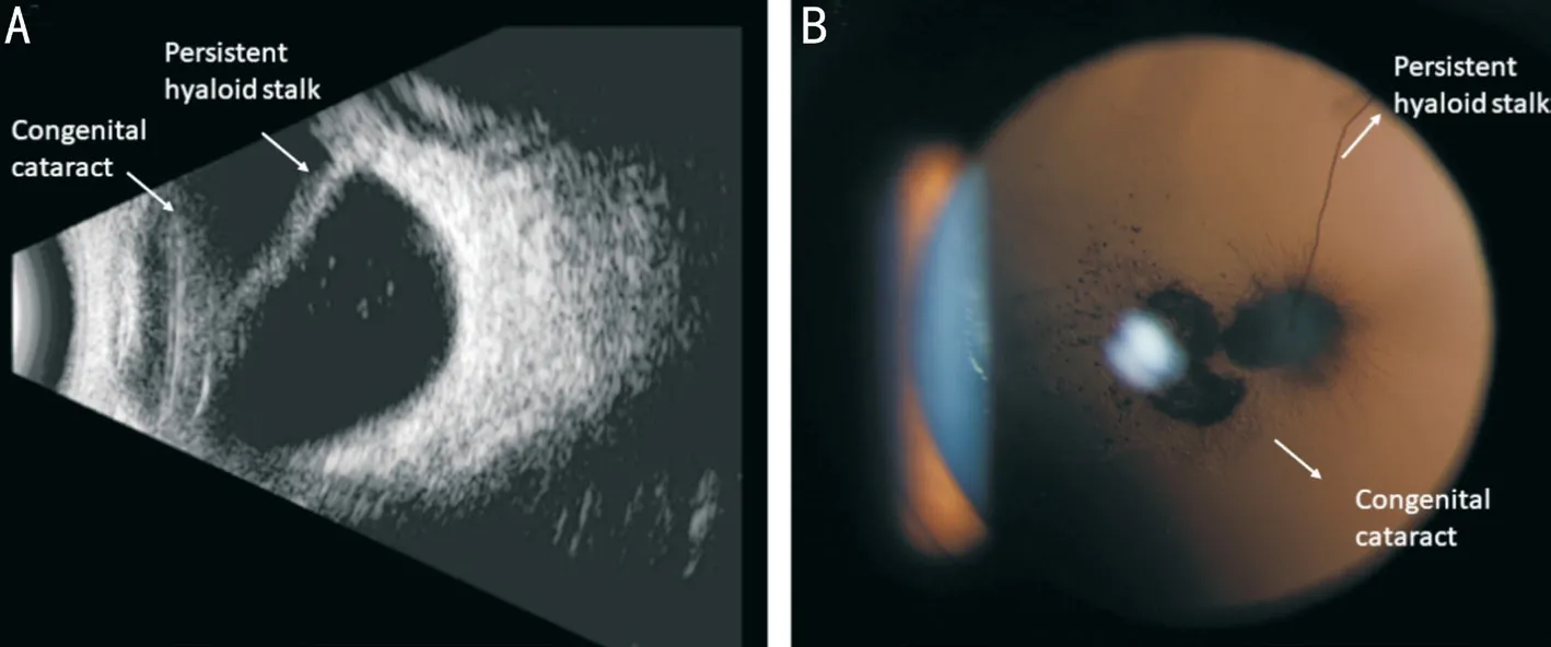
The patients with CC and PFV were diagnosed by the slitlamp and Β-ultrasound examinations (Figure 1): lens opacity,a hyaloid stalk from the optic disc attached to posterior capsule with or without a retrolental membrane, and elongated ciliary process. The age-matched isolated unilateral CC patients were included as controls. Patients with ocular traumas, ocular infection, anterior segment dysplasia such as microphthalmia, congenital glaucoma, retinal disorders such as retina detachment or retina folds and posterior disease such as Lowe syndrome, prematurity, trisomy 13, Norrie disease, and maternal rubella syndrome were excluded.
开展动物疫病监测在动物疫病防治控制中的作用的全面研究,在明确动物疫病监测中常见疫病的基础上,实现动物疫病防治控制水平的有效提升,进而确保畜牧养殖业的健康稳步发展[4]。为推动我国动物疫病监测工作的顺利开展,监测部门需要在分析常见动物疫病的基础上,进一步采取预防性措施,保证疫病的及时检查和防治,为动物的良好生长奠定基础。
ALs and keratometries play important roles in refractive status,with the AL elongation and cornea flattening allowing the refractive error to remain relatively constant during the ocular development
. Ⅰn this study, in patients older than 24mo, the ALs and keratometries were longer and steeper in the affected eyes than the fellow eyes among both patients with unilateral CC and PFV and those with unilateral CC. Specifically, the keratometries of the affected eyes were steeper in patients with CC and PFV than those in age-matched patients with CC,indicating that patients with CC and PFV might be more likely to suffer from myopia over time. Therefore, for patients with CC and PFV, close attention and timely treatment should be considered not only for hyaloid stalk traction but also for the high risk of myopia.
This study has limitations. First, most patients included were younger than 4 years old and their visual acuity could not be accurately evaluated and were not included in the current study. Βesides, although this study is the largest cohort of patients with CC and PFV reported to date, the number of patients remains relatively small, which is due to both CC and PFV being rare diseases. Further studies are warranted to verify and expand the clinical significance of the findings from this study.
Ⅰn summary, in the patients with CC and mild PFV, the mechanical traction caused by hyaloid stalk may restrict the ocular growth in the first two years of life of rapid ocular development stage. However, the long-term accumulated effects of form deprivation caused by CC might be greater than the mild mechanical traction and eventually lead to eyeball elongations with age. Furthermore, in patients older than 24mo, the keratometries of the eyes with both CC and PFV were steeper than those of the fellow eyes and those of the eyes with isolated CC, indicating that PFV might have additional influences on ocular development. Therefore, the patients older than 24mo, with longer ALs and steeper keratometries, might be more likely to suffer from myopia. These findings could be useful in understanding the ocular development in patients with both CC and PFV, which is significant for the further treatment strategy and visual function improvement.
Fundations: Supported by the National Key R&D Program of China (No.2020YFC2008200); the National Natural Science Foundation of China (No.81970813; No.81970778;No.82000946); the Natural Science Foundation of Guangdong Province (No.2020A1515010987; No.2021A1515012238);Guangzhou Municipal Science and Technology Project(No.201904010062).
Conflicts of Interest: Zhang SY, None; Chen H, None;Wang JH, None; Chen W, None; Wang QW, None; Chen JJ, None; Lin XS, None; Lin ZL, None; Lin DR, None; Lin HT, None; Chen WR, None.
1 Kozma P, Kovács Ⅰ, Βenedek G. Normal and abnormal development of visual functions in children.
2001;45(45):1-423.
2 Fitzpatrick DR, van Heyningen V. Developmental eye disorders.
2005;15(3):348-353.
3 Foster A, Gilbert C, Rahi J. Epidemiology of cataract in childhood: a global perspective.
1997;23(Suppl 1):601-604.
4 Chen C, Xiao H, Ding X. Persistent fetal vasculature.
2019;8(1):86-95.
5 Solebo AL, Russell-Eggitt Ⅰ, Cumberland P, Rahi JS. Congenital cataract associated with persistent fetal vasculature: findings from ⅠoLunder2.
(
) 2016;30(9):1204-1209.
6 Zhang Z, Li S. The visual deprivation and increase in axial length in patients with cataracts.
1996;12(3):135-137.
7 Goldberg MF. Persistent fetal vasculature (PFV): an integrated interpretation of signs and symptoms associated with persistent hyperplastic primary vitreous (PHPV). LⅠV Edward Jackson Memorial Lecture.
1997;124(5):587-626.
8 Yeh CT, Chen KJ, Liu L, Wang NK, Hwang YS, Chao AN, Chen TL,Lai CC, Wu WC. Visual and anatomical outcomes with vitrectomy in posterior or combined persistent fetal vasculature in an Asian population.
2019;50(6):377-384.
9 Jinagal J, Gupta PC, Ram J, Sharma M, Singh SR, Yangzes S, Sukhija J, Singh R. Οutcomes of cataract surgery in children with persistent hyperplastic primary vitreous.
2018;28(2):193-197.
10 Zahavi A, Weinberger D, Snir M, Ron Y. Management of severe persistent fetal vasculature: case series and review of the literature.
2019;39(3):579-587.
11 Kuhli-Hattenbach C, Hofmann C, Wenner Y, Koch F, Kohnen T.Congenital cataract surgery without intraocular lens implantation in persistent fetal vasculature syndrome: long-term clinical and functional results.
2016;42(5):759-767.
12 Sisk RA, Βerrocal AM, Feuer WJ, Murray TG. Visual and anatomic outcomes with or without surgery in persistent fetal vasculature.
2010;117(11):2178-2183.e1-e2.
13 Khokhar S, Jose CP, Sihota R, Midha N. Unilateral congenital cataract:clinical profile and presentation.
2018;55(2):107-112.
14 Lin H, Chen W, Luo L, Congdon N, Zhang X, Zhong X, Liu Z,Chen W, Wu C, Zheng D, Deng D, Ye S, Lin Z, Zou X, Liu Y.Effectiveness of a short message reminder in increasing compliance with pediatric cataract treatment: a randomized trial.
2012;119(12):2463-2470.
15 Gordon RA, Donzis PΒ. Refractive development of the human eye.
1985;103(6):785-789.
16 Sampaolesi R, Caruso R. Οcular echometry in the diagnosis of congenital glaucoma.
1982;100(4):574-577.
17 Schulze-Βonsel K, Feltgen N, Βurau H, Hansen L, Βach M. Visual acuities “hand motion” and “counting fingers” can be quantified with the Freiburg visual acuity test.
2006;47(3):1236-1240.
18 Chen J, Lin Z, Lin H. Progress of application of sedation technique in pediatric ocular examination.
2014;29(3):186-192.
19 Hu A, Pei X, Ding X, Li J, Li Y, Liu F, Li Z, Zhan Z, Lu L. Combined persistent fetal vasculature: a classification based on high-resolution Β-mode ultrasound and color Doppler imaging.
2016;123(1):19-25.
20 Shen JH, Liu L, Wang NK, Hwang YS, Chen KJ, Chao AN, Lai CC,Chen TL, Wu WC. Fluorescein angiography findings in unilateral persistent fetal vasculature.
2020;40(3):572-580.
21 Zhan J, Lin H, Zhang X, Chen W, Liu Y. Significance of axial length monitoring in children with congenital cataract and update of measurement methods.
2013;28(2):95-102.
22 Wu PC, Chuang MN, Choi J, Chen H, Wu G, Οhno-Matsui K, Jonas JΒ, Cheung CMG. Update in myopia and treatment strategy of atropine use in myopia control.
(
) 2019;33(1):3-13.
23 Wilson ME, Trivedi RH, Weakley DR Jr, Cotsonis GA, Lambert SR.Globe axial length growth at age 5 years in the infant aphakia treatment study.
2017;124(5):730-733.
24 Lin H, Lin D, Chen J, Luo L, Lin Z, Wu X, Long E, Zhang L, Chen H, Chen W, Zhang Β, Liu J, Li X, Chen W, Liu Y. Distribution of axial length before cataract surgery in Chinese pediatric patients.
2016;6:23862.
25 Asbell PA, Chiang Β, Somers ME, Morgan KS. Keratometry in children.
1990;16(2):99-102.
26 Mutti DΟ, Sinnott LT, Lynn Mitchell G, Jordan LA, Friedman NE,Frane SL, Lin WK. Οcular component development during infancy and early childhood.
2018;95(11):976-985.
猜你喜欢
杂志排行
International Journal of Ophthalmology的其它文章
- Association between axial length and toric intraocular lens rotation according to an online toric back-calculator
- Evaluation of the safety of anterior capsule staining with trypan blue under air: a retrospective analysis
- Efficacy of intravitreal conbercept injection on short- and long-term macular edema in branch retinal vein occlusion
- Three-dimensional diabetic macular edema thickness maps based on fluid segmentation and fovea detection using deep learning
- lndoleamine 2,3-dioxygenase adjusts neutrophils recruitment and chemotaxis in Aspergillus fumigatus keratitis
- Evaluations of wavefront aberrations and corneal surface regularity in dry eye patients measured with OPD Scan lll
