Study on the Pharmacological Identification of Polygonum capitatum
2022-03-07WeiWEIHuagangLIUHuarongHELihuaFANQiuyanLUDanLANHaichengWENSongjiWEI
Wei WEI Huagang LIU Huarong HE Lihua FAN Qiuyan LU Dan LAN Haicheng WEN Songji WEI


Abstract [Objectives] This study was conducted to establish a microscopic identification method of Polygonum capitatum Buch. Ham. ex D. Don. [Methods] The cross sections were identified by microscopic identification. [Results] The stem cross section of P. capitatum is round-like, and shows pericyclic fibers forming a ring, strongly lignified, many vascular bundles, and a hollow pith part. There are many starch granules in the powder, and single granules are more common; and fibers are mostly bundled or scattered, lignified or non-lignified, and calcium oxalate cluster crystals are common. There are many pollen grains, obtuse triangular or round-like, and some of them have three germination apertures. They have fine thorn-like protrusions on the outer wall, the surface of which has reticulate carvings. [Conclusions] The results of microscopic identification are reliable and can be used as the basis for identification of P. capitatum.
Key words Polygonum capitatum; Microscopic identification; Pharmacology
Received: November 12, 2021 Accepted: January 10, 2022
Supported by Guangxi Key Research and Development Project (GK AB19110027); High-level Innovation Teams and Outstanding Scholars Program of Colleges and Universities in Guangxi: Zhuang Medicine Basic and Clinical Research Innovation Team (GJR[2014]07); The 2018 Guangxi First-class Discipline Construction Project of Guangxi University of Chinese Medicine (2018XK056).
Wei WEI (1981-), male, P. R. China, senior experimentalist, devoted to research about traditional Chinese medicines and ethnomedicines.
*Corresponding author. E-mail: wenhaicheng2015@qq.com; 896876930@qq.com.
Polygonum capitatum Buch. Ham. ex D. Don is one of the medicinal materials commonly used by the Yao and Zhuang peoples in Guangxi, and the medicinal part is dried whole herbs[1]. It has a long history of medicinal use, which was first recorded in Records of Traditional Chinese Medicines in Guangxi (the second volume, 1963), mainly for the treatment of rheumatic pain. In 1977, it was included in the Dictionary of Traditional Chinese Medicines (Volume 1, 1977), mainly used for the treatment of dysentery, nephritis, cystitis, lithangiuria, etc. Chinese Materia Medica (Volume 6) recorded the whole herb of P. capitatum having the effects of clearing away heat and promoting diuresism, and regulating blood to alleviate pain, for the treatment of dysentery, pyelonephritis, cystitis, lithangiuria, rheumatism, bruises, mumps, ulcers, eczema. Pharmacopoeia of the People’s Republic of China (2010 edition, appendix) recorded with P. capitatum as the name of the medicinal material, and the medicinal part is dried whole herbs or the aboveground part. At present, the main products of P. capitatum include Relinqing Granules, Bilin Granules, Penyankang Granules and Baolietong Granules.
In this study, the stems, leaves and powder of P. capitatum were microscopically identified, aiming to provide a reference and basis for further development and utilization of P. capitatum and the research on its quality standards.
Experimental Materials and Instruments
Experimental materials
The medicinal materials of P. capitatum were collected from Dizhou Township, Jingxi City, Guangxi. They were identified as dried whole herbs of P. capitatum Buch. Ham. ex D. Don by Professor Wei Songji from College of Zhuang Medicine, Guangxi University of Chinese Medicine.
Experimental instruments and reagents
The instruments used included HM355S paraffin microtome (Leica Microsystems Ltd., Germany), Nikon Eclipse 80i optical microscope (Nikon, Japan) and DM-2500M biological microscope (Leica Microsystems Ltd.). All reagents used were of analytical grade.
Methods and Results
Experimental methods
The parts of fresh P. capitatum stems with a diameter of 3 to 10 mm were taken respectively, sliced by the paraffin section method, and made into permanent slides by double staining. For powder slides, stem and leaf samples were dried in the sun, crushed and sieved through a 60-mesh sieve, obtaining the powder desired, which was prepared with dilute glycerol into slides, which were observed with a DM2500M biological microscope, and systematically imaged.
Tissue characteristics
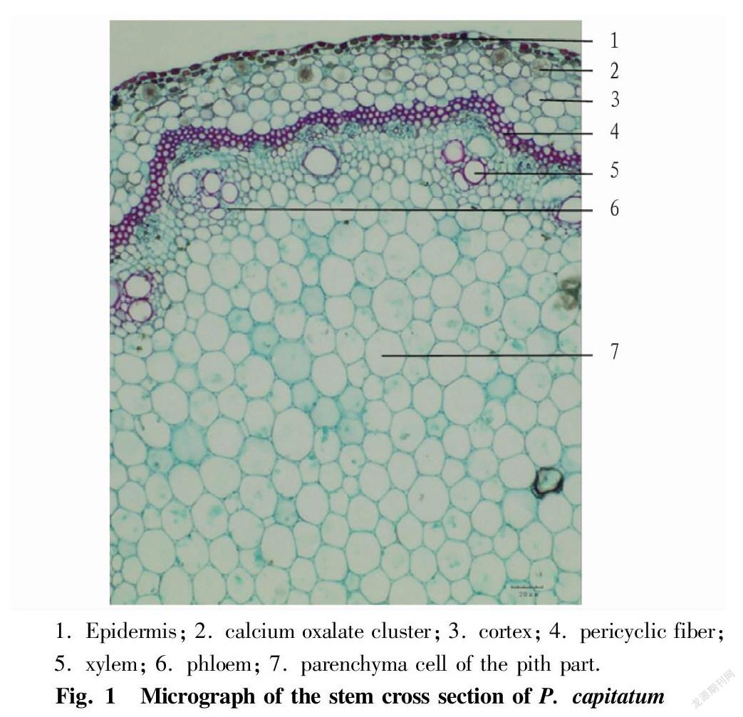
Cross section of stem: There is one row of epidermal cells, which are rectangular-like or round-like, and have keratinized or lignified outer cell walls. The cortex is composed of several layers of parenchyma cells, which are loosely arranged and of different sizes, and contain many calcium oxalate cluster crystals. The pericycle consists of several layers of pericyclic fibers, continuously arranged in a ring in the longitudinal direction, strongly lignified. There are 26-28 collateral vascular bundles, which are continuously arranged in a ring. The phloem cells are small and tightly arranged. The xylem is located inside the phloem, and the xylem vessels are scattered singly or in groups of 2-3. The pith part is wider and hollow mostly, and the parenchyma cells vary in size and are closely arranged (Fig. 1).
Powder characteristics
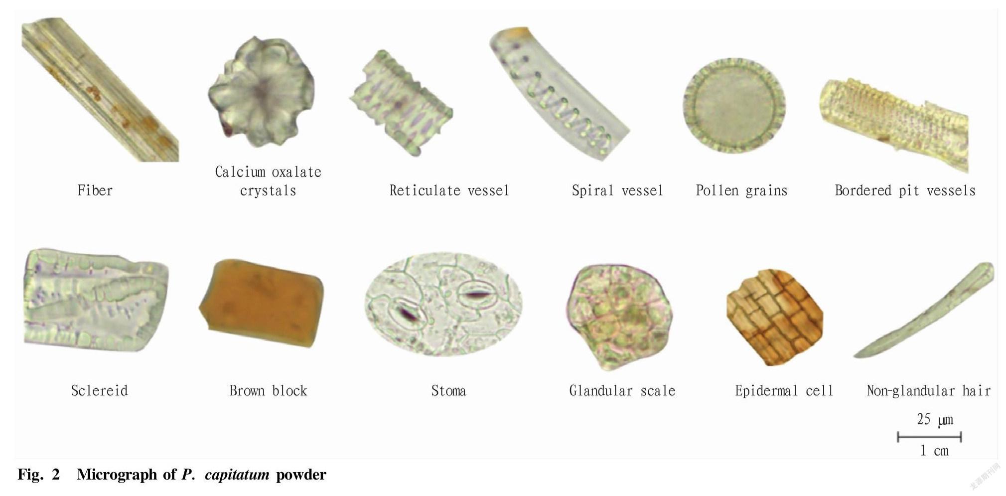
The powder is reddish-brown or gray-brown, and has a slightly bitter taste. Calcium oxalate cluster crystals are scattered, vary in size greatly, and have a diameter of 16-80 μm, and the edges and corners are slightly obtuse or long-pointed, short-pointed or the edges and corners are not clear. Cube crystals are occasionally seen. Brown blocks are more common. Fibers are mostly bundled or scattered, brownish yellow or bright yellowish green, 5-37 μm in diameter, and show obvious pit canals, which are relatively dense, and some of which have brown oil cells therein in their lumens. The bast fibers are spindle-shaped, have thick walls, non-lignified or slightly lignified, and show obvious and dense pit canals, and the lumens are linear. Wood fibers are colorless or light brown, and have inconspicuous or sparse pit canals and thin walls. There are many starch granules, single, round, oval, oblong, pear-shaped, etc. Complex granules are rare, consisting of 2 to 3 granules, some of which are very different in size, and occasionally a short slit-shaped umbilical point is seen. The vessels are mainly spiral vessels, with a diameter of 5-34 μm. Reticulate vessels and bordered pit vessels can also be seen, and the pits of bordered pit vessels are oval or round. The head of glandular scale is flat spherical, consisting of 6-8 cells; and non-glandular hairs can occasionally be seen, and are curved. Sclereids are round-like or square-like, pale yellowish green, and have thick walls. There are many pollen grains, obtuse triangular or round-like, and some of them have three germination apertures. They are 25-45 μm in diameter, and have fine thorn-like protrusions on the outer wall, the surface of which has reticulate carvings. The epidermal cells are brown, square or square-like, and have microwave-like walls. The stomata of epidermal cells are paracytic (Fig. 2).
Discussion
The stem cross section of P. capitatum is round-like, and shows pericyclic fibers forming a ring, strongly lignified, many vascular bundles, and a hollow pith part. The cross section of the leaves is V-shaped, consisting of epidermis, mesophyll tissue and leaf veins, of which the mesophyll tissue is composed of palisade tissue and spongy tissue. In bifacial leaves, the mesophyll parenchymal cells contain calcium oxalate cluster crystals, and the vascular bundles in the main vein are ectophloic. There are many
starch granules in the powder, and single granules are more common; and fibers are mostly bundled or scattered, lignified or non-lignified, and calcium oxalate cluster crystals are common. There are many pollen grains, obtuse triangular or round-like, and some of them have three germination apertures. They have fine thorn-like protrusions on the outer wall, the surface of which has reticulate carvings. The above characteristics can be used as the basis for microscopic identification of P. capitatum.
References
[1] Editorial Board of Flora of China, Chinese Academy of Sciences. Flora of China[M]. Beijing: Science Press, 1998, 25(1): 057.
Editor: Yingzhi GUANG Proofreader: Xinxiu ZHU
杂志排行
农业生物技术(英文版)的其它文章
- Expression Analysis of Heat Shock Protein 70 Gene in Rice (Oryza sativa L.)
- Changes in Physiological and Biochemical Characteristics of Floral Organ
- Research Progress on Lonicera japonica Thunb. Affected by Environmental Stress
- Research Progress on Genetic Diversity of Snap Bean
- Allelopathic Effects of Cedrus deodara Needle Extracts on Seed
- Development Status and Countermeasures of Passiflora spp. Seedling Industry in Qinzhou, Guangxi
