Characterization of Neopestalotiopsis clavispora, A New Etiological Agent of Leaf Spot Isolated from Banana
2022-02-17YanxiangQIHongZHAOHeZHANGYixianXIEFanyunZENGJunPENGQunfangYUXinZHANG
Yanxiang QI, Hong ZHAO, He ZHANG, Yixian XIE, Fanyun ZENG, Jun PENG, Qunfang YU, Xin ZHANG
1. Environment and Plant Protection Institute, Chinese Academy of Tropical Agricultural Sciences/Hainan Provincial Key Laboratory of Pests Detection and Control for Tropical Agriculture, Haikou 571101, China; 2. Center for Research and Development of Fine Chemicals, Guizhou University, Guiyang 550025, China
Abstract [Objectives] The study was to confirm the etiological agent of leaf spot on banana in Yunnan Province of China. [Methods] Fungus isolates were isolated from the diseased tissues, and cultured to observe the morphological characteristics of colony and spore. Furthermore, phylogenetic analyses and pathogenicity test were also conducted to confirm the pathogen. [Results] The fungus isolated from the diseased tissues was identified as Neopestalotiopsis clavispora. [Conclusions] N. clavispora is a new pathogen causing leaf spot of banana. This research provides the first description of N. clavispora as a causal agent on banana in China, and adds new insights related to the host range of N. clavispora.
Key words Musa acuminate; Neopestalotiopsis clavispora; Symptom; Morphology; Phylogeny; Pathogenicity
1 Introduction
Banana (MusaacuminateL.) is an important tropical fruit in China. Leaf spot disease has become the main reason limiting the high and stable yield of banana in China[1]. A new leaf spot disease was observed on banana plants at an orchard in Yunnan Province of China in October 2020. The disease incidence was about 1%. The leaf spots occurred sporadically and the percentage of leaf area covered by lesions was less than 5%. Symptoms on the leaves were initially small, irregular, reddish-brown spots that gradually expanded to fusiform-shaped lesions with a yellow halo and eventually became necrotic, dry, and cracked. This study aimed to identify the etiological agent causing leaf spot on banana based on colony and spore morphology, phylogenetic analyses and pathogenicity test.
2 Materials and methods
2.1 Fungal isolation and morphological observationTo isolate the pathogen, 30 symptomatic leaves (15 mm2) from 5 plants were surface-disinfected in 70% ethanol (10 s) and 0.8% NaClO (2 min), rinsed in sterile water 3 times, and transferred to potato dextrose agar (PDA) at 28 ℃ for 5 d. The fungal colonies that developed from the segments were subcultured on PDA. The colony and spore morphology on the media were observed.
2.2 Molecular identification and phylogenetic analysis
Representative isolates, morphologically matched the description ofNeopestalotiopsisspp., were grown on PDA at 28 ℃ for 7 d. Genomic DNA was extracted as described by Qietal.[2]. The internal transcribed spacer (ITS) region, translation elongation factor 1-α (TEF1-α) and β-tubulin (TUB2) genes of the two isolates were amplified with primers ITS1/ITS4[3], EF1-728/EF2[4-5]and T1/Bt2b[6-7], respectively. The PCR amplification was carried out in a 25 μL reaction mixture containing 0.5 μL of DNA sample, 12.5 μL of PremixTaqTM(TaKaRa Biotech, Dalian, China), 0.5 μL of each primer (20 μM), and 11 μL of nuclease-free sterile distilled water. The PCR conditions were as follows: pre-denaturating at 94 ℃ for 3 min; denaturating at 94 ℃ for 1 min, annealing for 1 min with the corresponding temperatures (55 ℃ for ITS, TEF-1α and TUB2), extension at 72 ℃ for 1 min, 30 cycles; final extension at 72 ℃ for 5 min. Amplified PCR production were gel-purified and sequenced directly by BGI Co., Ltd., Beijing, China. MEGA 7.0[8]was used to construct a maximum likelihood tree with 1 000 bootstrap replicates, based on a concatenation alignment of three gene sequences of the two isolates in this study as well as sequences of other Neopestalotiopsis species obtained from GenBank (Table 1).

Table 1 Collection details and GenBank accession numbers of isolates included in this study
2.3 Pathogenicity testThe pathogenicity of two isolates was tested on 6 banana seedlings at the 7-leaf stage. Two leaves from each potted plant were stab-inoculated by puncturing into 1 mm with a sterilized needle, and stabbed 3 points at both sides of leaf midrib. Afterwards, 10 μL of conidial suspension (1×10⁶conidia/mL) was inoculated to one side of wounded points, while sterile water was inoculated to the other side of wounded points as control. Inoculated plants were kept inside a plastic bag for 72 h and maintained in the greenhouse (12 h/12 h light/dark, 28 ℃, 90% relative humidity). The experiments were repeated twice.
3 Results and analysis
3.1 Fungal isolation and morphological observationTwenty-five colonies formed on the PDA were white with cottony aerial mycelia, and round and abundant black globular acervuli semi-immersed on PDA were observed after a week (Fig.1A, 1B). The conidia were straight or slightly curved, clavate to spindle, five-celled, with 4 septa with dimensions of (17.49-34.51) μm×(4.24-7.28) μm (avg. 23.83 μm×5.62 μm;n=50) (Fig.2C). The apical and basal cells were hyaline, whereas the three median cells were dark brown. Conidia had a single basal appendage with lengths of 2.95-17.7 μm (avg. 7.18 μm;n=50) and 2-3 apical appendages with lengths of 10.7-53.84 μm (avg. 17.36 μm;n=50) (Fig.1C). These morphological characteristics are consistent with those ofNeopestalotiopsisspp.[9].
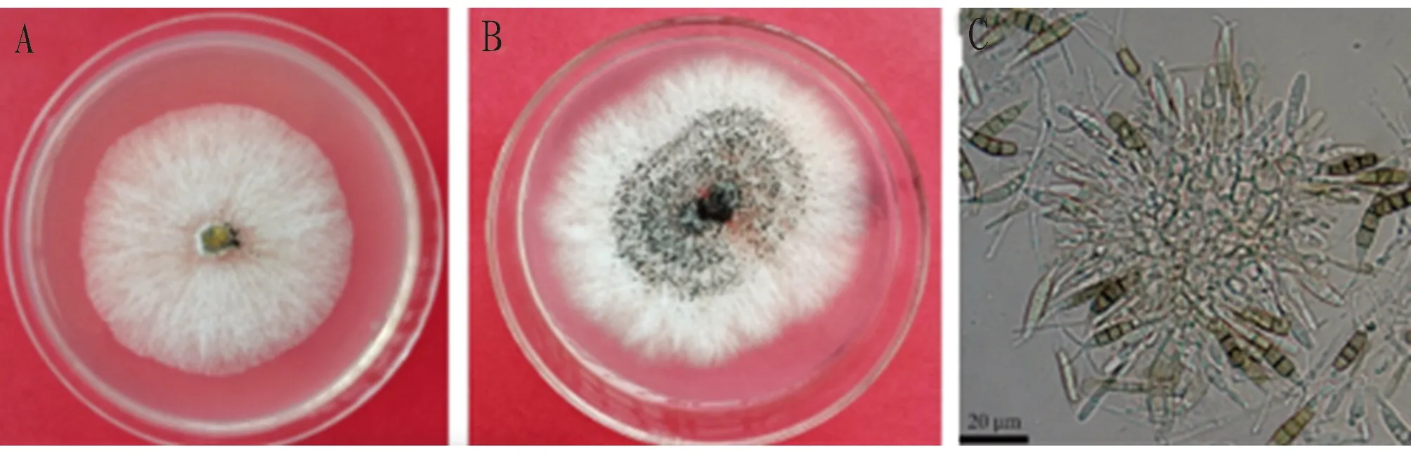
Fig.1 Colony, conidiogenous cells and conidia morphological characteristics of Neopestalotiopsis clavispora
3.2 Molecular identification and phylogenetic analysisTo confirm the species, single-spore cultures of two representative isolates CATAS-102001 and CATAS-102002 were selected for further identification. The ITS region, TEF-1α and TUB2 gene of two representative isolates (CATAS-102001 and CATAS-102002) were amplified and sequenced with the primers ITS1/ITS4, EF1-728/EF2 and T1/Bt2b. The sequences were deposited in GenBank (ITS: OM281005 and OM281006; TEF1-α: OM328820 and OM328821; TUB2: OM328818 and OM328819). A maximum likelihood phylogenetic tree was constructed using the MEGA 7.0 based on the concatenated sequences ITS region, EF1-α and TUB2 gene, and the cluster analysis placed the representative isolates CATAS-102001 and CATAS-102002 within a clade comprisingN.clavispora(Fig.2). The fungus was identified asC.pseudobrachysporaon the basis of colony and conidia morphology, and phylogenetic analyses.
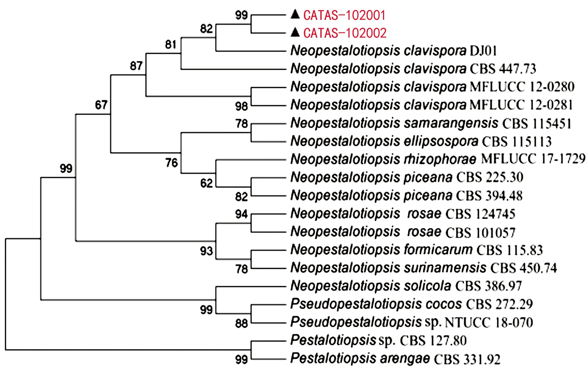
Note: Bootstrap values (1 000 replicates) greater than 50% are shown at the nodes. Isolates CATAS-102001 and CATAS-102002 in red were obtained in the present study.Fig.2 A maximum likelihood tree based on the concatenated sequences ITS, TEF-1α and TUB2 of Neopestalotiopsis, Pseudopestalotiopsis and Pestalotiopsis isolates
3.3 Pathogenicity testThe pathogenicity of this fungus on banana was confirmed by Koch’s postulates. Irregular necrotic lesions appeared on inoculated leaves at 7 d post inoculation, whereas the leaves in control were asymptomatic (Fig.3). The fungus was recovered from inoculated leaves, and its taxonomy was confirmed morphologically and molecularly, fulfilling Koch’s postulates.

Fig.3 Symptoms in pathogenicity test at 7 d post inoculation
4 Discussion
Neopestalotiopsisclavisporahas been reported on a wide range of hosts, includingPhedimusaizoonvar.latifolius[10],Photiniaserratifolia[11],Vacciniumspp.[12],Macadamiaintegrifolia[13],Ligustrumlucidum[14],Machilusthunbergii[15],Fragaria×ananassa[16],Mangiferaindica[17],Syzygiumcumini[18],Dendrobiumofficinale[19],Actinidiaargute[20],Taxusmedia[21]and ×Taxodiomeriapeizhongii[22], which was observed to be associated with leaf spot, canker and twig dieback, fruit rot, branch or leaf blight.
This study provides the first description ofN.clavisporaas a causal agent on banana in China, and adds new insights related to the host range ofN.clavispora. To our knowledge, this pathogen has not previously been associated with banana worldwide, and the identification ofN.clavisporaas the pathogen of the observed leaf spot disease on banana is critical to the prevention and control of this disease in the future.
杂志排行
植物病虫害研究(英文版)的其它文章
- Preparation and Characterization of Nano-ZnO Precursor for the Antibacterial Activity against Thielaviopsis paradoxa
- Applied Technology of Botanical Pesticides against Empoasca pirisuga Matumura
- Whether the Microemulsion is a Nano-pesticide: Exploration from the Perspective of Particle Size and Morphology